Article

Dermoscopy of Dermatofibroma

A dermatofibroma is a common benign bump in the skin.Dermatofibroma is usually harmless and typically appears on the lower legs. Dermatofibroma is easily mistaken from other skin tumors due to its appearance. This brings much difficult to diagnose. Hence, how to exactly diagnose dermatofibroma is special important to dermatology filed. What is dermatofibroma?Dermatofibroma, also known…

A dermatofibroma is a common benign bump in the skin.Dermatofibroma is usually harmless and typically appears on the lower legs. Dermatofibroma is easily mistaken from other skin tumors due to its appearance. This brings much difficult to diagnose. Hence, how to exactly diagnose dermatofibroma is special important to dermatology filed.

What is dermatofibroma?
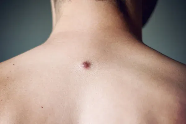
Dermatofibroma, also known as benign fibrous histiocytoma, is a type of benign growth of skin. It usually occur on the legs of adults, and it may also occur on arms, chest, elbow and upper back. Dermatofibroma can happen on anywhere on the body. Even though dermatofibroma is harmless and painless, but sometimes it’s itchy or tender. The exactly causes of dermatofibroma is not identified, but dermatofibroma is commonly caused by slight skin trauma, such as insect bites or scratches. Dermatofibroma usually appears as a small, hard bump on the skin that varies in color from pink to brown or black.

dermatofibroma picture

Why is dermoscopy needed for the diagnosis of dermatofibroma?
Dermatofibroma is a harmless and painless skin lesion, while it may erupt over a few months when people are in low immune system or have autoimmune diseases.Hence it is crucial to adopt dermoscopy to help for diagnosis. In our daily life, paying more attention to dermatofibroma is very necessary.

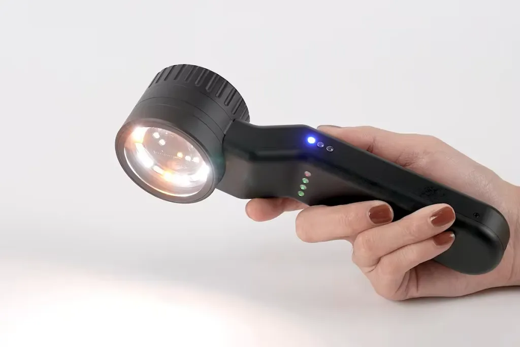
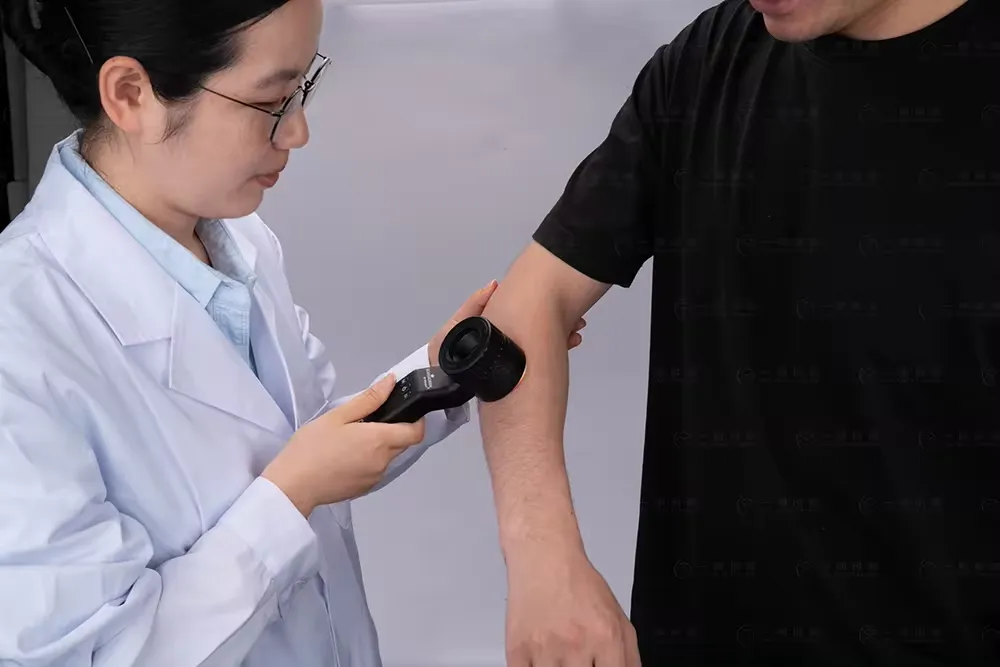
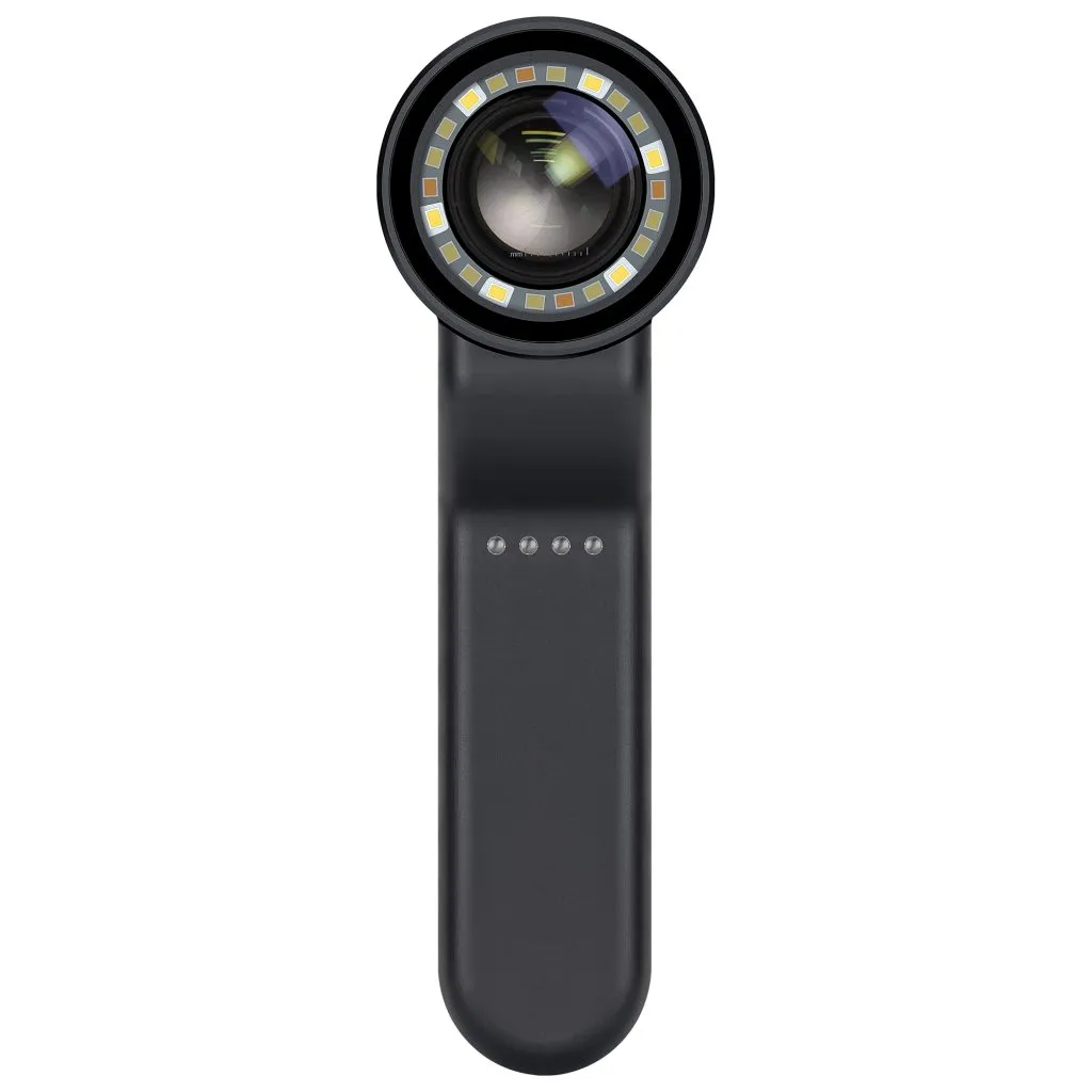
Dermatoscope is a very useful aiding device for skin doctors to detect and diagnose skin diseases, such as dermatofibroma. Dermatoscope combines a powerful optical system and big magnification to greatly increase the visual ranges and depth of the skin. It allows skin doctors inspect more details of structures, patterns and changes of skin lesion that is invisible for naked eyes. The using of dermatoscopy can reduce the unnecessary worry and surgery or biopsy.

What are dematoscopic features of dermatofibroma?
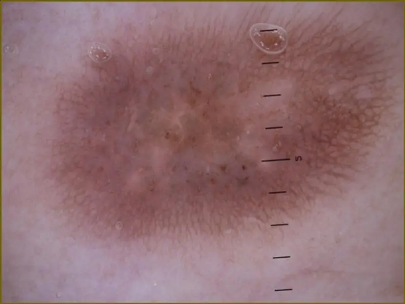
By using of dermoscopy, dermatofibroma is easily to diagnose due to brightened visual sights. Even signs in individuals are in vary, but the main typical features included like below:
Central white area: The central white area appears pale and amorphous, resembling a scar or resembling a plaque. The central white area occasionally has bright white linear stripes.
False network: The faint pigment network surrounding the central white region may sometimes resemble a network or “ring” spherical structure. The “ring” spherical structure is surrounded by white lines.
White lines and brown holes: White lines and brown holes may appear in the central white area (negative network).
Delicate pigment Network: Delicate pigment network with light to medium brown pigmentation. This network is usually located on the periphery of the lesion and gradually disappears into the surrounding skin.
Vascular structures: The most common vascular structures are punctate vessels, comma vessels, erythema, and hairpin vessels.

dermatofibroma dermoscopy

Treatment methods of dermatofibroma
Dermatofibromas are small, harmless growths that appear on the skin. Dermatofibromas are benign lesion of skin. Most of dermatofibromas are painless, and they do not cause obvious symptoms. Hence dermatofibromas generally do not require treatment. However, if need to remove dermatofibroma considering of aesthetic factors, the following treatment methods can be taken:
Surgical removal: If the dermatofibromas cause discomfort or affect aesthetics looks, the lesion part of the skin can be surgically removed.
Steroid injections: Local corticosteroid injections may be used to reduce pain or reduce the extent of the lesion.
Cryotherapy: Liquid nitrogen or other refrigerants can be used to freeze the growth of dermatofibroma, which helps to flatten them.
Laser treatment: Laser surgery can also be used to destroy the lesion part of dermatofibroma and encourage the growth of new growth.
Conservative treatment: Keep the lesion parts clean, and keep away from friction or irritation.

Dermatofibroma Dermoscopy in public self-examination
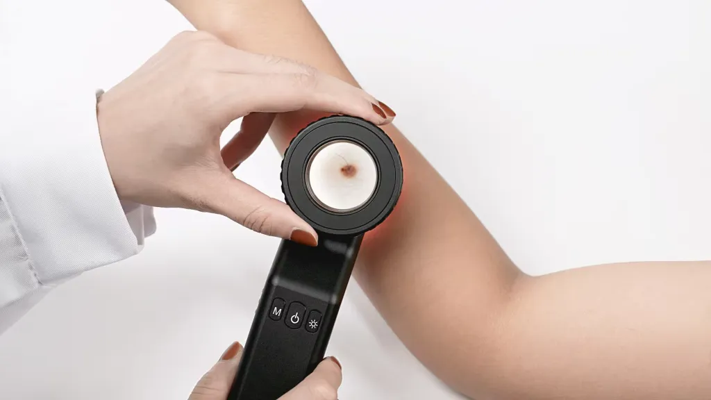
Dermoscopy is a reliable and useful aiding technique usually used by skin doctors at hospital or clinical for skin inspection. Dermoscopy is commenly designed into a handheld tool conveniently to hold by people. Except for that, skin doctors can take high definition pictures by dermoscopy combining with smart phone or tablet or camera for analysis and comparison. In professional hands, it is easy to detect dermatofibroma. People also can observe their dermatofibroma by themselves. When people use dermoscopy in self-examination, there are some factors in needed, such as:
Firstly, read and understand the function and uses of dermoscopy, then correctly operate dermoscopy.
Secondly, when use dermoscopy, keeping observations is very important.
Thirdly, any suspicious lesions are found by dermoscopy, it is necessary to have further checked by professional doctors in time and promptly.
Fourthly, after dermatofibroma detected or diagnosed by professional doctors, people should cooperate with doctors for suggestion and treatment in case it will relapse again.

skin examination by dermatoscope

Dermoscopy in the diagnosis of dermatofibroma
Dermatoscopy plays a very important role in the detection and diagnosis of skin lesions, the same as dermatofibroma. It is better to observe dermatofibroma regularly and timely by dermoscopy. There are certain advantages to use dermoscopy in the diagnosis of dermatofibroma include:
Observe conveniently: It is convenient to observe the lump if any changes at any place any time, even people can use dermoscopy for self-examination at home.
Supply More details:It is very clear and precise to find if the lesions worsen or become malignant through details of patterns, structures and vessels under the dermatoscopy.
Monitoring the treatment: Dermoscopy, as a monitor, can navigate the skin doctor to cure dermatofibroma.
Give feedback:Dermoscopy can give feedback of treatment on dermatofibroma in time. So that skin doctor will make an adjustment or a further step on it.

dermatoscope IBOOLO DE-4100

Other notes about dermatofibroma
Even a dermatofibroma is harmless and no need to cure. But when larger numbers of dermatofibromas are present, it may indicate an underlying condition will present, such as lupus or a weakened immune system, such as from leukemia or HIV. So accurate detection and diagnosis of dermatofbroma is really significant.

It is very helpful to use dermoscopy in the detection and diagnosis of dermatofibroma.Under the help of dermoscopy, it becomes more easily and conviently in these aspects, like observation and analysis, treatment, monitoring and feedback of dermatofibroma. In addition, skin doctors and patients become more confident and safety by using of the dermoscopy.
Hence it is necessary to use dermoscopy in right ways. Any suspicious findings, do not hesitate to ask skin doctors’ help. In daily life, people should keep regular skin examination in case of skin lesions and pay more attention to self-examination of dermatofibroma regularly.

Share this article

0

No products in the cart.

Have questions on gear or your order?

Our Gear Guides are here to help! Get personal advice from pro creatives

Name
Subject
Email address
How can we help?

Instant Answers

How can dermoscopy images be captured?

Dermoscopy images can be captured and stored in different ways, such as: • Using a smartphone or tablet with dermoscopic adapter, which consisted in the package.• Using a digital camera

Dermoscopy images can be captured and stored in different ways, such as:

• Using a smartphone or tablet with dermoscopic adapter, which consisted in the package.
• Using a digital camera with dermoscopic adapter, there’s 49mm screw size camera adapter available to order now.

Compatible phone/tablet models:
All iPhone models, 95% Android phones, 90% tablet. For phone/tablet size in 5.25-14mm

Compatible camera models:
All camera with built 49mm filter screw, such as Canon EOS 70D, 80D, 90D; Canon EOS R7, R10, R50, R100; Canon M100, M200, M50, Mark II; Canon G7X Mark III, Sony ZV-1

How can I connect my phone to my dermatoscope?

There’s universal phone adapter for all our dermoscopes. Please check the installation procedure bellow or watch operation guide. Smartphone Connector (1) Place phone adapter screw in the center of smartphone’s

There’s universal phone adapter for all our dermoscopes. Please check the installation procedure bellow or watch operation guide.

Smartphone Connector

(1) Place phone adapter screw in the center of smartphone’s main camera.
(2) Screw magnet attachment on phone adapter.
(3) Put dermoscope’s back ring and magnet attachment together

Take The Best Images

You need to adjust the focus ring after the dermoscpe connected on smartphone to get the best images.

How can I clean my dermoscopy after usage?

Cleaning your dermoscopy after usage is important to prevent cross-contamination and infection. The cleaning method may vary depending on the type and model of your dermoscopy, so you should always

Cleaning your dermoscopy after usage is important to prevent cross-contamination and infection. The cleaning method may vary depending on the type and model of your dermoscopy, so you should always follow the manufacturer’s instructions. However, some general steps are:

• Turn off and disconnect your dermoscopy from any power source or device.

• Wipe off any visible dirt or debris from the dermoscopy with a soft cloth or tissue.

• Disinfect the dermoscopy with an alcohol-based wipe or spray, or a disinfectant solution recommended by the manufacturer. Make sure to cover all surfaces, especially the lens and contact plate.

• Let the dermoscopy air dry completely before storing it in a clean and dry place.

• Do not use abrasive or corrosive cleaners, solvents, or detergents that may damage the dermoscopy.

• Do not immerse the dermoscopy in water or any liquid, unless it is waterproof and designed for immersion.

You should clean your dermoscopy after each use, or at least once a day if you use it frequently. You should also check your dermoscopy regularly for any signs of damage or malfunction, and contact the manufacturer or service provider if needed.

Polarized VS Non-polarized Dermoscopy

A dermoscopy is a device that allows the examination of skin lesions with magnificationand illumination. By revealing subsurface structures and patterns that are not visible tothe naked eye. It can

A dermoscopy is a device that allows the examination of skin lesions with magnificationand illumination. By revealing subsurface structures and patterns that are not visible tothe naked eye. It can improve the diagnose accuracy of skin lesions, such as melanoma,basal cell carcinoma, seborrheic keratosis, etc.

There are two main types of dermoscopy: Non polarized and polarized dermoscopy.We’ve fitted most of our dermoscopys with polarized and non-polarized light. They canbe used in multiple skin structures.

Non-polarized contact Mode

In non-polarized mode, the instrument can provide information about the superficialskin structures, such as milia-like cysts, comedo-like openings, and pigment in theepidemis.

The dermoscopy requires applying a liquid such as mineral oil or alcohol to the skin andplacing the lens in contact with the skin. This reduces surface reflection and enhancesthe view of subsurface structures.

Image with non-polarized light (DE-3100)

Polarized contact Mode

In polarized mode, the instrument allows for visualization for deeper skin structures,such as blood vessels, collagen, and pigment in the dermis.

The dermoscopy does not need to be in contact with the skin or use any liquid. Theirpolarized light can help to eliminate surface reflection and allow visualization ofvascular structures.

Image with polarized light (DE-3100)

Polarized non-contact Mode

The dermoscopy can also use polarized light to examine the skin without direct contact.

In polarized non-contact mode, the instrument allows for examination infected areasand lesions that are painful for the patient, or the difficult to contact pigmented lesions,such as nails and narrow areas.

The contact plate should be removed in this mode, and it does not require applying aliquid to the skin. As it doesn’t require pressure or fluid application on the skin, it canalso avoid cross-contamination and infection risk.

Image in polarized non-contact mode (DE-3100)

How effectiveness is dermoscopy

Compared with visual inspection, the dermoscopy can be used to capture and store skin lesion photos, which play an important role in early skin cancer examination. The dermoscopy allows the

Compared with visual inspection, the dermoscopy can be used to capture and store skin lesion photos, which play an important role in early skin cancer examination.

The dermoscopy allows the examination of skin lesions with magnification and illumination. This can be greatly avoiding the factors that cause interference to visual detection. Such as lighting, skin color, hair and cosmetics.

Several studies have demonstrated that dermoscopy is useful in the identification of melanoma, when used by a trained professional.

It may improve the accuracy of clinical diagnosis by up to 35%
It may reduce the number of harmless lesions that are removed
In primary care, it may increase the referral of more worrisome lesions and reduce the referral of more trivial ones

A 2018 Cochrane meta-analysis published the accuracy of dermoscopy in the detection.

Table 1. Accuracy of dermoscopy in the detection of melanoma in adults
Detection Method Sensitivity, % Specificity, % Positive Likelihood Ratio NegativeLikelihood Ratio
Visual inspection alone (in person) 76 75 3.04 0.32
Dermoscopy with visual inspection (in person) 92 95 18 0.08
Image-based visual inspection alone (not in person) 47 42 0.81 1.3
Dermoscopy with image-based visual inspection (not in person) 81 82 4.5 0.23
ROC—receiver operating characteristic. *Estimated sensitivity calculated on the summary ROC curve at a fixed specificity of 80%.

As we can see, the dermoscope can improve the accuracy of diagnosis of skin lesions, especially melanoma.

Table 1. Accuracy of dermoscopy in the detection of melanoma in adults
Detection Method Sensitivity, % Specificity, % Positive Likelihood Ratio NegativeLikelihood Ratio
Visual inspection alone (in person) 79 77 3.4 0.27
Dermoscopy with visual inspection (in person) 93 99 93 0.07
Image-based visual inspection alone (not in person) 85 87 6.5 0.17
Dermoscopy with image-based visual inspection (not in person) 93 96 23 0.07
ROC—receiver operating characteristic. *Estimated sensitivity calculated on the summary ROC curve at a fixed specificity of 80%.

Characteristics of the dermatoscopic structure of the skin lesions include:

• Symmetry or asymmetry
• Homogeny/uniformity (sameness) or heterogeny (structural differences across the lesion)
• Distribution of pigment: brown lines, dots, clods and structureless areas
• Skin surface keratin: small white cysts, crypts, fissures
• Vascular morphology and pattern: regular or irregular
• Border of the lesion: fading, sharply cut off or radial streaks
• Presence of ulceration

There are specific dermoscopic patterns that aid in the diagnosis of the following pigmented skin lesions:

• Melanoma
• Moles (benign melanocytic naevus)
• Freckles (lentigos)
• Atypical naevi
• Blue naevi
• Seborrhoeic keratosis
• Pigmented basal cell carcinoma
• Haemangioma

Hot Search Terms