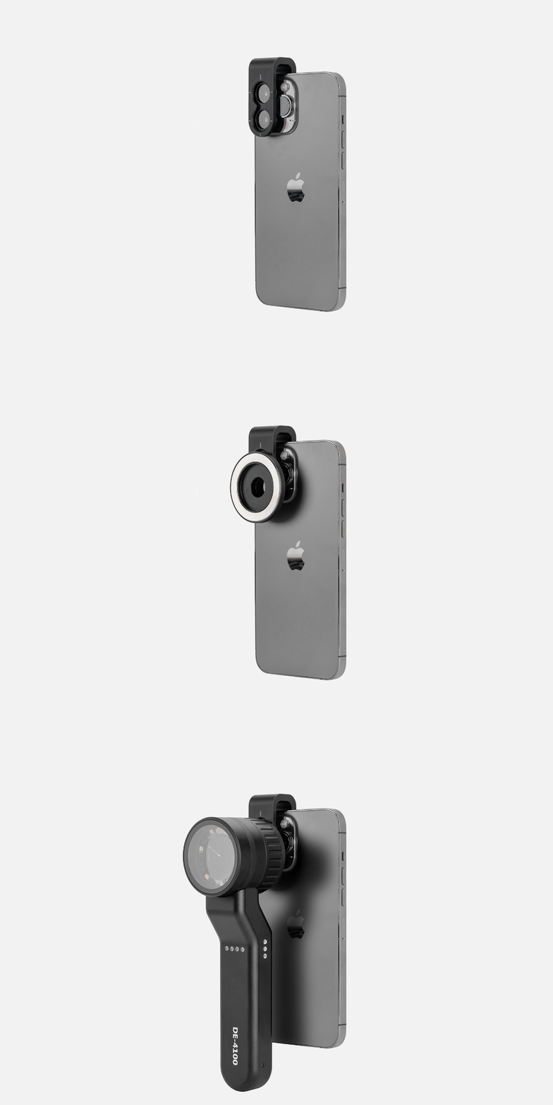
portable handheld dermatoscope precise skin lmaging - IBOOLO

IBOOLO DE-4100 handheld dermatoscope enables detailed magnified skin analysis anywhere with its lightweight, ergonomic design. customizable polarized lighting captures clear subsurface skin views.

The Convenience of Handheld and Smartphone Dermatoscopes

Regular skin self-examinations are crucial for detecting suspicious moles or lesions early, when skin cancers are most treatable. However, thoroughly inspecting hard-to-see areas like the scalp, back, and between toes can be challenging. This is where compact handheld dermatoscopes and smartphone dermatoscopes provide a convenient solution for at-home skin screening.

portable Dermoscopy power

Traditional dermatoscopes tend to be bulky desktop devices restricted to clinical settings. In contrast, today's handheld dermatoscopes enable portable dermoscopy anywhere. powered by batteries, these mobile phone dermatoscopes and compact handheld dermatoscopes free users from requiring an electrical outlet. Their lightweight, pocket-sized design allows easy whole-body examination in good lighting with a handheld dermatoscope.

Handheld dermatoscopes integrate quality optics like 10-20X magnification lenses combined with polarized LED lighting. This illuminates subsurface skin structures while minimizing surface glare for a clear view of pigmentation patterns and vasculature that can signify evolving moles when using a handheld dermatoscope. Many handheld dermatoscopes also provide different lighting modes to enhance contrast.

Smartphone Dermatoscope Versatility

Among the most versatile options are smartphone dermatoscopes designed as mobile phone attachments. Once coupled to your smartphone's camera, these smartphone dermatoscopes allow capturing dermoscopic photos and videos alongside regular photos using your phone's existing camera app. Dual lens smartphone dermatoscopes even enable simultaneous dermoscopy and regular image previewing.

Some smartphone dermatoscope accessories simply clip onto your mobile phone's camera lens. More advanced models integrate illumination with polarized lightings and high-powered 20-40X magnification optics. This enables clinical-quality skin examination and photo documentation rivaling professional dermatologist equipment with a smartphone dermatoscope.

Dermatoscope phone Apps

Accompanying mobile apps for smartphone dermatoscopes provide additional capabilities like annotation tools for labeling areas of concern on saved dermoscopic images. They often include skin mapping functions to log and track mole changes over time. Many apps also offer teleconsultation features to securely share images with dermatologists for remote evaluation of your handheld dermatoscope findings.

With their compact convenience and benefits like photo documentation, skin mapping, and teleconsultation enablement, smartphone dermatoscopes and handheld dermatoscopes empower meticulous self-screening between clinic visits. Having this clinical visualization power in your pocket with a handheld dermatoscope can lead to earlier detection when skin cancers are most treatable.

Consistent At-Home Screening

Easily incorporating mobile phone dermatoscope or pocket-sized handheld dermatoscope usage into a monthly self-exam routine allows frequently monitoring areas you can't easily see with the naked eye. Capturing baseline dermatoscopic photos with your handheld dermatoscope provides a record to compare against for identifying any subtle changes in existing spots and discovering new growths over time.

When used diligently for periodic at-home examinations, smartphone dermatoscopes and handheld dermatoscopes give individuals an enhanced tool for skin cancer prevention through earlier detection. Their portability with a handheld dermatoscope empowers more meticulous self-screening not possible with stationary dermoscopy equipment alone.

For many, a smartphone dermatoscope provides the ideal balance of powerful clinical optics and extreme convenience in a mobile solution. But a quality handheld dermatoscope achieves similar benefits in a dedicated, phone-free device. As telecommunications technology advances, integration of AI assistance and improved teleconsultation experiences will further enhance the screening capabilities of smartphone and handheld dermatoscopes. Untethered, highly visual dermoscopy with a portable handheld dermatoscope is poised to become a staple of proactive health monitoring.

Recommended reading

Download DE-215 User Manual – IBOOLO

is a High quality Download DE-215 User Manual supplier & manufacturer, information for Download DE-215 User Manual: DE-215 user manualDownload...

Dermatoscope – IBOOLO

Shenzhen Iboolo Optics Co.Ltd founded in 2012,is located in the beautiful city of Shenzhen, Guangdong. Our main products including Microscope, Dermatoscope, Macro lens and Woods Lamp, etc.

DE-215 Woods Lamp – IBOOLO

is a Custom DE-215 Woods Lamp supplier & factory, information for DE-215 Woods Lamp: Dermatology UV 365nm DE-215 Woods Lamp...

Professional Handheld 10X Magnify DE-4100 Dermatoscope

Info
Features & Compatibility
  • 10 x Magnification
  • 32mm wide field of view (Aperture)
  • Polarized, non-polarized, and amber lightillumination
  • Detachable protective glass
  • Automatic shutdown
  • Included adapter fits all phone
  • Bright LED illumination lights with 3 levels of brightness control
  • All-metal housing
Specs

Material

Optical & Aluminum

Optical Design

All glass, 4 elements, 3 groups

Lens Diameter

32mm

Magnification

10X

Distortion

8%

Resolution

300 LP/MM (Axis) 250 LP/MM (Edges)

Polarization

Cross Polarized

LED Type

SMD LED beads

Battery Capacity

1000mAh Lithium ion

Charge

USB-C

Close Focus

30mm

Dimensions

Φ55mm*H50mm*L195mm

Weight

325g

$799.00

$799.00
  • In Stock

  • Arrive in 5-7 days

  • Free Shipping Worldwide

  • 2 Years Warranty

How to Use

Check out our step-by step quick start guide of the device.

Play Video

Why Choose Our Dermatoscope

Exceptional Lens Design

Designed by optics engineers with over 20 years of experience making high-end camera lenses. Carefully crafted coatings, and hand-finished glass from our precision factory.

Made From Aerospace Grade Metal

It’s made of aerospace-grade metal instead plastic body design. The curved shape of the handle fit comfortably in your hand and provide a smooth and intuitive user experience.

Affordable Price

Our goal was to bring affordable and top quality into one product, while making the device durable for long time usage.

Sharp & Precise

Made from premium multi- coated and multi-element optics, the imaging performance is the best in the market.

LED Illumination

3 levels of brightness control, cross polarized, non polarized and amber light gives a lot choice to diagnose under different situations.

See Deeper

The polarized light can help to eliminate surface reflection and allow visualization of vascular structures. The instrument allows for visualization for deeper skin structures, such as blood vessels, collagen, and pigment in the dermis.

Easy to Install & Use

With magnet snap on and off, the dermatoscope can be installed on any smartphone in 5 seconds.

Under Naked Eye

Our dermoscopy is designed to support you in this endeavor - enhance a doctor’s view of the skin as much as possible.

Dermatoscopes are handy tools for spotting skin cancer, but they also help check out common skin issues like atopic eczema, psoriasis, rosacea, Grover’s disease or lichen ruber planus in daily practice.

A Picture Speaks a Thousand Words

Toggle to hold the "after" image

In The Box

1x DE-4100 Dermatoscope
1x Microfiber Cloth
1x Carrying Case
1x Charging Cable

Shot on Spot

Learn More

Comparison of Dermatoscopoi to Help You Choose the Right One

What is Dermoscopy (dermatoscopio)? Dermoscopy(dermatoscopio), is a valuable helpful technique usually used by skin doctors to inspect a variety of skin situations.Dermoscopy is also known as dermatoscopy, incident light microscopy,…

Dermoscopy Applications in the Diagnosis of Psoriasis

Dermoscopy of Psoriasis What is Psoriasis? Psoriasis is a long term skin disease which can grow anywhere but most commonly appear on the elbows, knees, scalp and trunk. Psoriasis is…

Dermascope vs Dermatoscope: Exploring the Diagnostic Power beyond the Magnifying Glass

Dermascope vs Dermatoscope What is a dermatoscope?Dermatoscope, also know as dermoscope or epiluminescence microscope. It is a useful and dependable hand-held microscope device for skin examination, such as melanoma, alopecia…

REVIEWS

1 review for DE-4100 Dermatoscope

  1. Marcia CostaMexico

    amei meu dermatoscopio.perfeita qualidade !!!! o vendedor super atencioso! rapidez na entrega do produto, chegou super bem embalado . super recomendo!!!!!

Only logged in customers who have purchased this product may leave a review.

0

No products in the cart.

Have questions on gear or your order?

Our Gear Guides are here to help! Get personal advice from pro creatives

Name
Subject
Email address
How can we help?

Instant Answers

How can dermoscopy images be captured?

Dermoscopy images can be captured and stored in different ways, such as: • Using a smartphone or tablet with dermoscopic adapter, which consisted in the package.• Using a digital camera

Dermoscopy images can be captured and stored in different ways, such as:

• Using a smartphone or tablet with dermoscopic adapter, which consisted in the package.
• Using a digital camera with dermoscopic adapter, there’s 49mm screw size camera adapter available to order now.

Compatible phone/tablet models:
All iPhone models, 95% Android phones, 90% tablet. For phone/tablet size in 5.25-14mm

Compatible camera models:
All camera with built 49mm filter screw, such as Canon EOS 70D, 80D, 90D; Canon EOS R7, R10, R50, R100; Canon M100, M200, M50, Mark II; Canon G7X Mark III, Sony ZV-1

How can I connect my phone to my dermatoscope?

There’s universal phone adapter for all our dermoscopes. Please check the installation procedure bellow or watch operation guide. Smartphone Connector (1) Place phone adapter screw in the center of smartphone’s

There’s universal phone adapter for all our dermoscopes. Please check the installation procedure bellow or watch operation guide.

Smartphone Connector

(1) Place phone adapter screw in the center of smartphone’s main camera.
(2) Screw magnet attachment on phone adapter.
(3) Put dermoscope’s back ring and magnet attachment together

Take The Best Images

You need to adjust the focus ring after the dermoscpe connected on smartphone to get the best images.

How can I clean my dermoscopy after usage?

Cleaning your dermoscopy after usage is important to prevent cross-contamination and infection. The cleaning method may vary depending on the type and model of your dermoscopy, so you should always

Cleaning your dermoscopy after usage is important to prevent cross-contamination and infection. The cleaning method may vary depending on the type and model of your dermoscopy, so you should always follow the manufacturer’s instructions. However, some general steps are:

• Turn off and disconnect your dermoscopy from any power source or device.

• Wipe off any visible dirt or debris from the dermoscopy with a soft cloth or tissue.

• Disinfect the dermoscopy with an alcohol-based wipe or spray, or a disinfectant solution recommended by the manufacturer. Make sure to cover all surfaces, especially the lens and contact plate.

• Let the dermoscopy air dry completely before storing it in a clean and dry place.

• Do not use abrasive or corrosive cleaners, solvents, or detergents that may damage the dermoscopy.

• Do not immerse the dermoscopy in water or any liquid, unless it is waterproof and designed for immersion.

You should clean your dermoscopy after each use, or at least once a day if you use it frequently. You should also check your dermoscopy regularly for any signs of damage or malfunction, and contact the manufacturer or service provider if needed.

Polarized VS Non-polarized Dermoscopy

A dermoscopy is a device that allows the examination of skin lesions with magnificationand illumination. By revealing subsurface structures and patterns that are not visible tothe naked eye. It can

A dermoscopy is a device that allows the examination of skin lesions with magnificationand illumination. By revealing subsurface structures and patterns that are not visible tothe naked eye. It can improve the diagnose accuracy of skin lesions, such as melanoma,basal cell carcinoma, seborrheic keratosis, etc.

There are two main types of dermoscopy: Non polarized and polarized dermoscopy.We’ve fitted most of our dermoscopys with polarized and non-polarized light. They canbe used in multiple skin structures.

Non-polarized contact Mode

In non-polarized mode, the instrument can provide information about the superficialskin structures, such as milia-like cysts, comedo-like openings, and pigment in theepidemis.

The dermoscopy requires applying a liquid such as mineral oil or alcohol to the skin andplacing the lens in contact with the skin. This reduces surface reflection and enhancesthe view of subsurface structures.

Image with non-polarized light (DE-3100)

Polarized contact Mode

In polarized mode, the instrument allows for visualization for deeper skin structures,such as blood vessels, collagen, and pigment in the dermis.

The dermoscopy does not need to be in contact with the skin or use any liquid. Theirpolarized light can help to eliminate surface reflection and allow visualization ofvascular structures.

Image with polarized light (DE-3100)

Polarized non-contact Mode

The dermoscopy can also use polarized light to examine the skin without direct contact.

In polarized non-contact mode, the instrument allows for examination infected areasand lesions that are painful for the patient, or the difficult to contact pigmented lesions,such as nails and narrow areas.

The contact plate should be removed in this mode, and it does not require applying aliquid to the skin. As it doesn’t require pressure or fluid application on the skin, it canalso avoid cross-contamination and infection risk.

Image in polarized non-contact mode (DE-3100)

How effectiveness is dermoscopy

Compared with visual inspection, the dermoscopy can be used to capture and store skin lesion photos, which play an important role in early skin cancer examination. The dermoscopy allows the

Compared with visual inspection, the dermoscopy can be used to capture and store skin lesion photos, which play an important role in early skin cancer examination.

The dermoscopy allows the examination of skin lesions with magnification and illumination. This can be greatly avoiding the factors that cause interference to visual detection. Such as lighting, skin color, hair and cosmetics.

Several studies have demonstrated that dermoscopy is useful in the identification of melanoma, when used by a trained professional.

It may improve the accuracy of clinical diagnosis by up to 35%
It may reduce the number of harmless lesions that are removed
In primary care, it may increase the referral of more worrisome lesions and reduce the referral of more trivial ones

A 2018 Cochrane meta-analysis published the accuracy of dermoscopy in the detection.

Table 1. Accuracy of dermoscopy in the detection of melanoma in adults
Detection Method Sensitivity, % Specificity, % Positive Likelihood Ratio NegativeLikelihood Ratio
Visual inspection alone (in person) 76 75 3.04 0.32
Dermoscopy with visual inspection (in person) 92 95 18 0.08
Image-based visual inspection alone (not in person) 47 42 0.81 1.3
Dermoscopy with image-based visual inspection (not in person) 81 82 4.5 0.23
ROC—receiver operating characteristic. *Estimated sensitivity calculated on the summary ROC curve at a fixed specificity of 80%.

As we can see, the dermoscope can improve the accuracy of diagnosis of skin lesions, especially melanoma.

Table 1. Accuracy of dermoscopy in the detection of melanoma in adults
Detection Method Sensitivity, % Specificity, % Positive Likelihood Ratio NegativeLikelihood Ratio
Visual inspection alone (in person) 79 77 3.4 0.27
Dermoscopy with visual inspection (in person) 93 99 93 0.07
Image-based visual inspection alone (not in person) 85 87 6.5 0.17
Dermoscopy with image-based visual inspection (not in person) 93 96 23 0.07
ROC—receiver operating characteristic. *Estimated sensitivity calculated on the summary ROC curve at a fixed specificity of 80%.

Characteristics of the dermatoscopic structure of the skin lesions include:

• Symmetry or asymmetry
• Homogeny/uniformity (sameness) or heterogeny (structural differences across the lesion)
• Distribution of pigment: brown lines, dots, clods and structureless areas
• Skin surface keratin: small white cysts, crypts, fissures
• Vascular morphology and pattern: regular or irregular
• Border of the lesion: fading, sharply cut off or radial streaks
• Presence of ulceration

There are specific dermoscopic patterns that aid in the diagnosis of the following pigmented skin lesions:

• Melanoma
• Moles (benign melanocytic naevus)
• Freckles (lentigos)
• Atypical naevi
• Blue naevi
• Seborrhoeic keratosis
• Pigmented basal cell carcinoma
• Haemangioma

Hot Search Terms