


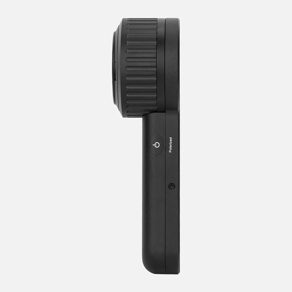
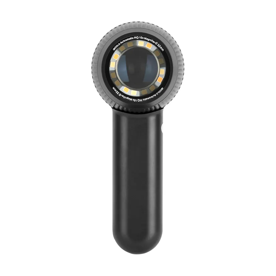
Polarized & 365nm UV LED DE-3100 PRO Dermatoscope
Polarized & 365nm UV LED DE-3100 PRO Dermatoscope


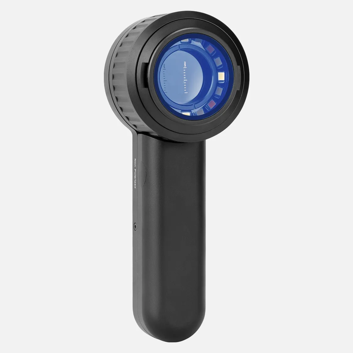


- 10 x Magnification
- 32mm wide field of view (Aperture)
- Polarized, non-polarized, and 365nm UV light illumination
- Detachable protective glass
- Automatic shutdown
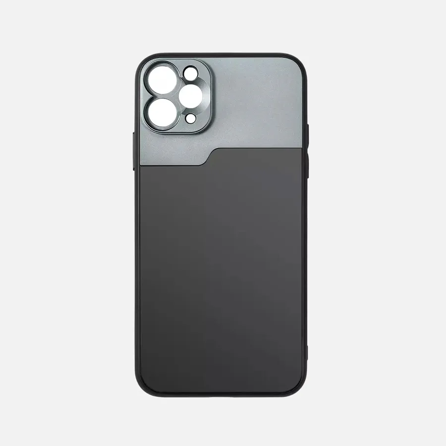
- Included adapter fits all phone
- Bright LED illumination lights
- All-metal housing
|
Material |
Optical & Aluminum |
|
Optical Design |
All glass, 4 elements, 3 groups |
|
Lens Diameter |
32mm |
|
Magnification |
10X |
|
Distortion |
8% |
|
Resolution |
300 LP/MM (Axis) 250 LP/MM (Edges) |
|
Polarization |
Cross Polarized |
|
Ultraviolet Warelength |
365nm UV |
|
Battery Capacity |
1000mAh Lithium ion |
|
Charge |
USB-C |
|
Close Focus |
30mm |
|
Dimensions |
Φ55mm*H50mm*L195mm |
|
Weight |
325g |
$599.00
-
In Stock
-
Arrive in 5-7 days
-
Free Shipping Worldwide $59+
-
2 Years Warranty


What Makes it Unique
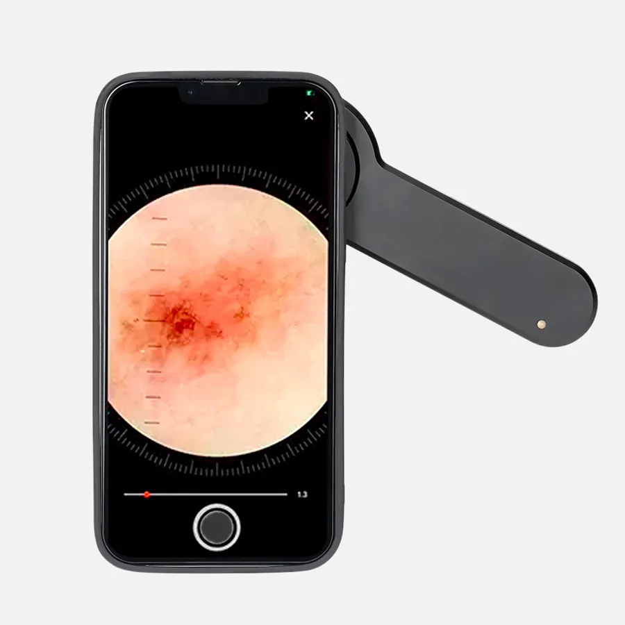
It’s a handheld dermatoscope, but it contains a universal phone adapter meaning convenient to connect with any smartphone or tablet to capture images. Build with 4K resolution optics, 10X magnification and 30mm wide field of view — the DE-3100 is perhaps the most super value dermatoscope we’ve ever seen.
Sharp & Precise
Made from premium multi-coated and multi-element optics, the imaging performance is the best in the market.
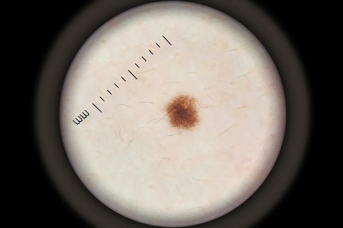
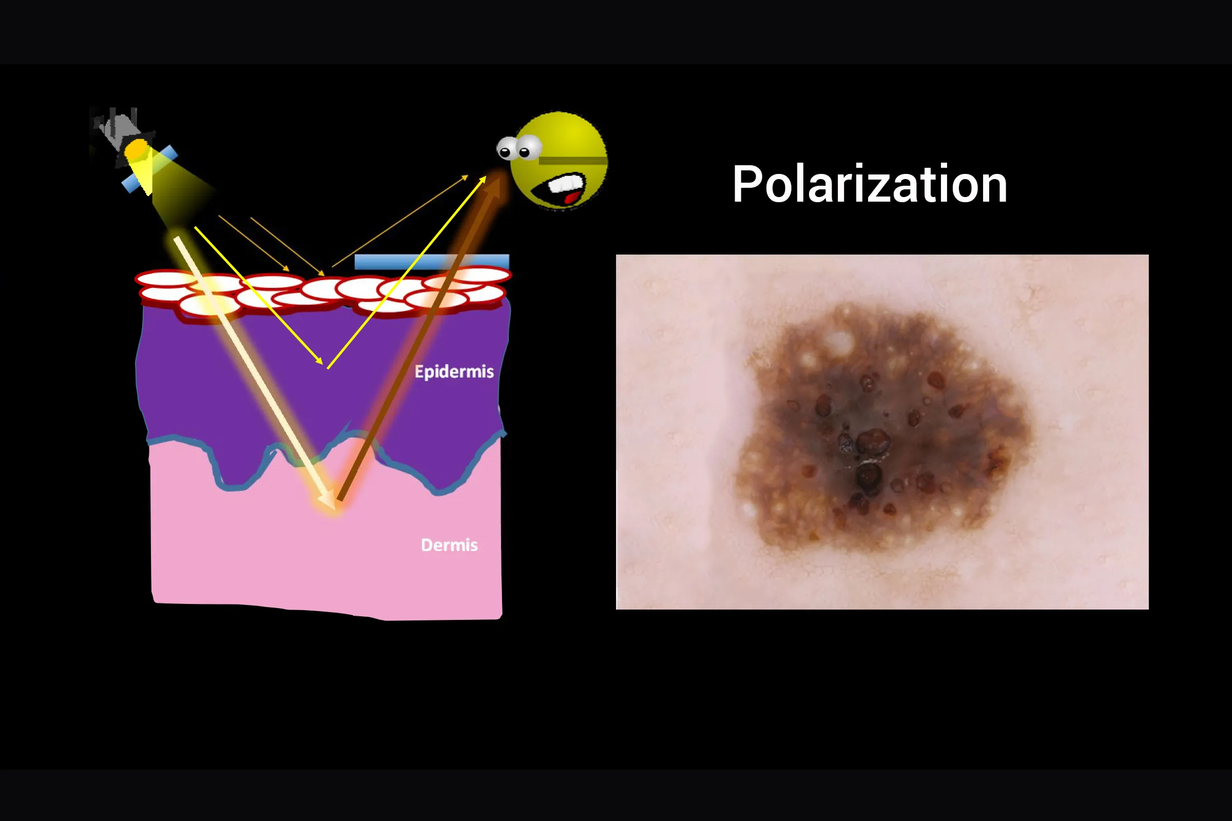
Cross Polarization
In polarization mode, the polarization filter becomes activated and absorbs the surface light reflection (no immersion fluid required). It allows you to examine the colours, shapes and texture features of the skin lesion more clearly, more precise and more detailed.
Efficient Lighting
Built with polarized, non-polarized and 365nm UV light, DE-3100 PRO offers two levels of color spectrum control to enhance imaging of deeper pigmentation.

Easy to Use
The magnet attachment gives the ability to attach on smartphone and take pictures in 5 seconds.
Best part? It compatible with all phone in the market, with a universal lens clip or a phone cover for easy on and off.

How Does it Compare
There’s an influx of dermatoscope on the market. Our devices are remarkable blend of pro-level features and affordable price. Their premium optics and efficient LED system delivers sharp & precise images.

DE-300
For Beginners
- 6X magnification
- Polarized &Non Polarized
- 32mm image diameter
- Phone camera detection
- 200mAh Battery

DE-400
For Enhanced
- 10X magnification
- Polarized &Non Polarized
- 45mm image diameter
- Phone camera detection
- 200mAh Battery

DE-3100
For Pros
- 10X magnification
- Polarized &Non Polarized
- 32mm image diameter
- Focus adjustable
- Phone camera detection
- 1000mAh Battery

DE-4100
For Pros
- 10X magnification
- Polarized &Non Polarized
- 32mm image diameter
- Focus adjustable
- Phone camera detection
- Naked eye detection
In The Box
1x DE-3100 Dermatoscope
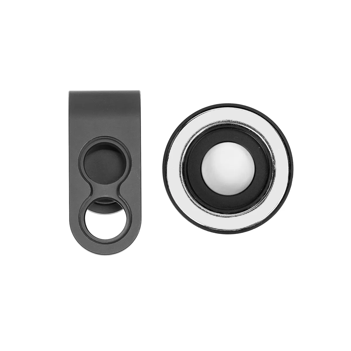
1x Universal Lens Mount
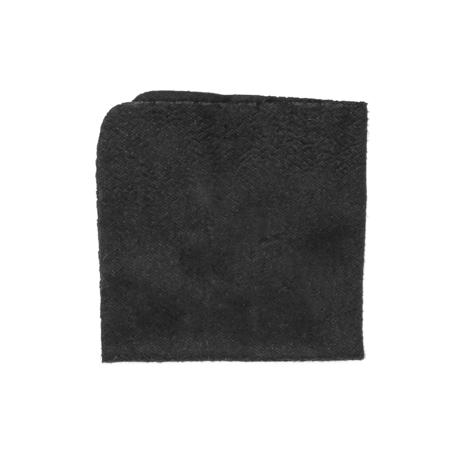
1x Microfiber Cloth
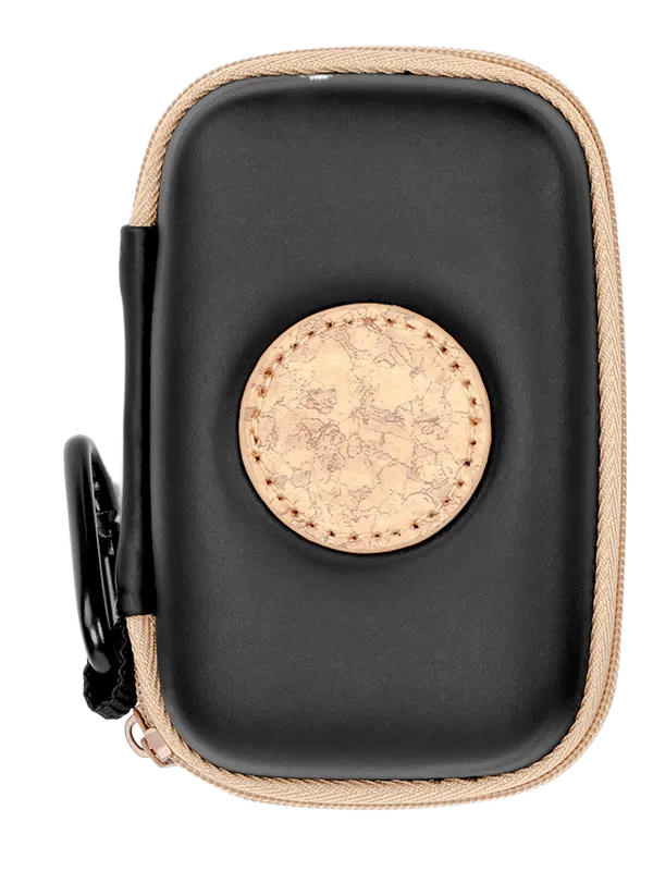
1x Carrying Case
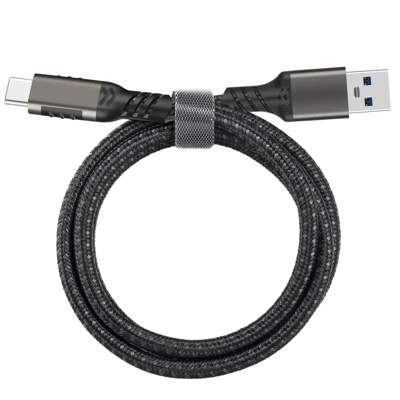
1x Charging Cable
Shot on Spot

Learn More

Medical Dermatoscopes
As a dermatologic diagnostic tool, dermoscopy is mainly used in dermatology to examine skin diseases, and can observe minute skin changes that are not detectable by the naked eye, helping…

Melanoma under Dermoscopy
Under dermoscopy, malignant melanoma can often be characterized by atypical pigment networks, irregular dot-ball shapes, and a bluish-white veil. For a clear view, the physician needs to clean the patient’s…

Dermatoscopes for Sale
As a non-invasive diagnostic tool, handheld dermatoscopes typically offer a magnification of around 10 times. Dermatoscopes are commonly equipped with built-in light sources to ensure adequate and even illumination during…
REVIEWS
Only logged in customers who have purchased this product may leave a review.


1 review for DE-3100 PRO Dermatoscope
There are no reviews yet.