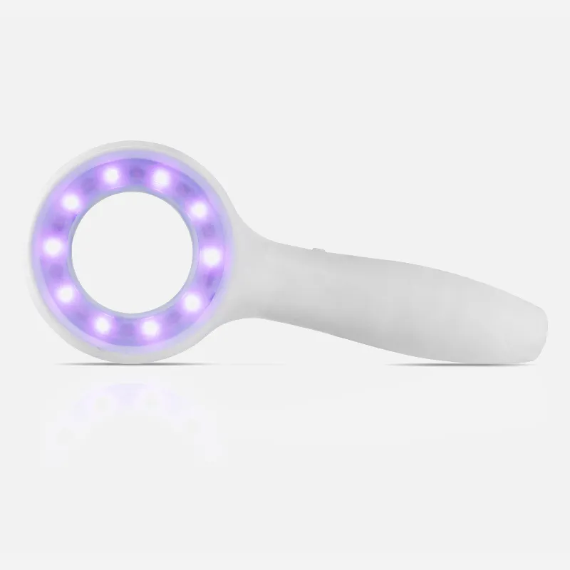
What Is A 365nm UV Lamp?

A 365nm UV lamp emits ultraviolet light at a wavelength of 365 nanometers. This wavelength falls within the near-ultraviolet or "black light" spectrum, just beyond what is visible to the human eye. 365nm UV light has a variety of uses due to its unique properties. It causes certain materials to fluoresce, allowing detection applications. It is also used to disinfect surfaces and as a curing lamp for UV-cured adhesives or inks. The invisibility and relatively long wavelength compared to shorter UV wavelengths make 365nm an ideal wavelength for special black light effects. Overall, a 365nm UV lamp produces a type of non-visible, near-UV light with key industrial, medical, and entertainment applications.

How Does A 365nm UV Lamp?

365 nm UV lamps emit a dull, bluish-white light that's part of the UVA wavelength range. This light is effective at killing bacteria because it falls between visible light and UVC, which are both more effective than either one alone. When these two wavelengths are combined, they cancel each other out, leaving only the desired effect.

365 nm UV lamps are used by medical professionals and for domestic use to:

  • Look for evidence of disease, fungi, and bacteria
  • Detect the presence of harmful molds that are not visible to the naked eye
  • perform inspections
  • Find cracks and leaks
  • UV cure adhesives and resin

UV radiation has enough energy to break chemical bonds. UV photons can cause ionization, which is a process where electrons break away from atoms. The resulting vacancy affects the chemical properties of the atoms and causes them to form or break chemical bonds that they otherwise would not.

People May Ask

What Nm Is Ideal for UV Light?

366 nmThe majority of fluorescence has a peak at 365 nm and is highest in the 320–380 nm range, as the chart below illustrates. For most blacklight applications, where maximum fluorescence is needed, we would therefore normally advise using a wavelength of 365 nm.

Which UV Radiation Is More Harmful?

UVC is short-wavelength. The UV radiation type that causes the most damage is UVC. But it never makes it to the surface of the earth because the atmosphere filters it out totally. Although it cannot penetrate past the outer layers of skin, medium-wavelength UVB is very physiologically active.

What Negative Impacts Do UV Lamps Have?

The most lethal type of skin cancer, melanoma, as well as squamous and basal cell skin cancers, are all known to be more common in those who are exposed to UV light from tanning beds and the environment." The same applies to UV manicure lights.

At 365 Nm, Is It UVA Or UVB?

At 395 nm and 365 nm, which are both in the UV-A band, UV LEDs are currently the most popular varieties. Although the specified wavelength represents the maximum wavelength that the LEDs emit, there is light at both lower and higher wavelengths.

Why Would A UV Lamp Be Used?

UV radiation serves a multitude of objectives in industrial operations as well as medical and dental practices. These include the destruction of microorganisms, the production of fluorescent effects, the curing of inks and resins, phototherapy, and suntanning.

Can Eyes Safely Be Exposed to UV Light at 365 Nm?

The reflected light from fluorescent objects under a UV light is acceptable for brief exposure periods, however prolonged exposure to 365nm UV will harm your eyes.

With A 365 Nm UV Flashlight, What Can You Detect?

Uses for 365nm UV Flashlight in Particular:Detection of Pet Urine (Note: Limited to dried urine stains in dimly lit areas.)Instead,Seeking Yooperlite Rocks through Rock Hunting.Locate Scorpions That Are Hard to Find.Track Down Tomato Hornworms.Ultraviolet Resin Curing.Verification of ID and Currency.Finding Leaks.

Is 395 Nm Preferable to 365 Nm?

The primary distinction is that the 365 nm LED emits far less visible light than the 395 nm LED. The 365 nm LED emits a dull, bluish-white light (the result of residual light energy that "tails" off into the visible spectrum), whereas the 395 nm LED emits a prominent violet-colored light.

Does Skin Damage Occur at 365 Nm UV?

Using 365nm light has no known negative effects. On the other hand, you might tan more deeply than usual if you end up sunburned after being out in the sun.

What Is The Purpose of The 365nm UV Light?

Using the FoxFury MF 365nm UV Forensic Light System, field crime scene investigators, lab workers, and medicolegal personnel (such as coroners and medical examiners) search for bite marks, bruises, bite fluids (such as urine and semen), accelerants (such as oil, gas, and turpentine), and trace evidence (such as hairs and...

365nm UV Lamp Products

China customized 365nm UV Lamp manufacturer & factory

【Suggested】Vansky 365nm and 51 LED 395nm Blacklight UV Flashlight Pet Urine/Cat Urine Detector

China portable 365nm UV Lamp

Rechargeable 365nM Ultraviolet Flashlight, Blue Light Flashlight for Pet Urine Detection, Blacklights for Bed Bugs, Ringworm, and Resin Curing: ESCO LITE UV Flashlight Black Light

365nm UV Lamp manufacturer & factory

Coospider-repta 4-Pack 32050 6 Watt Bulbs Replacement for DynaTrap DT2000XL and DT2000XLP, F6T5/BL 9 Inch Fluorescent Tube G5 Base Replacement Bulbs for Dyna Trap 1 Acre Mosquito & F Lying Insect Trap

China affordable 365nm UV Lamp manufacturer & factory

The Alonefire SV87 395nm UV Flashlight Zoom 5W Type C USB Rechargeable Black Light Money Detector is ideal for detecting resin, fish, minerals, leaks, pets, urine, and curing tape batteries.

China wholesale 365nm UV Lamp

Celiwace 12-Inch Fluorescent Tube Replacement BL368 UVA 365nm T5 8 Watt F8T5 BL Black Bulb BL368.

China high quality 365nm UV Lamp manufacturer & factory

SCIVIEW 1200 ALONEFIRE

China Professional 365nm UV Lamp manufacturer & factory

Two-pack 60-watt UV flood black light with fluorescent tape, IP67 waterproof high power LED black light, and a 4-foot cable and plug. It may be used for stage lighting, clubs, aquariums, car body paint, and other applications.

China wholesale 365nm UV Lamp manufacturer & factory

365nm+Red+White RovyVon E8 EDC Flashlight is USB C Rechargeable, has a 4000K High Color Rendering Index, and is a 500 Lumen Dual Power Keychain Flashlight that works with AAA batteries.

China portable 365nm UV Lamp manufacturer & factory

10 Long, 32W Black Replacement Bulb for BF190, Compatible with Flowtron BK-40D, FUL32T8/BL, 365nm U.V. A Lamp

China high quality 365nm UV Lamp

Compatible with Wall Sconce Models, Flowtron BK-40, BK-80D, Stinger b4040, FC7600, and Replacement Bulb BF-150 40W, Two packs of the FUL40T8/BL Black Bulb, 12 Inch U Shape U V A Light

Top Reviews

Advantage C.

This is an excellent UV lamp! So far, it's not giving me any trouble. After a few days of use, the battery life is currently good. (Not daily use, but frequent use)

San Mar

To utilize for nighttime fishing, I purchased the UV Beast. While UV light bothers picky fish less, regular flashlights frighten fish and draw bothersome insects (some of whom are drawn to UV light as well). When used in conjunction with UV fishing line and tackle, you can observe hookups hundreds of feet below the surface and see your line in total darkness. In my pursuit of trophy bucketmouths, the UV Beast changed the game. Strongly advised if, like me, you enjoy bass fishing. I had no idea there would be so many more uses for this light. It's possible to spot other fishermen's lost gear, such as beacons, by quickly scanning a rough shoreline in the dark. In just a few months, I've gathered equipment worth hundreds of dollars. It also highlights things that, for the most part, I'd prefer not to see. Scorpions. I was unaware of their quantity, even though I knew they existed in our area. Deep in Trinity National Recreation Area in Northern California, our favorite campground always seemed secure and comfortable. As I was preparing my fishing rods for the evening, I unintentionally saw a sco

bwdan2003

I occasionally work at night on "Cat in Tree" rescues. Knowing when my rope is over a branch is critical. Ordinary lights don't shine on the rope well enough to determine whether it is above a sturdy branch. It is absolutely fantastic and exactly where I want it to be thanks to the UV Beast. All that UV light can do is satisfy my urge. This light is enough bright to see my rope eighty feet up in the tree.Small things are frequently overlooked by us. But I got one for my wife and myself almost away after learning about the fascinating world of Myxomycetes from a friend who demonstrated the possibilities of this Tomlov digital microscope. Despite having a strong foundation, you can remove it from this microscope and, with a little patience, create stunning pictures and films similar to these.

0

Have questions on gear or your order?

Our Gear Guides are here to help! Get personal advice from pro creatives

Name
Subject
Email address
How can we help?

Instant Answers

How Can Dermoscopy Images Be Captured?

Dermoscopy images can be captured and stored in different ways, such as: • Using a smartphone or tablet with dermoscopic adapter, which consisted in the package.• Using a digital camera

Dermoscopy images can be captured and stored in different ways, such as:

• Using a smartphone or tablet with dermoscopic adapter, which consisted in the package.
• Using a digital camera with dermoscopic adapter, there’s 49mm screw size camera adapter available to order now.

Compatible phone/tablet models:
All iPhone models, 95% Android phones, 90% tablet. For phone/tablet size in 5.25-14mm

Compatible camera models:
All camera with built 49mm filter screw, such as Canon EOS 70D, 80D, 90D; Canon EOS R7, R10, R50, R100; Canon M100, M200, M50, Mark II; Canon G7X Mark III, Sony ZV-1

How Can I Connect My Phone to My Dermatoscope?

There’s universal phone adapter for all our dermoscopes. Please check the installation procedure bellow or watch operation guide. Smartphone Connector (1) Place phone adapter screw in the center of smartphone’s

There’s universal phone adapter for all our dermoscopes. Please check the installation procedure bellow or watch operation guide.

Smartphone Connector

(1) Place phone adapter screw in the center of smartphone’s main camera.
(2) Screw magnet attachment on phone adapter.
(3) Put dermoscope’s back ring and magnet attachment together

Take The Best Images

You need to adjust the focus ring after the dermoscpe connected on smartphone to get the best images.

How Can I Clean My Dermoscopy after Usage?

Cleaning your dermoscopy after usage is important to prevent cross-contamination and infection. The cleaning method may vary depending on the type and model of your dermoscopy, so you should always

Cleaning your dermoscopy after usage is important to prevent cross-contamination and infection. The cleaning method may vary depending on the type and model of your dermoscopy, so you should always follow the manufacturer’s instructions. However, some general steps are:

• Turn off and disconnect your dermoscopy from any power source or device.

• Wipe off any visible dirt or debris from the dermoscopy with a soft cloth or tissue.

• Disinfect the dermoscopy with an alcohol-based wipe or spray, or a disinfectant solution recommended by the manufacturer. Make sure to cover all surfaces, especially the lens and contact plate.

• Let the dermoscopy air dry completely before storing it in a clean and dry place.

• Do not use abrasive or corrosive cleaners, solvents, or detergents that may damage the dermoscopy.

• Do not immerse the dermoscopy in water or any liquid, unless it is waterproof and designed for immersion.

You should clean your dermoscopy after each use, or at least once a day if you use it frequently. You should also check your dermoscopy regularly for any signs of damage or malfunction, and contact the manufacturer or service provider if needed.

Polarized VS Non-Polarized Dermoscopy

A dermoscopy is a device that allows the examination of skin lesions with magnificationand illumination. By revealing subsurface structures and patterns that are not visible tothe naked eye. It can

A dermoscopy is a device that allows the examination of skin lesions with magnificationand illumination. By revealing subsurface structures and patterns that are not visible tothe naked eye. It can improve the diagnose accuracy of skin lesions, such as melanoma,basal cell carcinoma, seborrheic keratosis, etc.

There are two main types of dermoscopy: Non polarized and polarized dermoscopy.We’ve fitted most of our dermoscopys with polarized and non-polarized light. They canbe used in multiple skin structures.

Non-polarized contact Mode

In non-polarized mode, the instrument can provide information about the superficialskin structures, such as milia-like cysts, comedo-like openings, and pigment in theepidemis.

The dermoscopy requires applying a liquid such as mineral oil or alcohol to the skin andplacing the lens in contact with the skin. This reduces surface reflection and enhancesthe view of subsurface structures.

Image with non-polarized light (DE-3100)

Polarized contact Mode

In polarized mode, the instrument allows for visualization for deeper skin structures,such as blood vessels, collagen, and pigment in the dermis.

The dermoscopy does not need to be in contact with the skin or use any liquid. Theirpolarized light can help to eliminate surface reflection and allow visualization ofvascular structures.

Image with polarized light (DE-3100)

Polarized non-contact Mode

The dermoscopy can also use polarized light to examine the skin without direct contact.

In polarized non-contact mode, the instrument allows for examination infected areasand lesions that are painful for the patient, or the difficult to contact pigmented lesions,such as nails and narrow areas.

The contact plate should be removed in this mode, and it does not require applying aliquid to the skin. As it doesn’t require pressure or fluid application on the skin, it canalso avoid cross-contamination and infection risk.

Image in polarized non-contact mode (DE-3100)

How Effectiveness Is Dermoscopy

Compared with visual inspection, the dermoscopy can be used to capture and store skin lesion photos, which play an important role in early skin cancer examination. The dermoscopy allows the

Compared with visual inspection, the dermoscopy can be used to capture and store skin lesion photos, which play an important role in early skin cancer examination.

The dermoscopy allows the examination of skin lesions with magnification and illumination. This can be greatly avoiding the factors that cause interference to visual detection. Such as lighting, skin color, hair and cosmetics.

Several studies have demonstrated that dermoscopy is useful in the identification of melanoma, when used by a trained professional.

It may improve the accuracy of clinical diagnosis by up to 35%
It may reduce the number of harmless lesions that are removed
In primary care, it may increase the referral of more worrisome lesions and reduce the referral of more trivial ones

A 2018 Cochrane meta-analysis published the accuracy of dermoscopy in the detection.

Table 1. Accuracy of dermoscopy in the detection of melanoma in adults
Detection Method Sensitivity, % Specificity, % Positive Likelihood Ratio NegativeLikelihood Ratio
Visual inspection alone (in person) 76 75 3.04 0.32
Dermoscopy with visual inspection (in person) 92 95 18 0.08
Image-based visual inspection alone (not in person) 47 42 0.81 1.3
Dermoscopy with image-based visual inspection (not in person) 81 82 4.5 0.23
ROC—receiver operating characteristic. *Estimated sensitivity calculated on the summary ROC curve at a fixed specificity of 80%.

As we can see, the dermoscope can improve the accuracy of diagnosis of skin lesions, especially melanoma.

Table 1. Accuracy of dermoscopy in the detection of melanoma in adults
Detection Method Sensitivity, % Specificity, % Positive Likelihood Ratio NegativeLikelihood Ratio
Visual inspection alone (in person) 79 77 3.4 0.27
Dermoscopy with visual inspection (in person) 93 99 93 0.07
Image-based visual inspection alone (not in person) 85 87 6.5 0.17
Dermoscopy with image-based visual inspection (not in person) 93 96 23 0.07
ROC—receiver operating characteristic. *Estimated sensitivity calculated on the summary ROC curve at a fixed specificity of 80%.

Characteristics of the dermatoscopic structure of the skin lesions include:

• Symmetry or asymmetry
• Homogeny/uniformity (sameness) or heterogeny (structural differences across the lesion)
• Distribution of pigment: brown lines, dots, clods and structureless areas
• Skin surface keratin: small white cysts, crypts, fissures
• Vascular morphology and pattern: regular or irregular
• Border of the lesion: fading, sharply cut off or radial streaks
• Presence of ulceration

There are specific dermoscopic patterns that aid in the diagnosis of the following pigmented skin lesions:

• Melanoma
• Moles (benign melanocytic naevus)
• Freckles (lentigos)
• Atypical naevi
• Blue naevi
• Seborrhoeic keratosis
• Pigmented basal cell carcinoma
• Haemangioma

No products in the cart.

No products in the cart.

Hot Search Terms