


DE-400 Dermatoscope – IBOOLO
Unlocking The Microscopic Advantage: An Exploration of Dermatoscopy's Key Apparatus
Dermoscopy is a skin examination technique that uses a handheld device called a dermatoscope. It's also known as dermatoscopy, skin surface, and chemiluminescence microscopy.
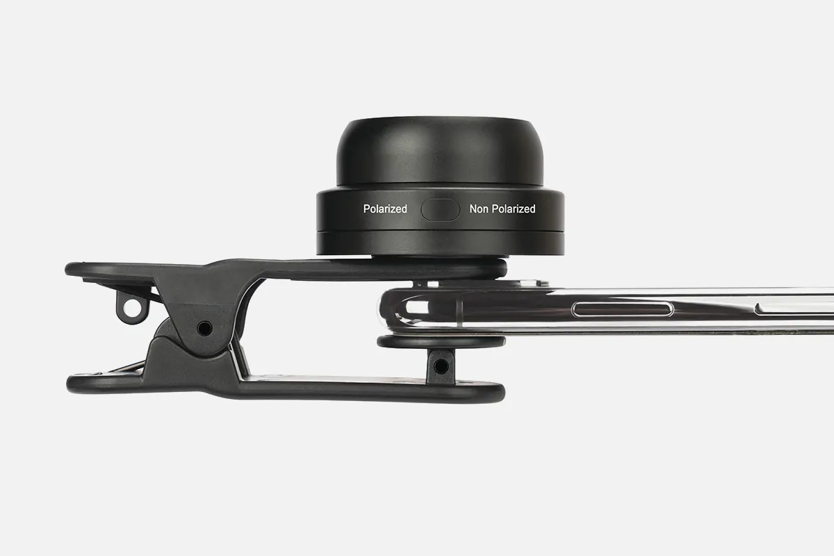
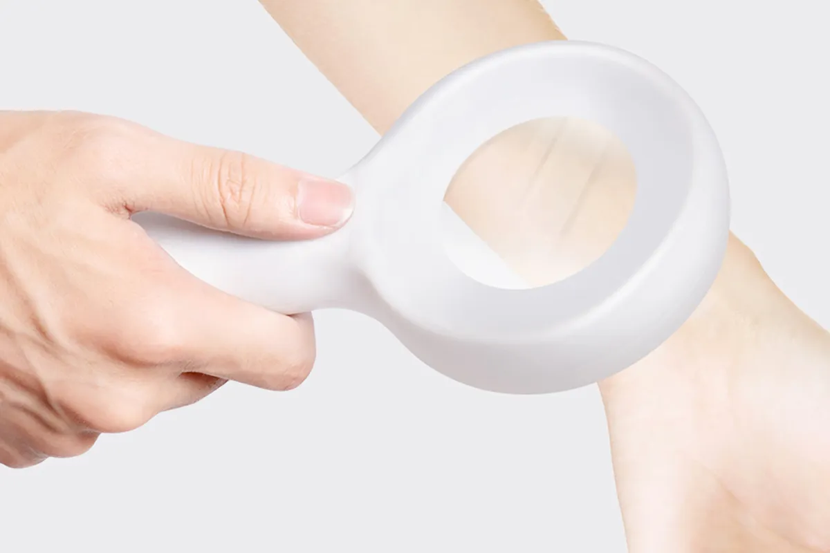
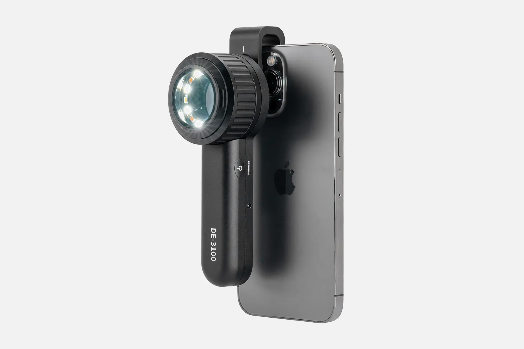
A technician holds the dermatoscopy onto the skin to examine it closely during dermoscopy. The dermatoscope has a high-quality magnifying lens and a powerful lighting system. It's similar to a camera, allowing for the inspection of skin lesions without being blocked by reflections from the skin's surface.
Dermoscopy is non-invasive and painless. It's most often used to help diagnose skin cancer and map moles. For example, sequential dermoscopy (mole mapping) imaging can help identify lesions.
Handheld dermatoscopes usually have a 10-fold magnification, which is usually sufficient for daily practice. Lower magnifications can provide a better overview of a large scalp area.
Uncovering The Functional Mechanics: Examining The Integral Components of Dermatoscopes
A dermatoscope is a handheld instrument that uses light and magnification to help dermatologists examine the skin. The basic principle of dermoscopy is transillumination, which involves shining a light through a lesion to study it with high magnification.
Here's how dermoscopy works:
- The patient stands in a designated location for full-body imaging.
- The dermatoscope is applied directly against the skin.
- A fluid interface makes the skin surface more transparent, allowing the visualization of subsurface structures.
- The dermatoscopy can assess structures to the depth of the reticular dermis.
- The camera sends the pictures to backend software, which compiles and generates a three-dimensional patient render.
- The three-dimensional model is often viewable in applications provided by the device.
Dermoscopy can help diagnose certain conditions by showing extra detail. For example, most skin cancers have a lot of blood vessel markings, which can be seen on a dermatoscope. Melanoma may be recognized when there are only 2-3 colors in the lesion on dermoscopy, while deeper melanomas reveal more colors.
People May Ask
La examinación cutánea is un componente esencial del examen físico estándar. All of the body's skin is meticulously examined as part of this examination. The assessment focuses on identifying abnormal signs in the skin, such as the cabelludo, orificios, uñas, and mucosal surfaces.
La examinación cutánea is un componente esencial del examen físico estándar. All of the body's skin is meticulously examined as part of this examination. The assessment focuses on identifying abnormal signs in the skin, such as the cabelludo, orificios, uñas, and mucosal surfaces.
Identify Your Skin SpotsSkin cancer comes in three different forms: melanoma, basal cell carcinoma, and squamous cell carcinoma. The ability to recognize every kind of skin cancer is a skill taught to dermatologists. Make an appointment with the dermatologist over the phone if you find anything strange during your self-examination.
Se aplica un gel líquido específico en el área de interés durante una dermatoscopia y coloca el dermatoscopio sobre la lesión para realizar el examen. This gel enables the physician to see structures beneath the skin's surface. The need to use the gel has been eliminated by the most recent dermatoscopy models.
It is possible to see a number of lunar structures that are invisible to the unaided eye thanks to this non-invasive technique. Las imágenes obtenidas se conservan digitalmente en la base de data fotográfica, lo que posibilita el detecto de cambios mínimos que pueden ser significativos de malignización en visitas a posteriori.
El diagnóstico dermatoscopio use fotografías de alta resolución para permitir un examen detallado de la piel estructura mediante una lente de aumento de alta calidad. El dermatoscopio permite evaluar las lesiones cutáneas según su forma, aspecto, color y pigmentación.
How long could you have melanoma without realizing it? It is contingent upon the kind of melanoma. Radial melanoma, on the other hand, might spread slowly over a decade, whereas nodular melanoma spreads quickly over a few weeks. Similar to a cavity, a melanoma can proliferate for years without showing any noticeable symptoms.
Dermatoscopes enable the user to do skin surface microscopy, and are commonly used by dermatologists, plastic surgeons, and general practitioners. The practitioner can now assess skin patterns and structures as a result.
The pigmented lesion must lack pattern symmetry and color uniformity in addition to at least one of the following characteristics in order to be diagnosed as melanoma: numerous brown dots, pseudopods, radial streaming, scar-like depigmentation, peripheral block spots/globules, five to six colors, a blue-white veil,...
Only potentially cancerous moles are seen visually on your skin. It is not able to confirm for you that you have it. A biopsy test is the sole method available for diagnosing the illness. Should your physician determine that a mole is a concern, they will administer a numbing injection and remove as much of the mole as feasible using a scrape.
Dermatoscopio Products
Featuring WF10x and WF20x eyepieces, 20X/40X/80X magnification, 2X and 4X objectives, upper and lower LED lighting, a reversible black/white stage plate, a pillar stand, and 120V or battery power, the AmScope SE306R-PZ-LED forward-mounted binocular stereo

A complete science kit that includes 10 microscope slide specimens, 5 blank slides, a 16-page user guide, and a phone adapter is the perfect choice for students studying science with a 40x–1000x microscope for adults.
GUVOP 50X-1000X Magnification WiFi Portable Handheld USB Microscopes with 8 LED & Stand: Wireless Digital Microscope Camera Compatible with iPhone, Android, Electronic Microscope for Kids and Adults - Blue

This AmScope PS25W prepared microscope slide set includes a fitted wooden case and 25 slides for teaching basic biological science.

20x/50x/100x Magnification, 17mm Field of View, Pen Light Included in AmScope H2510 Handheld Stand Measuring Microscope
Kila Scopes 专业听诊器 - 帓业单头心脏病和诊断听诊器 适用于医生和护士-带配件,K971 酒红

This product is a portable digital microscope with a 4 LCD screen, a 1500X pocket microscope for kids with LED lights, and a 32GB memory card for adults.Has a Camera and Video Function
Arsir 7.3*4.4 Large Folding Lighted Magnifier with 48 LED Lights (3 Modes), Rectangular Handhold, Full-Page 5X Magnifying Glass for Reading Black Magnifying Lens Gifts for Seniors Who Read Books and Prints
For iPhone, Android, and PC, Ninyoon 4K WiFi Microscope with Expert Stand, 50-1000X Digital USB Microscope Wireless Endoscope HD Camera Compatible with All Cellphones Windows Macintosh Tablet Android iPad Glimmer Windows

C-Mount for trinocular microscope, full coin view, 1200x magnification, MOYSUWE 2K HDMI Digital Microscope, 26MP Sony CMOS IMX Sensor Coin Soldering Microscope with 7.5 IPS Screen, Compatible with Windows and TV MDM-209
Hot Products


News & Blog


Top Reviews
emcgilto
One day, I was at work and noticed that one of my coworkers had one of them slung around her neck. My attention was drawn to the vivid color (hers is orange). When she informed me the pricing of this stethoscope, I was curious because I had been thinking about getting a Littman. I was even more interested when she said that it performed even better than her prior Littman stethoscope. It shocked me really nicely! This scope is amazing; it works better than any other scope I have ever owned, yet I have never paid less than $120 for a scope. Another coworker was astounded when I showed him my scope. He mentioned using a Kila as his next scope.
James W Pyle III
I examine plant stuff under this microscope. Although the image quality is far from 4k, it is still sharp enough to see a ton of details that are too small to be seen with the human eye. Get something different if you want to use this scope to capture pictures. There won't be enough quality in the pictures to be proud of. This scope is fantastic if all you're using it for is close-up viewing. Acquire it. Since I don't trust drivers from tiny businesses, this required NO drivers. The camera functioned flawlessly and Windows recognized it as a webcam.
Cindy M.
You "see" what this small lightweight endoscope picks up on its camera on your cell phone via an app. You don't actually look in the scope like your doctor does when examining you. This means you can easily look in your own ears and see a large image on your phone which is an added benefit. This unit came with a small card with two bar codes, one for downloading the app on your phone and a second to turn off ads on the app. Once the app is installed on your phone, you push the button to turn on the endoscope and find it via phone wifi settings to connect to it. Once you tell the app what kind of device it is, you are good to go. Clean a cone with alcohol, put it on the scope, and look in your ear (or wherever you need to look). You can capture photos with the touch of a button that you could easily send to your doctor if needed. The scope came fully charges, has a charger cord, little tools for use in your ear, and four starter alcohol swabs for cleaning before and after use. For the price, this will be a val


