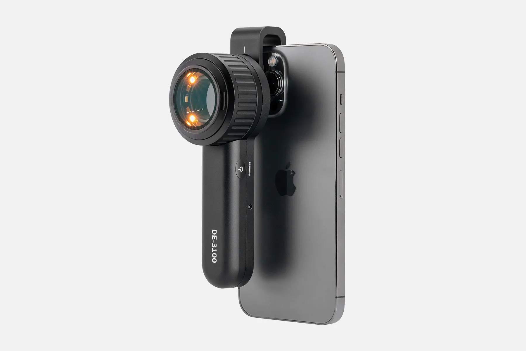
Explanation of What A Digital Dermatoscope

A digital dermatoscope is an advanced type of dermatoscope used to perform dermoscopy examinations of skin lesions. While a conventional dermatoscope employs optical magnification and lighting, digital versions integrate additional capabilities through electronic sensors and software.

These devices utilize a high-resolution digital imaging sensor coupled with specialized optics and illumination to capture precise dermoscopic photos and videos. This allows enhanced recording, tracking, comparison, and consultation around suspicious moles over time. Some systems also enable computer-assisted diagnostic features.

Key benefits of digital integration include mobile device connectivity for easy image captures, efficient electronic health record storage and retrieval, streamlined telemedicine consult workflows, and options for patient education and monitoring. These capabilities aim to improve clinical effectiveness.

Digital dermatoscopes enhance traditional dermoscopy through sophisticated imaging and data management. Quantifiable visual data grants doctors an expanded toolkit to identify diseases and communicate concerns. When combined with proper training, digital tools show promise for better screening and monitoring of melanoma and other skin cancers.

How Does A Digital Dermatoscope?

A digital dermatoscope uses magnification, lighting, and digital imaging technology to capture, store, and analyze images of the skin.

A dermatologist performs digital dermoscopy by:

1. Microscopic examination of the skin's surface

2. Locating lesions

3. photographing the lesions

4. Creating complete body maps of the photographs

Dermoscopes can be used with or without direct contact with the skin. The basic principle of dermoscopy is the transillumination of a lesion to study it with high magnification.

Digital dermoscopy allows physicians to:

  • Compare images at each follow-up visit
  • Provide an accurate diagnosis of an initially featureless melanoma

Dermoscopes are available as independent devices, cellular device attachments, or connections to a digital camera via coupling adaptors. The images captured by a dermatoscope can be recorded and analyzed on the device, mobile phone, or a PC/web-based software.

Dermatoscopy is a highly accurate, non-invasive diagnostic technique to evaluate the skin aspects that are non-accessible from the naked eye. The sensitivity of dermoscopy has been reported to range from 60% to 100%.

People May Ask

What Appearance Do Malignant Sun Spots Have?

melanoma. Although most melanomas are brown or black in color, some might have pink, tan, or even white hues. Not all melanomas are spherical like regular moles; some have colored sections. They could proliferate swiftly or even penetrate the skin around them.

A Dermatoscope Can See What?

With a dermatoscope, features can be evaluated down to the reticular dermis, and pictures can be taken for further analysis. Transillumination of a lesion for high magnification examination and visualization of minor details is the fundamental idea behind dermoscopy.

Are Melanomas Flat Or Elevated?

As they expand, melanomas may begin as flat patches. 4. If you can feel it, you should get it checked out, even if some moles can also be raised. Occasionally, when assessing melanoma, the "E" in the ABCDE guidance refers to "evolving." This is a result of the gradual changes in melanomas' size, shape, and color.

What Characteristics of The Skin Might Indicate Malignant Melanoma?

Thick pigmented lines around appendageal apertures, commonly referred to as rhomboidal structures, are the hallmarks of lentigo maligna's dermoscopic features.Unevenly colored follicular apertures.Instead,globules and dots of slate grey.

Can You Develop Cancer from A Mole You'Ve Had for Years?

Is melanoma possible from a common mole? Melanoma, the most dangerous kind of skin cancer, is extremely uncommon to develop from a common mole. Those with numerous little or multiple large moles are more likely to acquire melanoma, even though common moles are not malignant (1).

Can A Crusty Mole Not Develop Cancer?

Not all moles with scabs are malignant. But sometimes, scabby moles are malignant. If you are unable to link the scabbing to a recognized skin injury, it is crucial that you have them examined.

How Frequently Is A Malignant Melanoma Misidentified?

Initial clinical symptoms and misdiagnoses. In our study, the initial physician visit resulted in an erroneous diagnosis for 30% of the melanomas. This aligns with the outcomes of previous cohorts. Fortin and colleagues discovered a 25% rate of initial misdiagnosis, but Bristow and Acland reported a 33% rate of wrong diagnosis.

How Can A Mole's Malignant Status Be Determined?

pigment that has spread into the surrounding skin from the spot's edge. Redness or a fresh enlargement outside the mole's boundaries. Modification in feeling, including pain, sensitivity, or itching. A mole's surface may change, becoming scaly, leaking, bleeding, or developing a bulge or bump.

Is Mapping Mole Worthwhile?

An essential tool for melanoma early detection is melanoma mole mapping. Dermatologists may detect possible problems and offer prompt treatment by routinely checking your skin for changes.

How Do You Operate A Dermatoscope?

With a dermatoscope, features can be evaluated down to the reticular dermis, and pictures can be taken for further analysis. Transillumination of a lesion for high magnification examination and visualization of minor details is the fundamental idea behind dermoscopy.

Digital Dermatoscope Products

China high quality digital dermatoscope

Vividia Digital Borescope Microscope with 12mm Diameter OTG Android and USB PC Compatible with a Professional Multi-Functional Metal Stand

China customized digital dermatoscope

手持式 USB 数码显微敜带金属支枴,便携式高清 1000 倍放大检查摄像头,带 8 个 LED 灯,怂用于 Android Mac Windows 电脑

China handheld digital dermatoscope

Digital Portable Microscope with 8 LED Magnifier, Manually Adjustable for Computer Phone and Tablet, 50X-500X 0.3MP USB Magnification

China portable digital dermatoscope manufacturer & factory

The adult 4.3-inch IPS coin microscope with 1000X magnification is made by Hayve.Eight programmable LED coin collection suppliesCompatible with WindowsTF Card, 32GB

China customized digital dermatoscope manufacturer & factory

The VMS-007-DX2 is a USB digital microscope with 5 Megapixels, x300 magnification, the ability to capture and record photos and videos, up to 2592 x 1944 resolution, 8 LEDs, and an adjustable stand.

China smartphone digital dermatoscope manufacturer & factory

USB-C and Lightning Compatible Digital Microscope

China high quality digital dermatoscope manufacturer & factory

Digital video otoscope/earscope Firefly DE550 Wireless

China Professional digital dermatoscope

SupereyesB005+1~200X Portable USB Digital Microscope, Endoscope Loupe, Otoscope Magnifier with 11mm Health Kit Tripod Diameter

China affordable digital dermatoscope manufacturer & factory

Dino-Lite AM3113T USB Digital Microscope with 0.3 MP, 10x - 50x, 230x Optical Magnification, Microtouch, Measurement, and Discontinued

China Professional digital dermatoscope manufacturer & factory

Anykit Digital Otoscope with a gyroscope, 4.5-inch screen, 3.9mm ear scope camera with six lights, 32GB card, and an ear wax removal tool Allows for the capture of photos and videos

Top Reviews

Edmond D.

Fantastic watch, excellent numbers are visible at night, and Italian is a language that is easy to imitate. I really like this watch.

Rhisian

The lampshade really highlights the nursery motif. It's really gorgeous and precisely what we were looking for—the green is a little bit lighter than it appears in the photo, more like a light sage.

Mrs K Frampton

Though they appear smaller in the photo, these are quite adorable and a really affordable way to alter the appearance of a regular lightbulb. To add a special touch, I painted a couple of these as well???????

0

Have questions on gear or your order?

Our Gear Guides are here to help! Get personal advice from pro creatives

Name
Subject
Email address
How can we help?

Instant Answers

How Can Dermoscopy Images Be Captured?

Dermoscopy images can be captured and stored in different ways, such as: • Using a smartphone or tablet with dermoscopic adapter, which consisted in the package.• Using a digital camera

Dermoscopy images can be captured and stored in different ways, such as:

• Using a smartphone or tablet with dermoscopic adapter, which consisted in the package.
• Using a digital camera with dermoscopic adapter, there’s 49mm screw size camera adapter available to order now.

Compatible phone/tablet models:
All iPhone models, 95% Android phones, 90% tablet. For phone/tablet size in 5.25-14mm

Compatible camera models:
All camera with built 49mm filter screw, such as Canon EOS 70D, 80D, 90D; Canon EOS R7, R10, R50, R100; Canon M100, M200, M50, Mark II; Canon G7X Mark III, Sony ZV-1

How Can I Connect My Phone to My Dermatoscope?

There’s universal phone adapter for all our dermoscopes. Please check the installation procedure bellow or watch operation guide. Smartphone Connector (1) Place phone adapter screw in the center of smartphone’s

There’s universal phone adapter for all our dermoscopes. Please check the installation procedure bellow or watch operation guide.

Smartphone Connector

(1) Place phone adapter screw in the center of smartphone’s main camera.
(2) Screw magnet attachment on phone adapter.
(3) Put dermoscope’s back ring and magnet attachment together

Take The Best Images

You need to adjust the focus ring after the dermoscpe connected on smartphone to get the best images.

How Can I Clean My Dermoscopy after Usage?

Cleaning your dermoscopy after usage is important to prevent cross-contamination and infection. The cleaning method may vary depending on the type and model of your dermoscopy, so you should always

Cleaning your dermoscopy after usage is important to prevent cross-contamination and infection. The cleaning method may vary depending on the type and model of your dermoscopy, so you should always follow the manufacturer’s instructions. However, some general steps are:

• Turn off and disconnect your dermoscopy from any power source or device.

• Wipe off any visible dirt or debris from the dermoscopy with a soft cloth or tissue.

• Disinfect the dermoscopy with an alcohol-based wipe or spray, or a disinfectant solution recommended by the manufacturer. Make sure to cover all surfaces, especially the lens and contact plate.

• Let the dermoscopy air dry completely before storing it in a clean and dry place.

• Do not use abrasive or corrosive cleaners, solvents, or detergents that may damage the dermoscopy.

• Do not immerse the dermoscopy in water or any liquid, unless it is waterproof and designed for immersion.

You should clean your dermoscopy after each use, or at least once a day if you use it frequently. You should also check your dermoscopy regularly for any signs of damage or malfunction, and contact the manufacturer or service provider if needed.

Polarized VS Non-Polarized Dermoscopy

A dermoscopy is a device that allows the examination of skin lesions with magnificationand illumination. By revealing subsurface structures and patterns that are not visible tothe naked eye. It can

A dermoscopy is a device that allows the examination of skin lesions with magnificationand illumination. By revealing subsurface structures and patterns that are not visible tothe naked eye. It can improve the diagnose accuracy of skin lesions, such as melanoma,basal cell carcinoma, seborrheic keratosis, etc.

There are two main types of dermoscopy: Non polarized and polarized dermoscopy.We’ve fitted most of our dermoscopys with polarized and non-polarized light. They canbe used in multiple skin structures.

Non-polarized contact Mode

In non-polarized mode, the instrument can provide information about the superficialskin structures, such as milia-like cysts, comedo-like openings, and pigment in theepidemis.

The dermoscopy requires applying a liquid such as mineral oil or alcohol to the skin andplacing the lens in contact with the skin. This reduces surface reflection and enhancesthe view of subsurface structures.

Image with non-polarized light (DE-3100)

Polarized contact Mode

In polarized mode, the instrument allows for visualization for deeper skin structures,such as blood vessels, collagen, and pigment in the dermis.

The dermoscopy does not need to be in contact with the skin or use any liquid. Theirpolarized light can help to eliminate surface reflection and allow visualization ofvascular structures.

Image with polarized light (DE-3100)

Polarized non-contact Mode

The dermoscopy can also use polarized light to examine the skin without direct contact.

In polarized non-contact mode, the instrument allows for examination infected areasand lesions that are painful for the patient, or the difficult to contact pigmented lesions,such as nails and narrow areas.

The contact plate should be removed in this mode, and it does not require applying aliquid to the skin. As it doesn’t require pressure or fluid application on the skin, it canalso avoid cross-contamination and infection risk.

Image in polarized non-contact mode (DE-3100)

How Effectiveness Is Dermoscopy

Compared with visual inspection, the dermoscopy can be used to capture and store skin lesion photos, which play an important role in early skin cancer examination. The dermoscopy allows the

Compared with visual inspection, the dermoscopy can be used to capture and store skin lesion photos, which play an important role in early skin cancer examination.

The dermoscopy allows the examination of skin lesions with magnification and illumination. This can be greatly avoiding the factors that cause interference to visual detection. Such as lighting, skin color, hair and cosmetics.

Several studies have demonstrated that dermoscopy is useful in the identification of melanoma, when used by a trained professional.

It may improve the accuracy of clinical diagnosis by up to 35%
It may reduce the number of harmless lesions that are removed
In primary care, it may increase the referral of more worrisome lesions and reduce the referral of more trivial ones

A 2018 Cochrane meta-analysis published the accuracy of dermoscopy in the detection.

Table 1. Accuracy of dermoscopy in the detection of melanoma in adults
Detection Method Sensitivity, % Specificity, % Positive Likelihood Ratio NegativeLikelihood Ratio
Visual inspection alone (in person) 76 75 3.04 0.32
Dermoscopy with visual inspection (in person) 92 95 18 0.08
Image-based visual inspection alone (not in person) 47 42 0.81 1.3
Dermoscopy with image-based visual inspection (not in person) 81 82 4.5 0.23
ROC—receiver operating characteristic. *Estimated sensitivity calculated on the summary ROC curve at a fixed specificity of 80%.

As we can see, the dermoscope can improve the accuracy of diagnosis of skin lesions, especially melanoma.

Table 1. Accuracy of dermoscopy in the detection of melanoma in adults
Detection Method Sensitivity, % Specificity, % Positive Likelihood Ratio NegativeLikelihood Ratio
Visual inspection alone (in person) 79 77 3.4 0.27
Dermoscopy with visual inspection (in person) 93 99 93 0.07
Image-based visual inspection alone (not in person) 85 87 6.5 0.17
Dermoscopy with image-based visual inspection (not in person) 93 96 23 0.07
ROC—receiver operating characteristic. *Estimated sensitivity calculated on the summary ROC curve at a fixed specificity of 80%.

Characteristics of the dermatoscopic structure of the skin lesions include:

• Symmetry or asymmetry
• Homogeny/uniformity (sameness) or heterogeny (structural differences across the lesion)
• Distribution of pigment: brown lines, dots, clods and structureless areas
• Skin surface keratin: small white cysts, crypts, fissures
• Vascular morphology and pattern: regular or irregular
• Border of the lesion: fading, sharply cut off or radial streaks
• Presence of ulceration

There are specific dermoscopic patterns that aid in the diagnosis of the following pigmented skin lesions:

• Melanoma
• Moles (benign melanocytic naevus)
• Freckles (lentigos)
• Atypical naevi
• Blue naevi
• Seborrhoeic keratosis
• Pigmented basal cell carcinoma
• Haemangioma

No products in the cart.

No products in the cart.

Hot Search Terms