


DE-400 Dermatoscope – IBOOLO
Dermatoscopes for Enhanced Skin Lesion Visualization in Dermatology
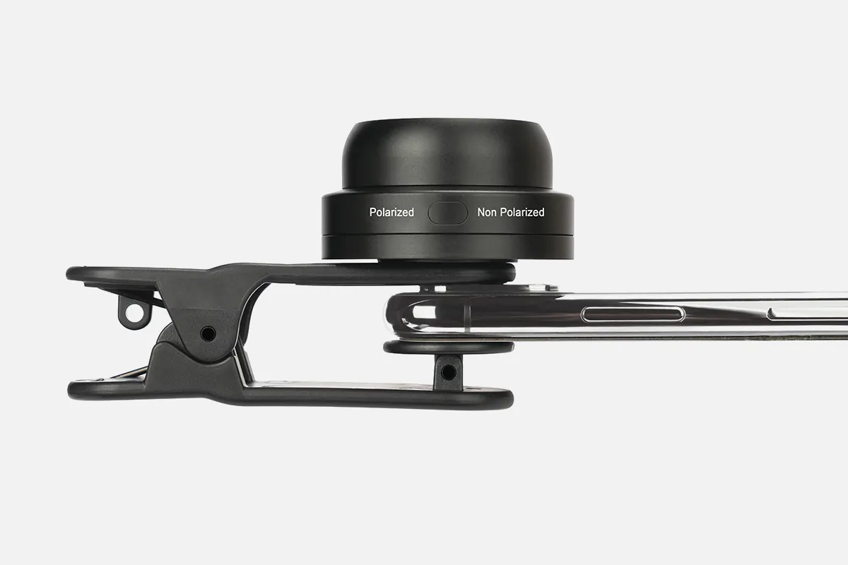
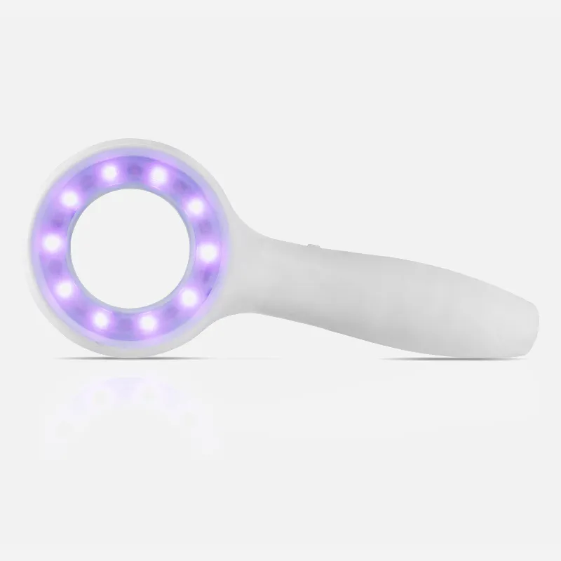
A dermatoscope is a specialized handheld medical device dermatologists use to evaluate skin lesions through optical magnification and lighting. It allows non-invasive visualization of subsurface skin structures normally invisible to the naked eye - including the epidermis, dermo-epidermal junction, and upper reticular dermis.
Specifically, dermatologists employ dermoscopy, applying the dermatoscope to transilluminate lesions. This reveals subtle visual morphological details related to structures and patterns like pigmentation, blood vessels, and skin texture. These insights assist in examining common growths such as
- Basal cell carcinomas
- Seborrheic keratoses
- Hemangiomas
- Benign lentigos
- Early stage melanomas
- Benign melanocytic nevi (moles)
Typical dermatoscopy models provide a 10x optical magnification level sufficient for routine skin evaluations and diagnostics. Higher-powered models also exist. Quality digital imagery enables tracking changes over time. Overall, dermatoscopes are invaluable visualization tools for dermatologists, improving detection accuracy and biopsy decisions through enhanced lesion insights.
How Dermatologists Use A Dermatoscope to Evaluate Skin Lesions
A dermatoscope is a small, specialized medical device used by dermatologists to visually inspect skin lesions up close utilizing light and magnification. Also known as a dermoscope, it assists with a technique called dermoscopy or dermatoscopy.
To perform an examination, gel or oil is first applied to the lesion. The dermatoscope's contact plate is placed directly on the skin and transilluminated at different angles using the device's embedded lighting. This allows seeing through the skin's surface to underlying structures at up to 10x magnification - the typical level required for routine evaluations. Subtle visual details related to patterns, blood vessels, and pigmentation changes become apparent.
Importantly, a dermatoscopy enables noninvasive visualization of subsurface skin characteristics in regions impossible to see with the naked eye - including the epidermis, dermo-epidermal junction, and upper dermis/papillary dermis. Storing images facilitates comparisons over time to observe lesion evolution.
In summary, dermatologists employ dermatoscopes to magnify and illuminate suspicious moles and lesions safely. Advanced insights can boost diagnostic accuracy over visual inspection alone and help guide biopsy decisions or treatment planning.
People May Ask
The following list of resources can assist dermatologists carry out their work responsibilities efficiently:Skin-cutting shear.Extractor for blackheads.A scalpel.Cutaneous biopsy punch.Microscope.Gloves.A laser dermabrasion apparatus.Bright, pulsating light.Additional things...
What Separates Dermatology from Dermatopathology? A medical student must be trained in either dermatology or pathology to become a dermatopathologist. Dermatopathologists acquire biopsy specimens, examine the tissue, and diagnose patients; dermatologists treat the patients.
Over the past few decades, advancements in dermatoscopy, skin imaging, immune-dermatology, lasers, and esthetic dermatology have expanded the field of dermatology into new areas. Dermatologists' approach to patient care is being completely transformed by new developments in rapid diagnostics and technology.
Lentigos, or accumulations of melanocytes in the cell's basal layer, are the most prevalent pigmented lesions. Although it is more frequently mistaken for a nevus, it can have the clinical look of a freckle. Although the margins are not clear, the lesion is not elevated.
Usually, pigmented lesions are not a reason for alarm. Nevertheless, some can progress to become distinct types of skin cancer. That's why it's critical that you and your physician keep a careful eye on them.
When recalling the warning indicators of melanoma, the "ABCDE" rule comes in handy:Unbalanced One part of the skin lesion is not the same shape as the other.Border. The edges are blurry, uneven, ragged, or notched.Vibrance. There could be tan, brown, and black tones present.Measurement of diameter.Evolving.
Dermoscopy has the potential to be a useful diagnostic tool for nail psoriasis since it can produce consistent clinical findings quickly and painlessly. This review outlines the most recent data on the distinct dermoscopic characteristics of nail psoriasis.
UV radiation from artificial sources is used by humans to initiate biologic processes that lower inflammation and slow down the growth of skin cells. This kind of light helps to cleanse the skin and reduce inflammation in the affected area when applied on a regular basis.
For a microscopic view of the cells, the dermatologist will cut a tiny slice of skin or the outer layer of the epidermis. For the light from the dermatologist microscope to travel through the sample and disclose any flaws or disease indicators, it must be thin.
A dermatoscope is a portable visual aid that a medical professional or layperson can use to inspect and diagnose diseases and skin lesions, including melanoma. It can also be useful for inspecting the nails, hair, and scalp. In a dermatologist's practice, a dermatoscope is usually present.
Dermatoscope for Dermatology Products
Fitzpatrick s Color Atlas and Clinical Dermatology Synopsis, Ninth Edition,
1–1000X Magnification Zoom, 4.3 Inch 1080P LCD Digital Microscope 10MP Wireless USB Camera Video Recorder with HD Screen, Stereo Microscope

Compatible with iPhone, iPad, Android, Windows, Mac computer, this wireless digital microscope is the SKYBASIC 50X–1000X Magnification WiFi USB HD Portable Handheld Pocket Microscopes Camera with 8 Adjustable LEDs.

First Edition of Dermoscopy Guide
USB Digital Microscope: SKYEAR 50X-1600X Magnification Handheld Digital Microscope with Adjustable Stand, 8 LED Lights, and Portable Microscope Camera for Kids and Adults – Compatible with iOS & Android Devices
Aopick Handheld USB 1440P HD Inspection Digital Microscope Camera with 50x–1600x Magnification Carry-around Pocket Microscopes featuring 8 LEDs and a stand that work with iPhone and Android devices

Featuring a stand, the Bysameyee 4K 3840x2160P Wireless Digital Microscope is a portable high-definition USB inspection camera borescope with a 50x–1000x magnification that works with iPhone, iPad, Android, Tablet, Windows, and Mac computers.
Dermatoscopy: Examining pigmented and non-pigmented lesion patterns
This coin PCB soldering repair plant features an LCD digital USB microscope, a 4.3-inch screen with 1000x magnification, an adjustable stand, a rechargeable battery, and eight LED lights.
Wireless 5.5-inch 1080P 10 million pixel LCD digital microscope with wireless zoom and magnification up to 1000x A USB stereo microscope camera with a 10 million pixel camera and an HD screen video recorder
Hot Products


News & Blog


Top Reviews
Kindle Customer
I've tried jewelers loupes, but they simply kept breaking. This works pretty well for me. The program was simple to use and straightforward. Works on your wifi, eliminating the need for cords when in use, and it seems to keep a charge well. A surgeon's hand is required to obtain an excellent photo, but if you use the included stand and lay the material underneath, you can easily zoom in and out and operate the device with great ease. I tried to upload a picture, but it wouldn't let me.
TheHandiCapperGeneral
I couldn't get any response from my first one. About my unfavorable rating and the unit I had, the owner of this business emailed me. She gave me a replacement right away and then checked in to make sure I had received the high-quality item she claimed to be selling. This time, the second was a lot clearer. I like a business that values its clients and goes above and beyond to meet their needs!
S
I don't usually write reviews, but I'm happy that I bought this item. The focus is easily adjustable using the huge knob, and the picture is crisp and clear. Merely observing the fibers of an article of clothing makes me feel like a child once more. What degree of magnification would be appropriate for various purposes was one of my concerns while choosing which type to purchase. Having any kind of sense about what 60x actually means in real life is difficult. The universe of clothes fibers and phone screen pixels, which is otherwise invisible, may be seen by magnifying things 20 times, as I have discovered. (*Yes, unsolvable, but not truly invisible.) Allow your children to play with this after you've had a great time.


