


DE-400 Dermatoscope – IBOOLO
Demystifying The Clinical Dermatoscope: Understanding This Critical Medical Apparatus
A medical dermatoscope is a specialized dermatoscope designed for healthcare applications like skin cancer screenings, clinical diagnosis, and therapeutic monitoring.
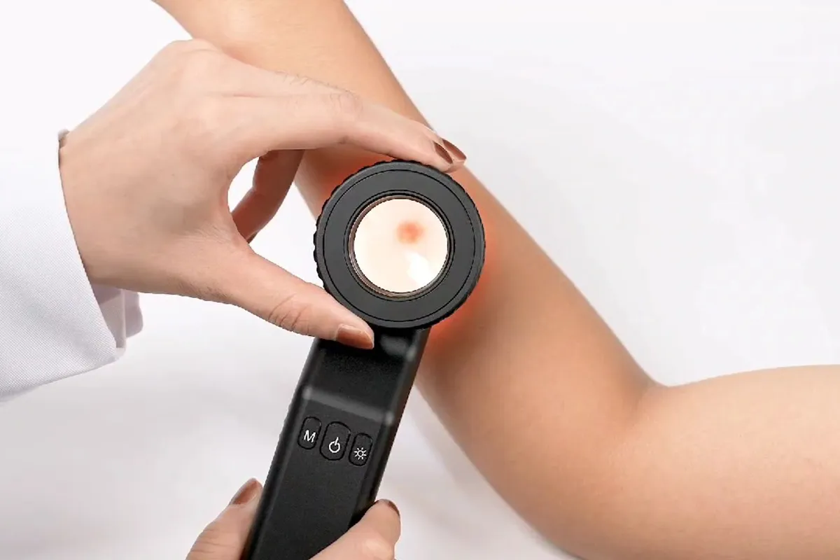
Compared to basic models, medical dermatoscopes feature robust, ergonomic construction optimized for frequent professional use. They provide consistent, high-quality visualization through calibrated optics like 10x to 40x magnification and polarized lighting modes that reduce surface reflections.
Strict validation testing also ensures accurate color rendition, precise structural imaging capabilities, and reliable long-term performance - vital for clinical procedures. Measurement grids, image storage, and camera connectors offer added examination tools as well.
By meeting stringent healthcare device standards, medical dermatoscopes qualify as regulated Class I diagnostic instruments approved for integration with hospital systems and medical documentation workflows.
Medical dermatoscopes are purpose-built solutions to serve clinician needs during patient skin evaluations, assist in the detection of melanoma, and visualize characteristics of various dermatologic conditions. Their specialized design and functionality aim to facilitate reliable skin analysis.
Examining The Operational Principles of Diagnostic Dermatoscopes
Medical dermatoscopes utilize specialized optics and lighting techniques to provide enhanced magnification and visualization of skin lesions for accurate clinical diagnosis.
To perform a dermoscopic exam, the gel is first applied to the skin to enable light transmission to subsurface layers. The high magnification lens (typically 10-40x) is steadied over the area of interest while adjusting internal lighting modes.
Inside the precision device, features like polarized, non-polarized, and UV lighting configurations transform scattered light returning from the skin into a highly detailed microscopic perspective that surpasses the capabilities of the naked human eye alone.
The amplified subsurface structural data emerging from the skin is carefully analyzed by trained dermatologists against known morphological patterns that may indicate malignancy or other conditions. Concerning findings can warrant biopsy for further investigation.
Medical dermatoscopes leverage optimized optics and illumination techniques to grant physicians a vital augmented view of the skin’s subsurface anatomy. This facilitates clinical evaluations and assists in the accurate detection of melanoma as well as other skin cancers.
People May Ask
Dermoscopy is a non-invasive, in-vivo technique that has been traditionally helpful for the examination of suspected skin lesions. It is sometimes referred to as dermatoscopy, epiluminescence microscopy, or skin surface microscopy.
Dermoscopy can help diagnose melanoma more accurately, but it cannot take the place of a histopathologic examination. Even with dermoscopy, many lesions-particularly early melanomas-may be difficult to diagnose because they lack distinct dermoscopic characteristics.
Generally speaking, moles are benign. They might develop wrinkles, be elevated, or have hairs on them. Consult your physician if a mole changes in size or color, or if you have any discomfort, bleeding, swelling, or itching.
Dermoscopy can be used to evaluate nonpigmented skin disorders such as inflammatory diseases, in addition to improving the visualization of pigmented structures. It can also be used to identify subtle structures like vascular structures and hemorrhagic areas [2,9].
The process of dermoscopy is easy, quick, and painless. Before a dermoscopy, there is nothing you need to do. Please contact your doctor if you have any questions regarding the necessity of this test or your results.
A melanoma diagnosis is strongly suggested by a total dermoscopic score of > 5.45; this is calculated as follows: 1.3 (A score) + 0.1 (B score) + 0.5 (C score) + 0.5 (D score).
Dermoscopy can help diagnose melanoma more accurately, but it cannot take the place of a histopathologic examination. Even with dermoscopy, many lesions-particularly early melanomas-may be difficult to diagnose because they lack distinct dermoscopic characteristics.
According to research, GPs may be able to more precisely identify and prioritize skin lesions, including possible skin malignancies, when they utilize dermoscopy. In the UK, however, only a small percentage of GPs use dermoscopy.
The ABCDE ruleInstead,Unbalance. One part of the skin lesion does not resemble the other in shape.Instead,border. There are jagged, notch, uneven, or fuzzy edges.Instead,Shade. There might be variations in tones of tan, brown, and black.In diameter. Typically, the diameter has increased in size or is greater than six millimeters (mm).Changing.
Dermoscopy dates back to the middle of the modern era, with significant contributions by Ernst Karl Abbe, Unna, Muller, Saphier, and others, as well as Borel's discovery (1655–1656) that lay the groundwork.
Medical Dermatoscope Products

Diagnostic Instruments for Skin Examination in Black Color: DDP Dermatology Dermatoscope Set
Woods Lamp Ringworm Detection Light, Magnifying Wood Lamp, Wood S Lamp for Skin Testing, I0DO Woods Lamp Skin Analyzer Professional

Zyrev ZetaLife Otoscope: Lighted Otoscope, Pocket-Sized Ear Infection Detector, Available in More Than Ten Colors! (Inky)

The Best Quality Dermal Instruments for Professional Dermatology Skin Diagnosis with G.S. New Professional Set

Zyrev Otoscope Oph Diagnostic Set: 34-piece Educational and Professional Setting Otoscope/Opthalmoscope Kit for Students in Medicine and Nursing with Plastic Case (Advanced)
3Gen Lumio Wooden Examination Lamp for Skin Dermatology and UV Polarization
a brand-new, expert dermatological skin examination Skin instrument: dermoscope set
Dermatoscope DELTA 20 W / handle and charger EA k-256.20.420tl (1043782) Heine Made in the USA LTD., sold separately

ODM examination tools, odontomed2011 ® dermatalogy dermatoscope ^肤
With its 8 LED magnifying endoscope camera and carrying case, this USB digital microscope can magnify objects 40X to 1000X. It is compatible with Android, Windows 7, 8, 10, 11, Linux, and Mac.
Hot Products


News & Blog


Top Reviews
TOMH
many applications. Professional grade laryngoscope, opthalmoscope, and otoscope
K. Mullaney
It took some getting used to. In an instant, the ear picking tool transitioned from a blind pick to an image screen. I can see exactly what's happening in my ear through the screen, even if I'm not comfortable using the gadget just yet. Unlike an endoscope, which can only detect the depth of an ear's discomfort, this device can detect my ear congestion in advance. Even if the earspoon has a light, I'm used to caring for my ears during the day. ????Note: There is an integrated screen that can be used by pushing buttons. ????If the display was touch-enabled and full-screen, this visual ear cleaner would have greater power.
Yaneira
I haven't use it yet, but looks good, seems good materials and quality, I'll come back to this review when I try it.


