Article

Dermatoscope of Basal Cell Carcinoma

Dermatoscope of Basal Cell Carcinoma Basal cell carcinoma (BCC) is the most common skin cancer in humans. In all over the world, the incidence of basal cell carcinoma accounts for at least 80% of skin cancers besides melanoma and 32% of all cancers. BCC mainly occurs in Caucasians and is very rare in people with…

Dermatoscope of Basal Cell Carcinoma Basal cell carcinoma (BCC) is the most common skin cancer in humans. In all over the world, the incidence of basal cell carcinoma accounts for at least 80% of skin cancers besides melanoma and 32% of all cancers. BCC mainly occurs in Caucasians and is very rare in people with dark skin. The incidence of BCC is increasing globally. The number of reported cases in the United States has also steadily increased, with an estimated 3.6 million Americans diagnosed with BCC each year.

Basal cell carcinoma is caused by abnormal, uncontrolled growth of basal cells. Due to cell carcinomas grow slowly, so most of them are curable. Although it is rarely fatal, basal cell carcinoma can be highly destructive and destroyed in local tissues if treatment is inadequate or delayed. It needs to be detected early and treated aggressively so that the damage caused by basal cell carcinoma is minimal. So understanding the genesis, risk factors, and warning signs of basal cell carcinoma can help us detect them early. Let basal cell carcinoma get early treatment and cure.

Basal cell carcinoma

What is basal cell carcinoma?
Basal cell carcinoma (BCC) is a kind of skin cancer that starts in the basal cells, which are located in the deepest layer of the epidermis. Basal cell carcinoma occurs most often when exposure to ultraviolet (UV) radiation from the sun or indoor tanning triggers DNA damage that causes changes in the basal cells of the outermost layer of the skin, leading to uncontrolled growth. Therefore, basal cell carcinoma most often occurs in areas of the skin that are exposed to a lot of strong sunlight. Exposed areas like the face, neck, arms, legs, etc. Otherwise, it is rarely on the palms and soles of the feet. Basal cell carcinoma usually appears as small shiny bumps or pink growths on the skin.

What are the clinical features of basal cell carcinoma?
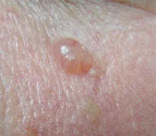
Basal Cell Carcinoma (BCC) is the most common skin cancer and has specific clinical features, including:

  • Chronic growth: Basal cell carcinomas generally grow slowly, usually over months to years. Lesions of basal cell carcinoma usually enlarge gradually over months to years.
  • Rare transfer: Basal cell carcinoma rarely transfer far away, but it can invade surrounding tissues and organs if not treated promptly.
  • Presentation: BCC usually appears as skin colored, pink, or pigmented mass that may be accompanied by bleeding, erosion, or ulcers.
  • Marginal features: The margins of basal cell carcinoma are usually well-defined, semi-circular or oval in shape.
  • Central depression: BCC often has a central depression or ulcer.
  • Common Sites: Basal cell carcinoma is most common in areas with high exposure to ultraviolet light, such as the face, scalp, neck and back.
BCC symptom

Basal cell carcinoma dermoscopy

What is dermoscope?
Dermoscope is also known as transmittance microscope of skin.It can remove the reflection from the surface of skin lesions through polarization or nonpolarized mode, and observe subtle pigmentation loss deep in the skin that is invisible to the naked eye, as well as changes in skin texture and blood vessel structure. Under dermoscopy, skin doctors can comprehensively
detect and diagnose the skin lesions of skin diseases. Just like a physician’s stethoscope, dermoscope is a non-invasive examination tool commonly used in dermatology.
Such as dermoscope can help detect early cancers that is difficult to detect with the naked eyes. Especially, dermoscopy is useful for observing key features like lesion boundaries and vascular distribution of basal cell carcinoma. So that dermatologists can diagnose and treat basal cell carcinoma accurately and timely. Basal cell carcinoma dermoscope means greatly in medical field.

What are the characteristics of basal cell carcinoma dermoscopy?
There are some key characteristics of basal cell carcinoma dermoscopy like belowing:

  • Border: Irregular edge
  • Color: Translucent, pearl white, skin color or pink
  • Shape: Lumps, papules, spots, balls, scars
  • Distribution: Various morphological vascular distribution, such as linear and dendritic
  • Others: Localized ulceration, basal cells may ooze, itch, bleed or crust
basal cell carcinoma dermoscopy

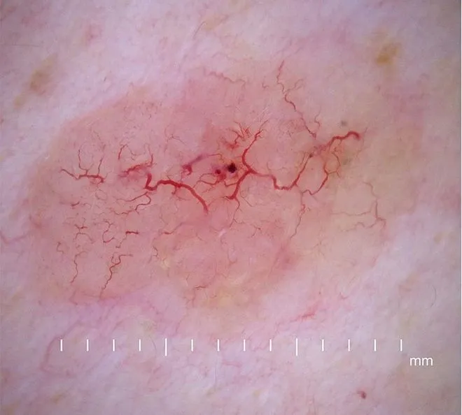
It’s important to note that everyone’s features of basal cell carcinoma dermoscopy may look different. Dermoscopy indeed plays a very important role in the analysis and diagnosis of basal cell carcinoma.

Application of basal cell carcinoma dermoscopy

A dermoscope is a microscope used to examine skin lesions. In the diagnosis and treatment of basal cell carcinoma, basal cell carcinoma dermoscopy is widely used in the following aspects:

Early diagnosis: By magnifying and permeating the details of skin lesions, dermoscopy allows doctors to identify the characteristics of basal cell carcinoma more clearly, so that lesions can be detected and treated as early as possible. It can help doctors accurately detect and diagnose basal cell carcinoma at its early stage.

Treatment monitoring: For patients who have already been diagnosed with basal cell carcinoma, dermoscopy can be used to monitor the whole changes in the lesion at any time.

Feedback comparison: Physicians can compare dermoscopic images at different time to assess the effectiveness of treatment and then determine whether treatment needs to be adjusted.

Surgical aid: When surgically treating basal cell carcinoma, dermoscopy can help the doctor confirm the boundaries and depth of the lesion, not only allowing for more precise removal of the tumor but also less damage to healthy skin.

Precautions for basal cell carcinoma dermoscope in daily life
Dermoscopy is a necessary and reliable technique for skin analysis in daily life, while there some main matters need attention:

  • Correct operation: Know how to use the dermoscope correctly when detect the basal cell carcinoma. In addition,make sure to clean and disinfect the equipment to avoid cross infection or the spread of bacteria before using the dermoscope.
  • Proper light environment: Ensure the working environment of the dermoscope is under appropriate light, too strong or too weak light both will reduce the effect of basal cell carcinoma dermoscopy.
  • Photo documentation: Know how to take photos and keep records for later analysis and evaluation, and comparison of before and after the basal cell carcinoma dermoscopy.
  • Communicate with dermatologist: While a dermoscope can help you detect skin lesions better, any suspicious skin lesions noted, please have them evaluated by a dermatyologist.

It is very helpful to use basal cell carcinoma dermoscopy at its early stage. Dermoscopy plays a significant role in the diagnosis, treatment, and monitoring of basal cell carcinoma. Dermatoscope not only improves the accuracy of diagnosis, guides treatment decisions, but also enhance confidence for skin doctors and patience. People should pay more attention to self-examination of basal cell carcinoma dermoscopy regularly.

Share this article

0

No products in the cart.

Have questions on gear or your order?

Our Gear Guides are here to help! Get personal advice from pro creatives

Name
Subject
Email address
How can we help?

Instant Answers

How can dermoscopy images be captured?

Dermoscopy images can be captured and stored in different ways, such as: • Using a smartphone or tablet with dermoscopic adapter, which consisted in the package.• Using a digital camera

Dermoscopy images can be captured and stored in different ways, such as:

• Using a smartphone or tablet with dermoscopic adapter, which consisted in the package.
• Using a digital camera with dermoscopic adapter, there’s 49mm screw size camera adapter available to order now.

Compatible phone/tablet models:
All iPhone models, 95% Android phones, 90% tablet. For phone/tablet size in 5.25-14mm

Compatible camera models:
All camera with built 49mm filter screw, such as Canon EOS 70D, 80D, 90D; Canon EOS R7, R10, R50, R100; Canon M100, M200, M50, Mark II; Canon G7X Mark III, Sony ZV-1

How can I connect my phone to my dermatoscope?

There’s universal phone adapter for all our dermoscopes. Please check the installation procedure bellow or watch operation guide. Smartphone Connector (1) Place phone adapter screw in the center of smartphone’s

There’s universal phone adapter for all our dermoscopes. Please check the installation procedure bellow or watch operation guide.

Smartphone Connector

(1) Place phone adapter screw in the center of smartphone’s main camera.
(2) Screw magnet attachment on phone adapter.
(3) Put dermoscope’s back ring and magnet attachment together

Take The Best Images

You need to adjust the focus ring after the dermoscpe connected on smartphone to get the best images.

How can I clean my dermoscopy after usage?

Cleaning your dermoscopy after usage is important to prevent cross-contamination and infection. The cleaning method may vary depending on the type and model of your dermoscopy, so you should always

Cleaning your dermoscopy after usage is important to prevent cross-contamination and infection. The cleaning method may vary depending on the type and model of your dermoscopy, so you should always follow the manufacturer’s instructions. However, some general steps are:

• Turn off and disconnect your dermoscopy from any power source or device.

• Wipe off any visible dirt or debris from the dermoscopy with a soft cloth or tissue.

• Disinfect the dermoscopy with an alcohol-based wipe or spray, or a disinfectant solution recommended by the manufacturer. Make sure to cover all surfaces, especially the lens and contact plate.

• Let the dermoscopy air dry completely before storing it in a clean and dry place.

• Do not use abrasive or corrosive cleaners, solvents, or detergents that may damage the dermoscopy.

• Do not immerse the dermoscopy in water or any liquid, unless it is waterproof and designed for immersion.

You should clean your dermoscopy after each use, or at least once a day if you use it frequently. You should also check your dermoscopy regularly for any signs of damage or malfunction, and contact the manufacturer or service provider if needed.

Polarized VS Non-polarized Dermoscopy

A dermoscopy is a device that allows the examination of skin lesions with magnificationand illumination. By revealing subsurface structures and patterns that are not visible tothe naked eye. It can

A dermoscopy is a device that allows the examination of skin lesions with magnificationand illumination. By revealing subsurface structures and patterns that are not visible tothe naked eye. It can improve the diagnose accuracy of skin lesions, such as melanoma,basal cell carcinoma, seborrheic keratosis, etc.

There are two main types of dermoscopy: Non polarized and polarized dermoscopy.We’ve fitted most of our dermoscopys with polarized and non-polarized light. They canbe used in multiple skin structures.

Non-polarized contact Mode

In non-polarized mode, the instrument can provide information about the superficialskin structures, such as milia-like cysts, comedo-like openings, and pigment in theepidemis.

The dermoscopy requires applying a liquid such as mineral oil or alcohol to the skin andplacing the lens in contact with the skin. This reduces surface reflection and enhancesthe view of subsurface structures.

Image with non-polarized light (DE-3100)

Polarized contact Mode

In polarized mode, the instrument allows for visualization for deeper skin structures,such as blood vessels, collagen, and pigment in the dermis.

The dermoscopy does not need to be in contact with the skin or use any liquid. Theirpolarized light can help to eliminate surface reflection and allow visualization ofvascular structures.

Image with polarized light (DE-3100)

Polarized non-contact Mode

The dermoscopy can also use polarized light to examine the skin without direct contact.

In polarized non-contact mode, the instrument allows for examination infected areasand lesions that are painful for the patient, or the difficult to contact pigmented lesions,such as nails and narrow areas.

The contact plate should be removed in this mode, and it does not require applying aliquid to the skin. As it doesn’t require pressure or fluid application on the skin, it canalso avoid cross-contamination and infection risk.

Image in polarized non-contact mode (DE-3100)

How effectiveness is dermoscopy

Compared with visual inspection, the dermoscopy can be used to capture and store skin lesion photos, which play an important role in early skin cancer examination. The dermoscopy allows the

Compared with visual inspection, the dermoscopy can be used to capture and store skin lesion photos, which play an important role in early skin cancer examination.

The dermoscopy allows the examination of skin lesions with magnification and illumination. This can be greatly avoiding the factors that cause interference to visual detection. Such as lighting, skin color, hair and cosmetics.

Several studies have demonstrated that dermoscopy is useful in the identification of melanoma, when used by a trained professional.

It may improve the accuracy of clinical diagnosis by up to 35%
It may reduce the number of harmless lesions that are removed
In primary care, it may increase the referral of more worrisome lesions and reduce the referral of more trivial ones

A 2018 Cochrane meta-analysis published the accuracy of dermoscopy in the detection.

Table 1. Accuracy of dermoscopy in the detection of melanoma in adults
Detection Method Sensitivity, % Specificity, % Positive Likelihood Ratio NegativeLikelihood Ratio
Visual inspection alone (in person) 76 75 3.04 0.32
Dermoscopy with visual inspection (in person) 92 95 18 0.08
Image-based visual inspection alone (not in person) 47 42 0.81 1.3
Dermoscopy with image-based visual inspection (not in person) 81 82 4.5 0.23
ROC—receiver operating characteristic. *Estimated sensitivity calculated on the summary ROC curve at a fixed specificity of 80%.

As we can see, the dermoscope can improve the accuracy of diagnosis of skin lesions, especially melanoma.

Table 1. Accuracy of dermoscopy in the detection of melanoma in adults
Detection Method Sensitivity, % Specificity, % Positive Likelihood Ratio NegativeLikelihood Ratio
Visual inspection alone (in person) 79 77 3.4 0.27
Dermoscopy with visual inspection (in person) 93 99 93 0.07
Image-based visual inspection alone (not in person) 85 87 6.5 0.17
Dermoscopy with image-based visual inspection (not in person) 93 96 23 0.07
ROC—receiver operating characteristic. *Estimated sensitivity calculated on the summary ROC curve at a fixed specificity of 80%.

Characteristics of the dermatoscopic structure of the skin lesions include:

• Symmetry or asymmetry
• Homogeny/uniformity (sameness) or heterogeny (structural differences across the lesion)
• Distribution of pigment: brown lines, dots, clods and structureless areas
• Skin surface keratin: small white cysts, crypts, fissures
• Vascular morphology and pattern: regular or irregular
• Border of the lesion: fading, sharply cut off or radial streaks
• Presence of ulceration

There are specific dermoscopic patterns that aid in the diagnosis of the following pigmented skin lesions:

• Melanoma
• Moles (benign melanocytic naevus)
• Freckles (lentigos)
• Atypical naevi
• Blue naevi
• Seborrhoeic keratosis
• Pigmented basal cell carcinoma
• Haemangioma

Hot Search Terms