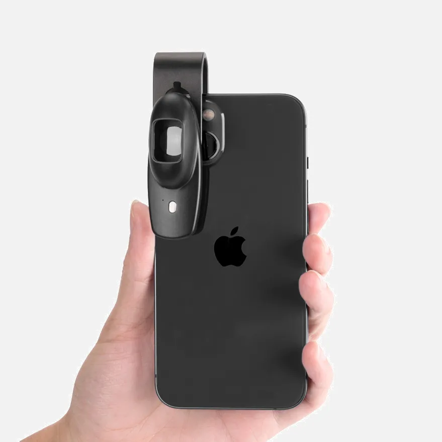
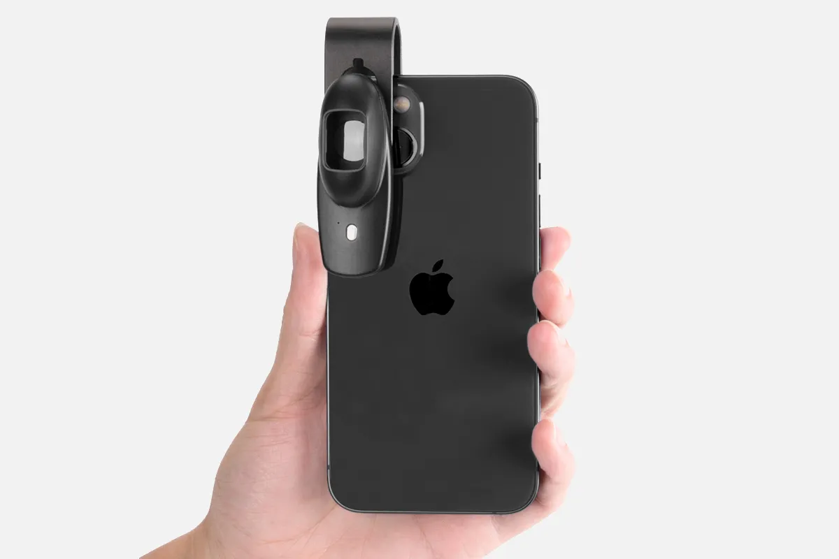
What Is The Dermoscopy Price?

Dermoscope prices range depending on included features and quality. Basic manual models with fixed magnification may cost $200-$400. More advanced digital dermoscopes with enhanced visualization modes, connectivity, storage, and software range from $500 up to $5000+.

Some example price points include:

  • DermLite DL100: A leading affordable manual option at $395
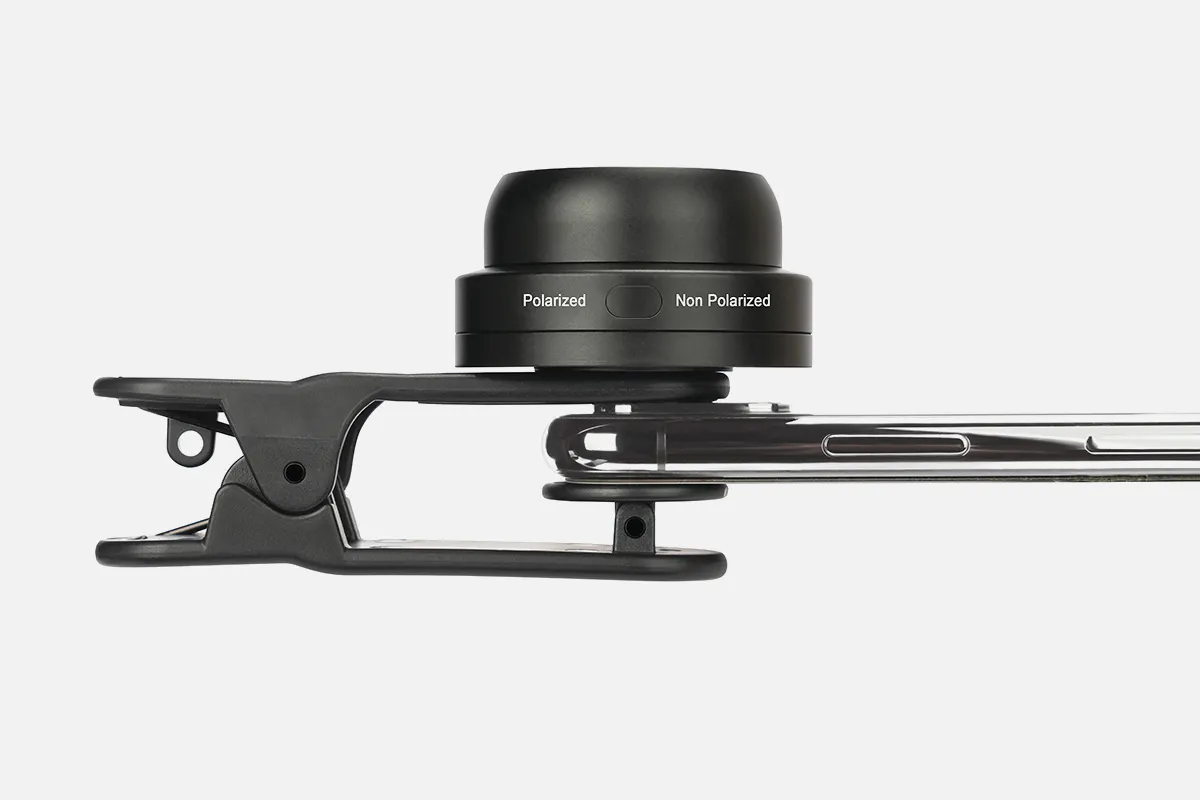
  • Lumio Cross polarized Dermatoscope: $595
  • DermLite DL200 Hybrid: Digital model with smartphone connectivity at $1025
  • Lumio2 With UV Imaging: $995
  • 3Gen DermLite IV DL4: Advanced digital model at $1350

Pricing is impacted by optics like magnification power, lighting type (polarized vs. non-polarized), attachment accessories, image/video capture capabilities, durability, and bundled software.

Reputable dermoscopy models start around $300. Mid-range options with improved optics and digital add-ons generally span $500-$1500. Top-tier advanced digital systems with the highest quality components, maximum versatility, and software ecosystem integration are priced at over $1500.

Explanation of Dermoscopy Pricing Considerations

Dermoscope prices vary greatly depending on the functionality and features provided. Basic manual models with fixed magnification and illumination may cost $200-400. More sophisticated digital dermascopes range from $500 up to $5000.

Key variables that impact pricing typically include:

  • Magnification power and optical lens quality
  • Type of illumination (polarized, non-polarized, UV, etc.)
  • Digital sensor resolution for image/video capture capabilities
  • Accompanying mobile device connectivity and software
  • Storage capacity, data tracking, computer-assisted analysis
  • Durability and construction quality

High-end dermoscopes leveraging the latest CMOS/CCD technology, multi-spectral lighting, and cloud analytics can provide advanced digital capabilities for thousands more than a basic model. Reputable brands also command premium pricing in the market relative to generics.

Purchasing cost depends greatly on the clinical setting's required imaging, diagnostic, and integration needs. Medical facilities should evaluate use cases to determine suitable solutions balancing price, performance, and usability.

People May Ask

What Is Scheduled for A Dermoscopy?

The Hadley Wood Hospital in High Barnet is where we offer our private dermoscopy treatments. Dermoscopy is a skin examination process that allows a dermatologist to examine skin markings up close using a device called a dermatoscope, which is a special magnifying glass.

Does A Mole's Appearance Allow A Doctor to Determine Whether It Is Cancerous?

In order to diagnose skin cancer, a skin biopsy is always necessary.A skin biopsy is what your dermatologist will do to get rid of the spot. It is imperative to have a skin biopsy. It's the sole method for determining if you have skin cancer. There is no other way to be certain.

What Does A Dermoscopy Aim to Achieve?

A handheld device known as a dermatoscope is used to perform dermoscopy. Subsurface skin structures in the epidermis, papillary dermis, and dermoepidermal junction-structures that are often invisible to the unaided eye-can be seen thanks to this method [2-4].

Could A Dermatologist Determine Whether It's Cancer?

The dermatologist will examine your skin and inquire about any changes you may have seen. To examine the skin up close, they might use a magnifying glass. In order to send a small portion of the afflicted skin to a lab for a cancer screening, the specialist might also advise having it removed.

Which Time Is Best for A Dermoscopy?

A basic skin exam is called a dermoscopy. Your doctor might do a dermatoscopy if you have a mole or pigmented skin lesion that is alarming. With a dermoscopy, your doctor can more precisely diagnose pigmented skin lesions, sometimes preventing the need for a skin sample or needless mole excision.

Can Melanoma Be Ruled out with Dermoscopy?

Melanoma lesions lacking traditional characteristics on both dermoscopy and visual inspection are referred to as featureless melanoma. Menzies (1996), a dermatologist who was a pioneer in dermoscopy, acknowledged this shortcoming while demonstrating that 8% of melanomas escaped detection using dermoscopy.

Is Dermoscopy Secure?

Dermoscopy is a non-invasive procedure that carries little risk of consequences. The rare chance of cross-infection between patients is the only problem, particularly when using contact dermoscopy.

Does Skin Cancer Show up during A Dermoscopy?

It is very helpful in separating melanoma from other pigmented lesions and in the early detection of malignant melanoma.

Is Dermatoscopy Applicable to General Practitioners?

According to research, GPs may be able to more precisely identify and prioritize skin lesions, including possible skin malignancies, when they utilize dermoscopy.

Is A Dermascope A Consumable?

The DocCheck Shop is the ideal location for you! We provide an extensive range of premium dermatoscopes from leading brands, including LUXAMED, Spengler, and HEINE Optotechnik. An overview of our goods and their details may be found below, allowing you to select the one that best meets your needs.

Dermoscopy Price Products

China smartphone dermoscopy price

Dermoscopy 1 Har/Psc第一 版本: A Useful Guide

China smartphone dermoscopy price suppliers & manufacturers

An Atlas of Dermoscopy, Third Edition

China affordable dermoscopy price suppliers & manufacturers

2018 版本 Comprehensive Atlas of Dermatoscopy Cases, First Ed.

China affordable dermoscopy price

A Conspectus in the Skin of Color: Dermoscopy - Histopathology Correlation, First Edition, 2021 版本

China portable dermoscopy price

First edition, 2023 版本 Clinical and Dermoscopic Atlas of Non-Neoplastic Dermatoses: Variability According to Phototypes

China customized dermoscopy price suppliers & manufacturers

Dermoscopy, an Edition of Dermatologic Clinics (The Clinics: Dermatology Book 31) 1欬一 퉈本, Kindle电子乘

dermoscopy price suppliers & manufacturers

Kindle book Quick Guide to Dermoscopy in Laser and IPL Treatments, 1st ed., 2020 扈本

China portable dermoscopy price suppliers & manufacturers

Paperback version of Atlas de dermoscopie - October 30, 2013

China Professional dermoscopy price suppliers & manufacturers

GLEAVI 1 PC Metal Cleaner and Plastic Trash Can Stainless Steel Pan Cleaner with Comfort Grip Dust Tray for Desktop and Household Cleaning, Garbage Shovel for Kitchen Dust Tray Cleaning

China wholesale dermoscopy price suppliers & manufacturers

100 pieces Starburst Indications Luminous Indicators Neon Retail Sales Labels with Starburst Signs Signs for garage sales and yards Announcements with Burst Paper Signs 4.7 x 6.3-inch poster board decoration

Top Reviews

Customer

It was a really pleasant experience dealing with IBOOLO; great shipping time, clear instructions, wonderful quality papers, and excellent images.

Jonathan Jeter

Very nicely written. Comparable to the second version, but with enough new content to justify the price.

Boutros Ghassibi

By Orit Markowitz, DERMOSCOPY: Excellent in every way, both technically and in terms of teaching this craft. The paper is beautiful, the images and explanation drawings are crystal clear. The author is, in my opinion, a very skilled and successful teacher. The book is well worth the amount it costs. Production of it has involved a great deal of work. Pierre Ghassibi, M.D.

0

Have questions on gear or your order?

Our Gear Guides are here to help! Get personal advice from pro creatives

Name
Subject
Email address
How can we help?

Instant Answers

How Can Dermoscopy Images Be Captured?

Dermoscopy images can be captured and stored in different ways, such as: • Using a smartphone or tablet with dermoscopic adapter, which consisted in the package.• Using a digital camera

Dermoscopy images can be captured and stored in different ways, such as:

• Using a smartphone or tablet with dermoscopic adapter, which consisted in the package.
• Using a digital camera with dermoscopic adapter, there’s 49mm screw size camera adapter available to order now.

Compatible phone/tablet models:
All iPhone models, 95% Android phones, 90% tablet. For phone/tablet size in 5.25-14mm

Compatible camera models:
All camera with built 49mm filter screw, such as Canon EOS 70D, 80D, 90D; Canon EOS R7, R10, R50, R100; Canon M100, M200, M50, Mark II; Canon G7X Mark III, Sony ZV-1

How Can I Connect My Phone to My Dermatoscope?

There’s universal phone adapter for all our dermoscopes. Please check the installation procedure bellow or watch operation guide. Smartphone Connector (1) Place phone adapter screw in the center of smartphone’s

There’s universal phone adapter for all our dermoscopes. Please check the installation procedure bellow or watch operation guide.

Smartphone Connector

(1) Place phone adapter screw in the center of smartphone’s main camera.
(2) Screw magnet attachment on phone adapter.
(3) Put dermoscope’s back ring and magnet attachment together

Take The Best Images

You need to adjust the focus ring after the dermoscpe connected on smartphone to get the best images.

How Can I Clean My Dermoscopy after Usage?

Cleaning your dermoscopy after usage is important to prevent cross-contamination and infection. The cleaning method may vary depending on the type and model of your dermoscopy, so you should always

Cleaning your dermoscopy after usage is important to prevent cross-contamination and infection. The cleaning method may vary depending on the type and model of your dermoscopy, so you should always follow the manufacturer’s instructions. However, some general steps are:

• Turn off and disconnect your dermoscopy from any power source or device.

• Wipe off any visible dirt or debris from the dermoscopy with a soft cloth or tissue.

• Disinfect the dermoscopy with an alcohol-based wipe or spray, or a disinfectant solution recommended by the manufacturer. Make sure to cover all surfaces, especially the lens and contact plate.

• Let the dermoscopy air dry completely before storing it in a clean and dry place.

• Do not use abrasive or corrosive cleaners, solvents, or detergents that may damage the dermoscopy.

• Do not immerse the dermoscopy in water or any liquid, unless it is waterproof and designed for immersion.

You should clean your dermoscopy after each use, or at least once a day if you use it frequently. You should also check your dermoscopy regularly for any signs of damage or malfunction, and contact the manufacturer or service provider if needed.

Polarized VS Non-Polarized Dermoscopy

A dermoscopy is a device that allows the examination of skin lesions with magnificationand illumination. By revealing subsurface structures and patterns that are not visible tothe naked eye. It can

A dermoscopy is a device that allows the examination of skin lesions with magnificationand illumination. By revealing subsurface structures and patterns that are not visible tothe naked eye. It can improve the diagnose accuracy of skin lesions, such as melanoma,basal cell carcinoma, seborrheic keratosis, etc.

There are two main types of dermoscopy: Non polarized and polarized dermoscopy.We’ve fitted most of our dermoscopys with polarized and non-polarized light. They canbe used in multiple skin structures.

Non-polarized contact Mode

In non-polarized mode, the instrument can provide information about the superficialskin structures, such as milia-like cysts, comedo-like openings, and pigment in theepidemis.

The dermoscopy requires applying a liquid such as mineral oil or alcohol to the skin andplacing the lens in contact with the skin. This reduces surface reflection and enhancesthe view of subsurface structures.

Image with non-polarized light (DE-3100)

Polarized contact Mode

In polarized mode, the instrument allows for visualization for deeper skin structures,such as blood vessels, collagen, and pigment in the dermis.

The dermoscopy does not need to be in contact with the skin or use any liquid. Theirpolarized light can help to eliminate surface reflection and allow visualization ofvascular structures.

Image with polarized light (DE-3100)

Polarized non-contact Mode

The dermoscopy can also use polarized light to examine the skin without direct contact.

In polarized non-contact mode, the instrument allows for examination infected areasand lesions that are painful for the patient, or the difficult to contact pigmented lesions,such as nails and narrow areas.

The contact plate should be removed in this mode, and it does not require applying aliquid to the skin. As it doesn’t require pressure or fluid application on the skin, it canalso avoid cross-contamination and infection risk.

Image in polarized non-contact mode (DE-3100)

How Effectiveness Is Dermoscopy

Compared with visual inspection, the dermoscopy can be used to capture and store skin lesion photos, which play an important role in early skin cancer examination. The dermoscopy allows the

Compared with visual inspection, the dermoscopy can be used to capture and store skin lesion photos, which play an important role in early skin cancer examination.

The dermoscopy allows the examination of skin lesions with magnification and illumination. This can be greatly avoiding the factors that cause interference to visual detection. Such as lighting, skin color, hair and cosmetics.

Several studies have demonstrated that dermoscopy is useful in the identification of melanoma, when used by a trained professional.

It may improve the accuracy of clinical diagnosis by up to 35%
It may reduce the number of harmless lesions that are removed
In primary care, it may increase the referral of more worrisome lesions and reduce the referral of more trivial ones

A 2018 Cochrane meta-analysis published the accuracy of dermoscopy in the detection.

Table 1. Accuracy of dermoscopy in the detection of melanoma in adults
Detection Method Sensitivity, % Specificity, % Positive Likelihood Ratio NegativeLikelihood Ratio
Visual inspection alone (in person) 76 75 3.04 0.32
Dermoscopy with visual inspection (in person) 92 95 18 0.08
Image-based visual inspection alone (not in person) 47 42 0.81 1.3
Dermoscopy with image-based visual inspection (not in person) 81 82 4.5 0.23
ROC—receiver operating characteristic. *Estimated sensitivity calculated on the summary ROC curve at a fixed specificity of 80%.

As we can see, the dermoscope can improve the accuracy of diagnosis of skin lesions, especially melanoma.

Table 1. Accuracy of dermoscopy in the detection of melanoma in adults
Detection Method Sensitivity, % Specificity, % Positive Likelihood Ratio NegativeLikelihood Ratio
Visual inspection alone (in person) 79 77 3.4 0.27
Dermoscopy with visual inspection (in person) 93 99 93 0.07
Image-based visual inspection alone (not in person) 85 87 6.5 0.17
Dermoscopy with image-based visual inspection (not in person) 93 96 23 0.07
ROC—receiver operating characteristic. *Estimated sensitivity calculated on the summary ROC curve at a fixed specificity of 80%.

Characteristics of the dermatoscopic structure of the skin lesions include:

• Symmetry or asymmetry
• Homogeny/uniformity (sameness) or heterogeny (structural differences across the lesion)
• Distribution of pigment: brown lines, dots, clods and structureless areas
• Skin surface keratin: small white cysts, crypts, fissures
• Vascular morphology and pattern: regular or irregular
• Border of the lesion: fading, sharply cut off or radial streaks
• Presence of ulceration

There are specific dermoscopic patterns that aid in the diagnosis of the following pigmented skin lesions:

• Melanoma
• Moles (benign melanocytic naevus)
• Freckles (lentigos)
• Atypical naevi
• Blue naevi
• Seborrhoeic keratosis
• Pigmented basal cell carcinoma
• Haemangioma

No products in the cart.

No products in the cart.

Hot Search Terms