


DE-400 Dermatoscope – IBOOLO
Unveiling The Secrets of The Skin: The Vital Role of Dermatoscopes for Modern Dermatology
The Skin's Roadmap
Every person's skin is like a unique map, chronicling the journey of our lives. However, sometimes this "map" may reveal suspicious landmarks—moles, bumps, or other abnormalities. It is then that dermatologists need to carefully decipher the skin's code to detect potential health risks. Their indispensable tool for this task is the dermatoscope.
A Window into The Skin's Microscopic World
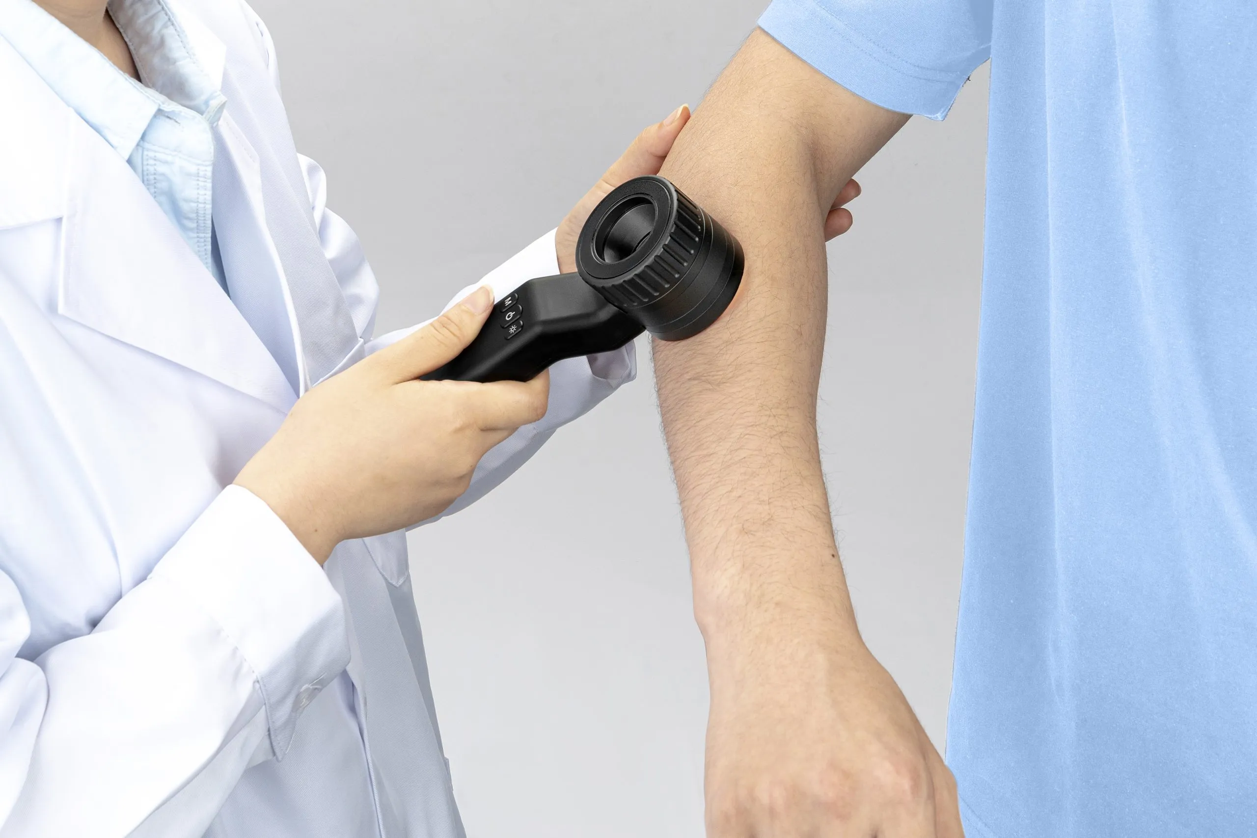
Dermatoscopes, with their optical lenses and polarized light sources, magnify minute skin structures that are invisible to the naked eye, opening a window into the skin's microscopic world. Through this window, doctors can observe features like pigment distribution and capillary patterns, enabling them to make more accurate assessments of benign and malignant lesions.
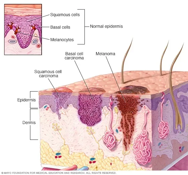
Uncovering Melanoma's Clues
Take malignant melanoma, for instance. In its early stages, this dangerous skin cancer often appears as an innocuous mole to the naked eye. However, under the dermatoscopy's scrutiny, these cancerous growths frequently exhibit irregular borders, multiple colors, and abnormal vasculature—telltale signs that prompt timely detection and treatment. Conversely, benign moles tend to exhibit more uniform and symmetrical patterns when viewed through the dermatoscope, allowing doctors to differentiate them from suspicious lesions requiring further examination.
Beyond Cancer Detection
The dermatoscope's utility extends far beyond the realm of cancer detection. In inflammatory skin conditions like psoriasis, it can reveal pathological changes such as epidermal scaling and capillary dilation, helping doctors evaluate disease activity and tailor treatment plans accordingly. Even in cosmetic dermatology, dermatoscopes can precisely delineate the boundaries of lesions, minimizing damage to healthy tissue during surgical excisions.
Mastering The Art of Dermatoscopy
Harnessing the full potential of dermatoscopes, however, requires specialized training. Doctors must not only master the proper operation techniques but also memorize the distinctive "faces" of various skin conditions under magnification. To this end, some medical centers are exploring the integration of artificial intelligence into dermatoscopic diagnosis, leveraging deep learning algorithms to intelligently identify microscopic image patterns and assist doctors in their interpretations, ultimately enhancing diagnostic efficiency.
The Future of Dermatoscopy: Mobile And Accessible
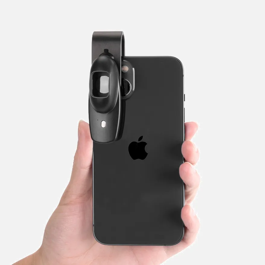
Notably, dermatoscopes are not confined to clinical settings. Micro-dermatoscopes integrated into smartphones and other mobile devices are bringing this technology into homes worldwide. Individuals can now perform preliminary self-examinations and share images with professionals for remote guidance, fostering greater public awareness of skin health issues.
A Pivotal Tool in Modern Dermatology
Without a doubt, dermatoscopes are becoming indispensable tools in modern dermatology. Looking ahead, as instrument capabilities continue to improve and emerging technologies like artificial intelligence and mobile healthcare converge, this "vision enhancer" promises to deliver even more precise, efficient, and personalized diagnostic and treatment experiences for patients, ensuring healthy and radiant skin for all.
People May Ask
The following list of resources can assist dermatologists carry out their work responsibilities efficiently:Skin-cutting shear.Extractor for blackheads.A scalpel.Cutaneous biopsy punch.Microscope.Gloves.A laser dermabrasion apparatus.Bright, pulsating light.Additional things...
What Separates Dermatology from Dermatopathology? A medical student must be trained in either dermatology or pathology to become a dermatopathologist. Dermatopathologists acquire biopsy specimens, examine the tissue, and diagnose patients; dermatologists treat the patients.
Over the past few decades, advancements in dermatoscopy, skin imaging, immune-dermatology, lasers, and esthetic dermatology have expanded the field of dermatology into new areas. Dermatologists' approach to patient care is being completely transformed by new developments in rapid diagnostics and technology.
Lentigos, or accumulations of melanocytes in the cell's basal layer, are the most prevalent pigmented lesions. Although it is more frequently mistaken for a nevus, it can have the clinical look of a freckle. Although the margins are not clear, the lesion is not elevated.
Usually, pigmented lesions are not a reason for alarm. Nevertheless, some can progress to become distinct types of skin cancer. That's why it's critical that you and your physician keep a careful eye on them.
When recalling the warning indicators of melanoma, the "ABCDE" rule comes in handy:Unbalanced One part of the skin lesion is not the same shape as the other.Border. The edges are blurry, uneven, ragged, or notched.Vibrance. There could be tan, brown, and black tones present.Measurement of diameter.Evolving.
Dermoscopy has the potential to be a useful diagnostic tool for nail psoriasis since it can produce consistent clinical findings quickly and painlessly. This review outlines the most recent data on the distinct dermoscopic characteristics of nail psoriasis.
UV radiation from artificial sources is used by humans to initiate biologic processes that lower inflammation and slow down the growth of skin cells. This kind of light helps to cleanse the skin and reduce inflammation in the affected area when applied on a regular basis.
For a microscopic view of the cells, the dermatologist will cut a tiny slice of skin or the outer layer of the epidermis. For the light from the dermatologist microscope to travel through the sample and disclose any flaws or disease indicators, it must be thin.
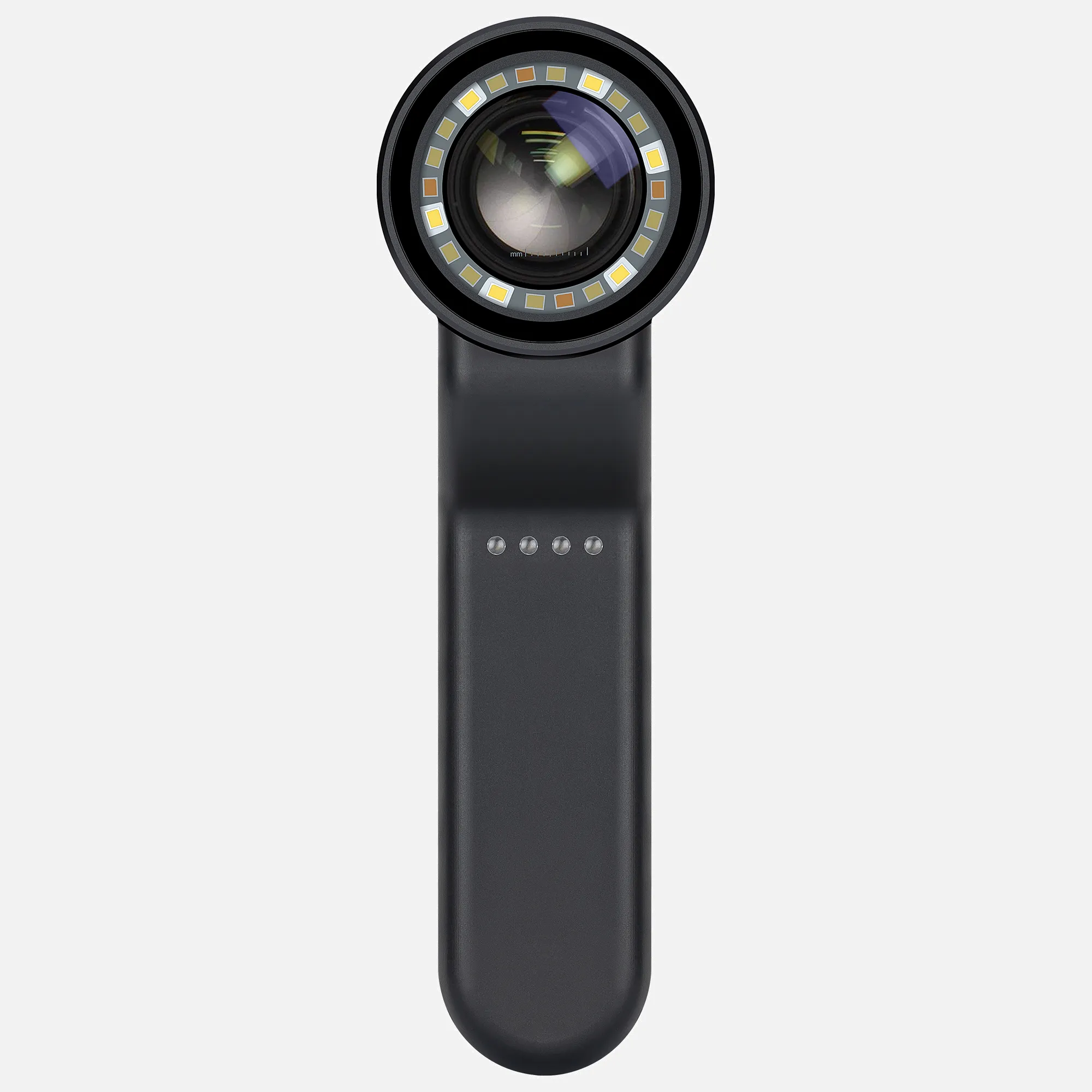
A dermatoscope is a portable visual aid that a medical professional or layperson can use to inspect and diagnose diseases and skin lesions, including melanoma. It can also be useful for inspecting the nails, hair, and scalp. In a dermatologist's practice, a dermatoscope is usually present.
Dermatoscope for Dermatology Products
Wireless 5.5-inch 1080P 10 million pixel LCD digital microscope with wireless zoom and magnification up to 1000x A USB stereo microscope camera with a 10 million pixel camera and an HD screen video recorder
This coin PCB soldering repair plant features an LCD digital USB microscope, a 4.3-inch screen with 1000x magnification, an adjustable stand, a rechargeable battery, and eight LED lights.
Dermatoscopy: Examining pigmented and non-pigmented lesion patterns
Featuring a stand, the Bysameyee 4K 3840x2160P Wireless Digital Microscope is a portable high-definition USB inspection camera borescope with a 50x–1000x magnification that works with iPhone, iPad, Android, Tablet, Windows, and Mac computers.
Aopick Handheld USB 1440P HD Inspection Digital Microscope Camera with 50x–1600x Magnification Carry-around Pocket Microscopes featuring 8 LEDs and a stand that work with iPhone and Android devices
USB Digital Microscope: SKYEAR 50X-1600X Magnification Handheld Digital Microscope with Adjustable Stand, 8 LED Lights, and Portable Microscope Camera for Kids and Adults – Compatible with iOS & Android Devices
First Edition of Dermoscopy Guide
Compatible with iPhone, iPad, Android, Windows, Mac computer, this wireless digital microscope is the SKYBASIC 50X–1000X Magnification WiFi USB HD Portable Handheld Pocket Microscopes Camera with 8 Adjustable LEDs.
1–1000X Magnification Zoom, 4.3 Inch 1080P LCD Digital Microscope 10MP Wireless USB Camera Video Recorder with HD Screen, Stereo Microscope

Fitzpatrick s Color Atlas and Clinical Dermatology Synopsis, Ninth Edition,
Hot Products


News & Blog


Top Reviews
Kindle Customer
I've tried jewelers loupes, but they simply kept breaking. This works pretty well for me. The program was simple to use and straightforward. Works on your wifi, eliminating the need for cords when in use, and it seems to keep a charge well. A surgeon's hand is required to obtain an excellent photo, but if you use the included stand and lay the material underneath, you can easily zoom in and out and operate the device with great ease. I tried to upload a picture, but it wouldn't let me.
TheHandiCapperGeneral
I couldn't get any response from my first one. About my unfavorable rating and the unit I had, the owner of this business emailed me. She gave me a replacement right away and then checked in to make sure I had received the high-quality item she claimed to be selling. This time, the second was a lot clearer. I like a business that values its clients and goes above and beyond to meet their needs!
S
I don't usually write reviews, but I'm happy that I bought this item. The focus is easily adjustable using the huge knob, and the picture is crisp and clear. Merely observing the fibers of an article of clothing makes me feel like a child once more. What degree of magnification would be appropriate for various purposes was one of my concerns while choosing which type to purchase. Having any kind of sense about what 60x actually means in real life is difficult. The universe of clothes fibers and phone screen pixels, which is otherwise invisible, may be seen by magnifying things 20 times, as I have discovered. (*Yes, unsolvable, but not truly invisible.) Allow your children to play with this after you've had a great time.


