


DE-400 Dermatoscope – IBOOLO
Enhanced Visualization of Skin Structures: Understanding The Dermatoscopic Technique
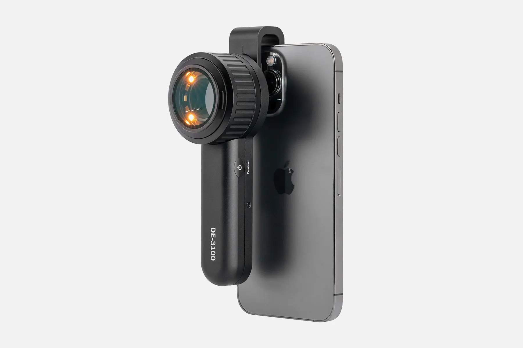
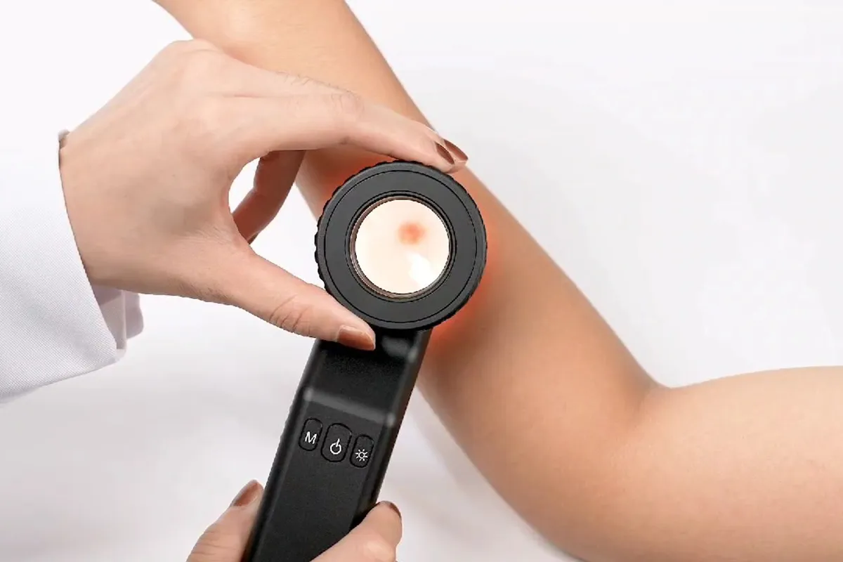
Dermoscopy, also referred to as dermatoscopy or chemiluminescence microscopy, is a non-invasive medical imaging technique that utilizes optical magnification and light to visualize subtle subsurface structures in skin lesions that are not visible to the naked human eye. It is performed using a specialized handheld device called a dermatoscope or dermoscope.
During a dermoscopic exam, a gel is first applied to the skin. Then, the lens of the dermatoscope is held firmly against the lesion to examine it under magnification, typically 10x power or greater. Features like polarized lighting optimize visualization of delicate morphological patterns and blood vessel arrangements in the epidermis, dermo-epidermal junction, and upper dermis.
This enhanced in vivo microscopic perspective of skin structures facilitates expert medical evaluation of lesions - especially important for detecting melanoma and ruling out malignant skin cancers. Dermoscopy improves diagnostic accuracy and sensitivity compared to plain visual skin inspection. It helps identify areas needing biopsy versus benign lesions. Overall, dermoscopy grants physicians a vital window beneath the skin's surface through precision optics.
Decoding Dermatoscopic Imaging: Understanding The Science behind This Diagnostic Tool
Dermoscopy, or epiluminoscopy, is a non-invasive technique for examining the skin's surface. It uses light and magnification to study skin lesions at high magnification.
Dermoscopy works by transilluminating a lesion. This means light reflects, refracts, diffracts, and/or absorbs off the skin. The physical properties of the skin affect these phenomena.
During a dermoscopy assessment, a specialist applies oil or gel to the skin and holds a dermatoscope close to it. The dermatoscope is an instrument that uses light and magnification to evaluate skin lesions.
The stratum corneum reflects light, which reduces the ability to see structures under the skin. Applying alcohol gel to the skin can overcome this refractive property.
According to a 2018 Cochrane review, dermatoscopes are more accurate in diagnosing melanomas than the naked eye alone when used by a trained professional.
People May Ask
A skin examination looks for birthmarks, moles, or other pigmented areas that don't seem normal in terms of size, shape, color, or texture. Your doctor may remove all or part of the abnormal skin during a biopsy, along with a tiny portion of surrounding normal tissue. Under a microscope, a pathologist examines the tissue to hunt for cancerous cells.
A benign tumor has edges that are smooth, regular, and distinct. In comparison to a benign tumor, a malignant tumor grows more quickly and has uneven borders. Moreover, a malignant tumor has the potential to spread to other bodily parts. Although a benign tumor can grow to be rather large, it won't travel to other parts of your body or infiltrate surrounding tissue.
The Use of Dermoscopy in BCC ManagementFor the assessment of residual tumor in BCC patients, dermoscopic examination following nonablative treatment has also shown promise. Remaining dermoscopic characteristics such as arborizing telangiectasias, ulceration, and pigmented formations are good markers of tumor persistence.
relative mass confinement.There is varying degrees of epidermal or follicular attachment.Large nuclear palisade-surrounded basaloid lobules.Because of the overproduction of mucin, loculules may be solid or exhibit central cyst development.Scleromyxoid stroma.A cleft forms in the stroma between the tumor lobules.Instead,Additional things...
How does a BCC look? Open sores, red spots, pink growths, shiny bumps, scars, or growths with slightly elevated, rolling edges and/or a center indentation are some of the appearances of BCCs. BCCs have the potential to leak, crust, itch, or bleed. The lesions usually appear on body parts that are exposed to the sun.
Using a dermascope gives a skin cancer specialist an additional advantage even if they can still detect malignant indications with their eyes. a color and structural clarity that would be invisible to the unaided eye or even a low-powered microscope. Color, particularly the distribution of pigments, can serve as a diagnostic tool in and of itself.
polarized light Non-polarized illuminationInstead,When light is polarized, the electric field only oscillates in one direction. Its electric field is oscillating in all directions.2. Light that is polarized is coherent by nature. Unpolarized light cannot be coherent in the natural world.
Dermoscopy is a non-invasive procedure that carries little risk of consequences. The rare chance of cross-infection between patients is the only problem, particularly when using contact dermoscopy.
Biopsy of the SkinYour dermatologist can typically identify psoriasis simply by looking at your skin. In the event that additional information is required to validate the diagnosis and exclude alternative etiologies of symptoms, such as cutaneous lupus or eczema, a skin biopsy might be conducted.
Your doctor will typically check your nails, scalp, and skin for symptoms of psoriasis in order to make the diagnosis. Inquiries concerning your health, medical history, and family history could also be made, including whether you: Feel sensations like skin that is burning or itching. had a recent illness or undergone a stressful event.
Dermoscopy Products

MEDCASE Radiance Otoscope with Light German Fiber Optic Otoscopes: Excellent for Professional Use - Ear Scope with LED Light and Speculum for Ear Examination and Diagnosis

Cancer (Astrology) - Discovering Love and Harmony in Every Relationship: Boxed Set of Cancer Horoscopes (Relationship Books for Dating Couples) Kindle攵子书

Cancer: A Special Gift for Cancer Patients And 121 Pages of Notepad/Journal with Matte Finish, 6 x 9 平装 – 2020年 1月 6日
Skin Cancer Diagnosis Made Simple: Over 220 full-color case discussions and a tutorial-based approach to skin cancer diagnosis for novices and experts alike. Kindle攵子书
The 30-Day Raw Vegan Plant-Based Detoxification & Regeneration Journal & Tracker: Healing Nevoid Basal Cell Carcinoma Syndrome A Reversing Conditions Journal and Tracker. Journal 2 - February 28, 2020 - Paperback
A Clear Guide to Medical Conditions: Diagnosis and Treatment of Basal Cell Carcinoma Kindle攵子书
The socio-economic significance of PGMs, PGM preconcentration and spectroscopy, chemistry and spectroscopy, adsorption and preconcentration, and PGM separation and determination are all covered in this section.
Ebook Enhanced Segmentation-Based Method Utilizing PSO and K-MEANS Algorithm for Basal Cell Carcinoma Identification from Skin Lesions
Diagnosis and Treatment of Common Disorders in Clinical Dermatology, Second Edition 2第二 版本
Dermoscopy, an Edition of Dermatologic Clinics (The Clinics: Dermatology Book 31) 1欬一 퉈本, Kindle电子乘
Related Products
Hot Products


News & Blog


Top Reviews
ampm247
My second tube of Sunspot Es is this one. Since my first tube's results were so good, I bought a second one after a friend convinced me not to use it for her issue. Regrettably, the FDA is refusing to let the firm disclose that its true benefit is treating minor skin malignancies. I mean business. My personal experience was with a basal cell carcinoma on my ankle that the doctor wanted to remove. Despite my hesitation, I tried this Sunspot. Es product. Over the course of two weeks, it performed flawlessly except from occasional stinging and itching. On his cheek, my spouse had a large area of keratoses. It looked terrible, and it bled every now and again. Reluctantly, he gave the Sunspot Es a try, and was astonished to discover how powerful it was. The spot took around a month to vanish since he neglected to apply a bandage. When applied correctly, it is an excellent product for treating kerotases, squamous cell, or basal cell carcinomas. I was aware that removing a malignancy using traditional techniques results in significant harm or destruction of the surrounding tissue, which
Dawn McKenna
I was pleasantly surprised to find this book contained much more than the dull, elementary content I had assumed based on its cover. This book has an easy-to-read, pleasant, and sympathetic tone, but its substance is jam-packed with reliable scientific data and practical advice. Rather than merely wearing sunscreen and waiting for my next skin check, I'm so happy to finally have information that I can truly utilize to improve myself. I now have a ton of good actions that I can do to strengthen my immunity and maybe stop future eruptions, in addition to the therapeutic alternatives that this book explained. For the first time, I've been informed of alternatives to avoiding the sun, and I find that information to be really valuable.
Cliente
Written for the BEST dermatoscopist, it's an excellent book with appropriate content, good photos, and helpful outlines. It's essential, in my opinion, for anybody with an interest in dermoscopy.


