






Article
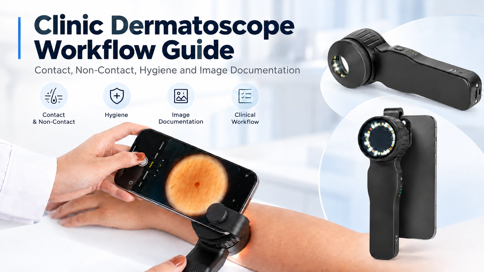
Clinic Dermatoscope Workflow Guide Contact, Non-Contact, Hygiene and Image Documentation
A dermatoscope may be a compact handheld instrument, but using it efficiently in a clinic involves more than switching on the light and examining a lesion. A practical clinic workflow dermatoscope setup needs to consider how the device is prepared, whether contact or non-contact observation is appropriate, how different illumination modes are used, how images…
A dermatoscope may be a compact handheld instrument, but using it efficiently in a clinic involves more than switching on the light and examining a lesion. A practical clinic workflow dermatoscope setup needs to consider how the device is prepared, whether contact or non-contact observation is appropriate, how different illumination modes are used, how images are captured, and how accessories are handled between patients.
At IBOOLO, we believe an easy-to-use dermatoscope should support this complete workflow rather than only provide magnification. For doctors working through multiple consultations each day, accessories such as removable contact plates, non-contact attachments, disposable protective covers and smartphone adapters can be just as relevant as the optical system itself.
This guide looks at how a handheld medical dermatoscope can fit into a structured clinical workflow, with particular attention to contact technique, non-contact observation, hygiene management and image documentation.
What Are the Main Steps in a Clinical Dermatoscope Examination?
A consistent workflow helps clinicians move from visual examination to dermoscopic observation and image documentation without unnecessary interruptions. The exact procedure will depend on the device, the lesion being examined and the clinic’s own infection-control protocol, but a typical workflow can be organized into eight stages.
(1) Prepare the Dermatoscope
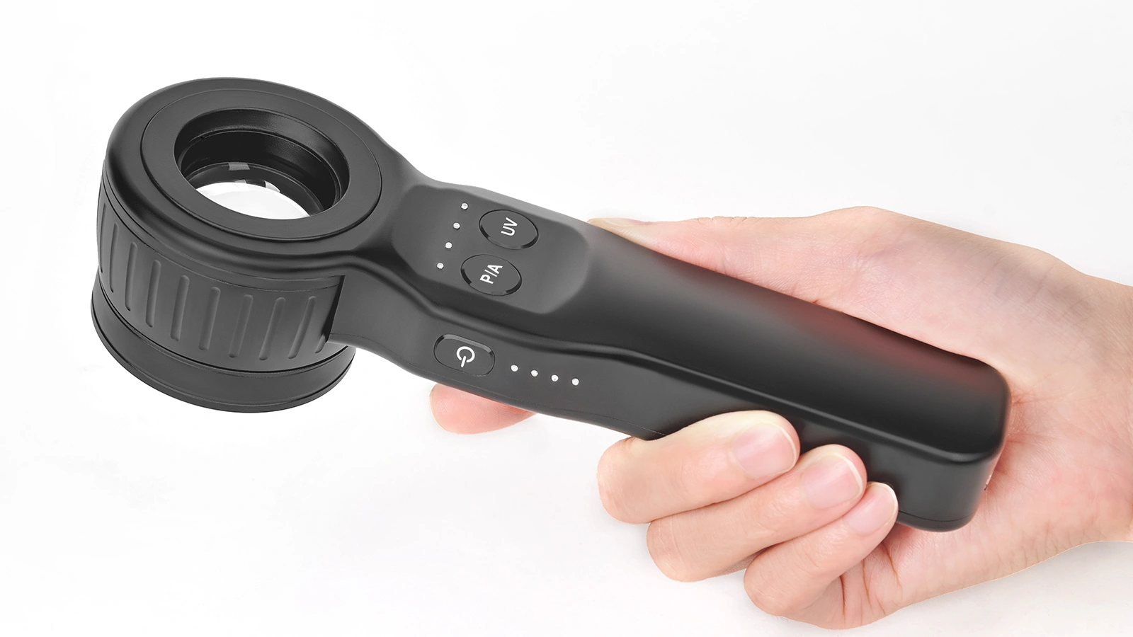
Before beginning the examination, check that the dermatoscope has sufficient battery power and that the required accessories are available. Depending on the planned examination, this may include a standard contact plate, a small contact plate, a non-contact attachment, a disposable protective cover or a smartphone adapter.
(2) Select the Examination Method
The clinician should decide whether the lesion will be examined using contact or non-contact dermoscopy. This choice can depend on the anatomical location, the structures of interest, whether surface contact is desirable and which illumination mode is being used.
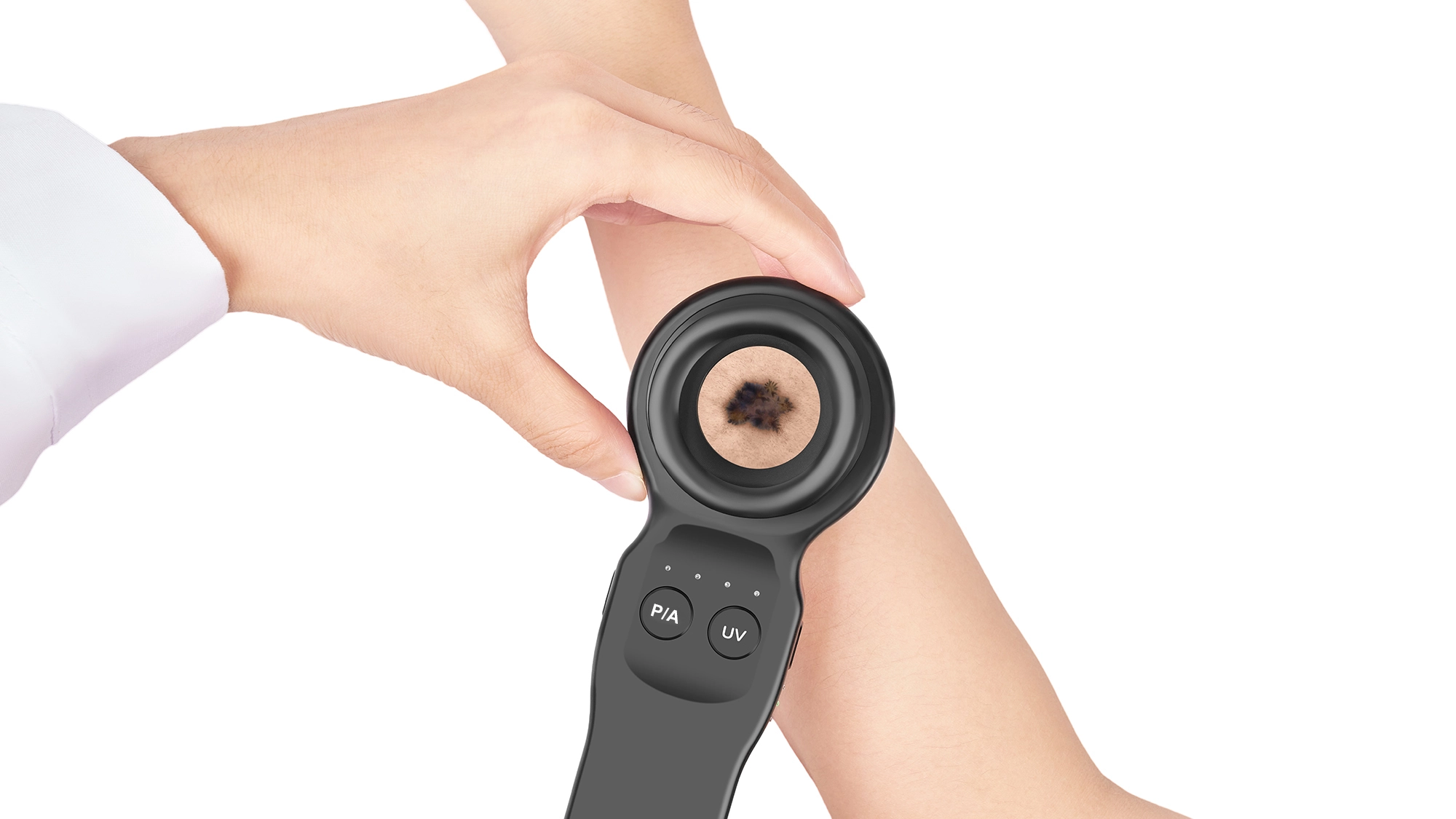
(3) Observe the Lesion
The dermatoscope should be positioned so that the area of interest is clearly visible and in focus.
(4) Compare Illumination Modes When Appropriate
A multi-mode clinical dermatoscope for doctors can provide complementary views of the same area. Polarized illumination can reduce much of the surface glare and make subsurface information easier to observe, while non-polarized contact dermoscopy provides a different view of more superficial structures.
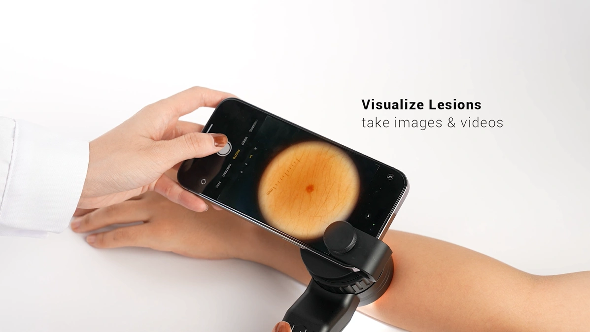
(5) Capture the Image
Once the lesion is correctly positioned and the desired lighting mode has been selected, the clinician can capture a dermoscopic image if documentation is required.
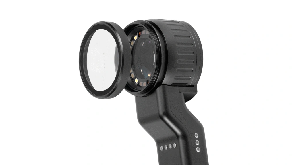
(6) Remove or Process Contact Accessories
Disposable protective components should be discarded after use when designed for single use.
(7) Clean the Device and Prepare It for the Next Patient
The parts of the dermatoscope that may have been contaminated should be cleaned or disinfected according to the healthcare facility’s infection-control policy.
(8) Label and Store the Images
After the examination, images should be associated with the correct patient, anatomical location, date and, where useful, the lighting mode used.
A simple clinical workflow can therefore be summarized as:
Prepare → Select Mode → Observe → Compare Lighting → Capture → Handle Accessories → Clean → Record
What Is the Difference Between Contact and Non-Contact Dermoscopy in Clinical Use?
Contact and non-contact dermoscopy are not simply two ways of holding the same device. They change how the dermatoscope interacts with the skin and can influence both the image and the workflow.
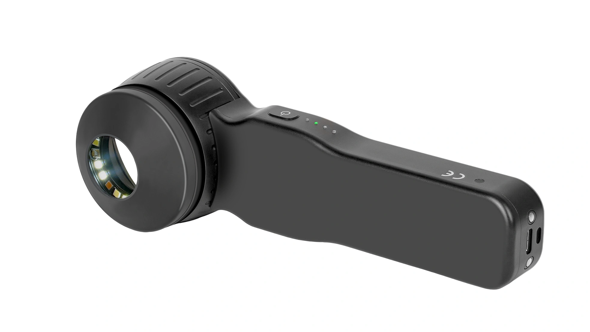
| Workflow Factor | Contact Dermoscopy | Non-Contact Dermoscopy |
| Skin Contact | Contact plate touches the skin | Optical system remains above the skin |
| Pressure | Some pressure may be introduced | No direct plate pressure |
| Optical Coupling | May use an interface fluid | Usually no coupling fluid required with polarized imaging |
| Vascular Appearance | Pressure may compress superficial vessels | No direct compression |
| Hygiene | Contact surfaces require processing | Direct contact reduced but cleaning is still required |
| Typical Use | Stable close observation | Sensitive areas, vascular observation, rapid scanning |
Why Can Contact Pressure Change the Dermoscopic Image?
Pressure is easy to overlook during contact dermoscopy.
When a contact plate is pressed against the skin, it does more than stabilize the dermatoscope. Mechanical pressure can compress superficial tissue and blood vessels. As a result, vascular structures that were visible before contact may appear less prominent or may temporarily disappear.
For examinations where vascular morphology is important, minimal pressure is generally preferable. Non-contact polarized observation can also be useful because it avoids direct compression from the contact plate. Contact technique may also influence the appearance of superficial structures because the contact plate, interface medium and illumination mode together create a different optical condition.
This does not mean that contact dermoscopy is incorrect. It simply means that contact pressure, optical coupling and illumination mode can all influence the final image.
How Can Clinics Manage Cross-Contamination Risk During Dermoscopy?
Any device repeatedly used on different patients requires appropriate hygiene management, and dermatoscopes are no exception. This becomes particularly important when the contact plate directly touches the patient’s skin.
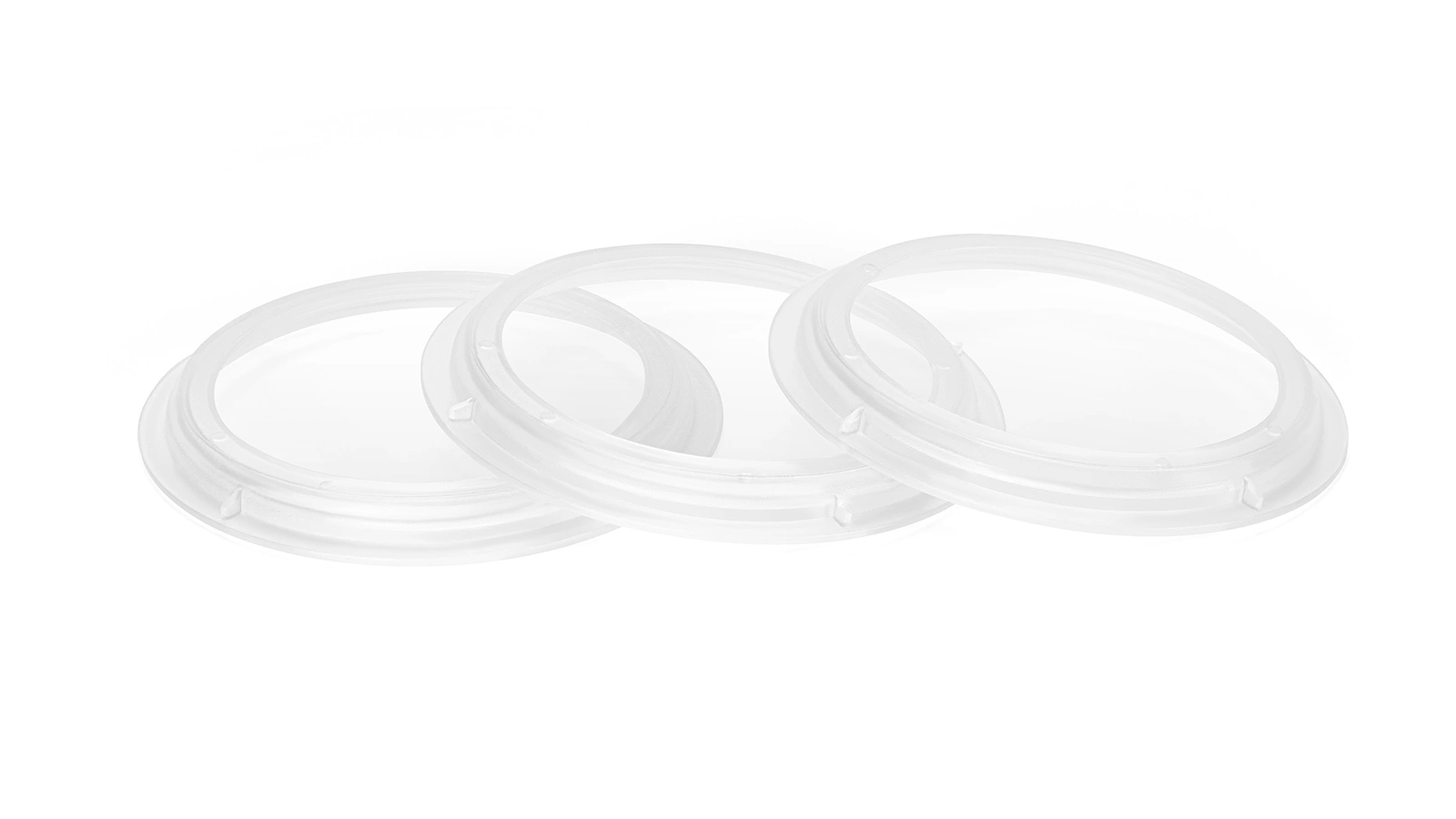
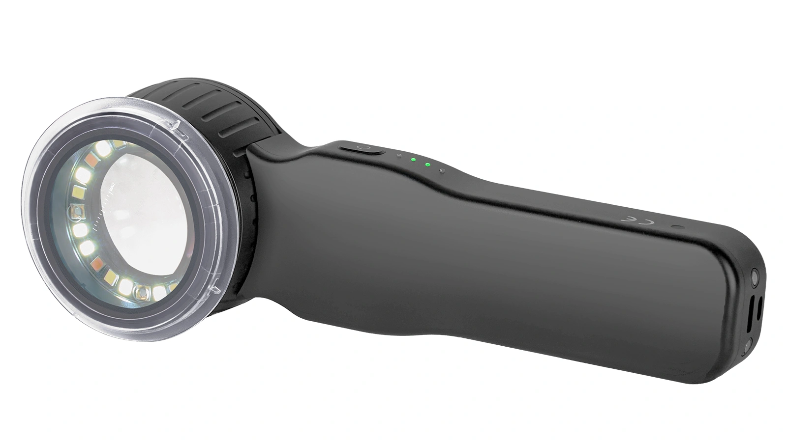
Clinics can incorporate several measures into their dermoscopy workflow:
- Use disposable protective covers where appropriate.
- Remove reusable contact plates for appropriate cleaning and disinfection.
- Clean relevant device surfaces between patients.
- Follow the clinic’s established infection-control protocol.
- Avoid reusing accessories that are designated for single use.
- Consider non-contact polarized dermoscopy when direct contact is unnecessary.
Non-contact observation can reduce the number of surfaces that touch the patient, but it should not be regarded as a complete solution to cross-contamination. Parts of the device may still accidentally touch the skin, so routine cleaning and hygiene procedures remain necessary. For IBOOLO, this is also why interchangeable accessories are an important part of dermatoscope design. A combination of removable contact plates, non-contact attachments and disposable protective components gives clinics more flexibility when building their own hygiene workflow.
What Accessories Are Useful for Nails, Narrow Areas or Raised Lesions?
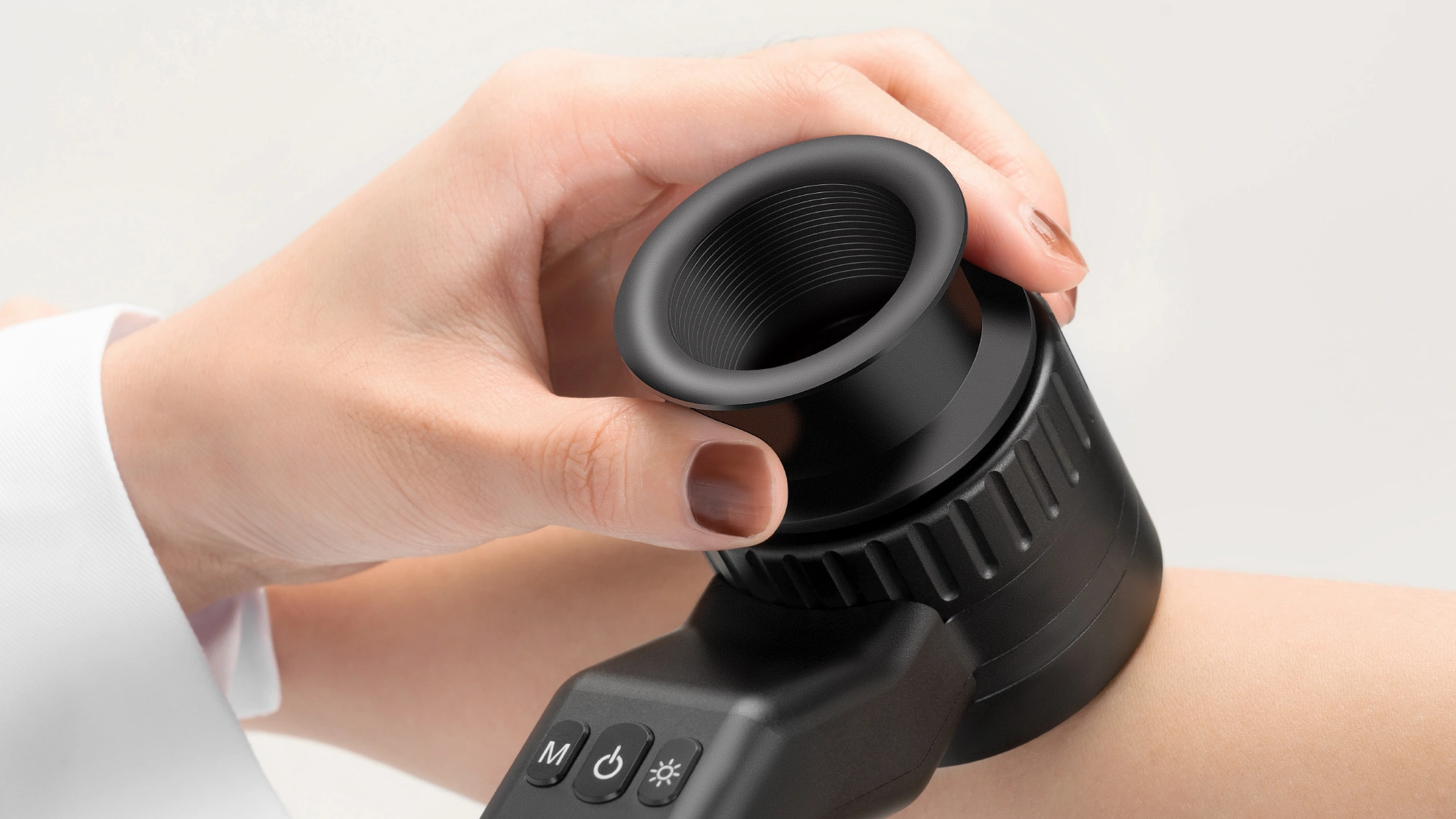
A full-size contact plate works well for many routine examinations, but not every anatomical area is flat or easy to access. A small contact plate can make positioning easier when examining narrow or localized areas. Depending on the examination, this can include areas around the nail, curved anatomical surfaces or small raised areas.
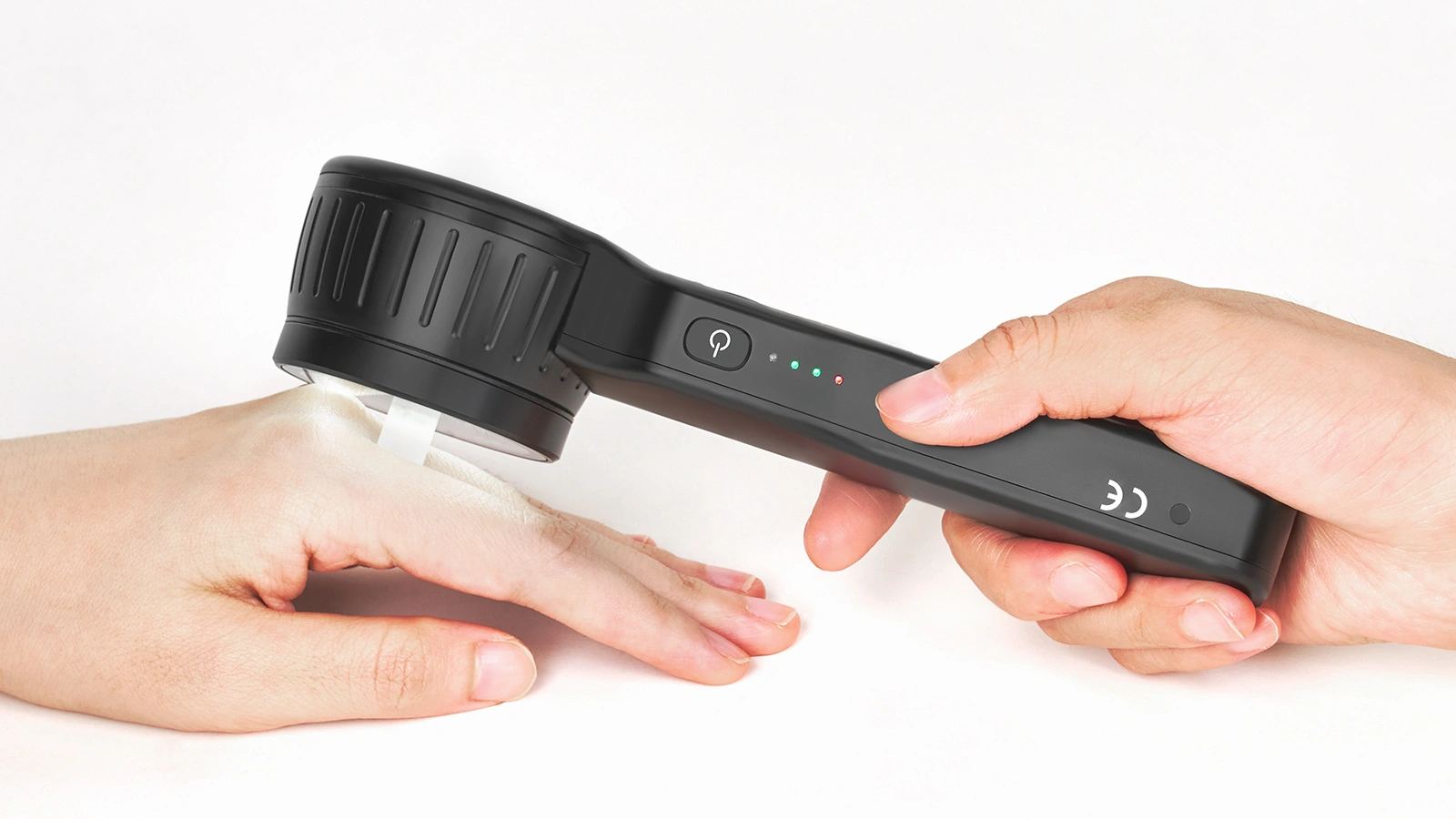
A non-contact attachment is another option when direct pressure on the observation area is undesirable. Because polarized dermoscopy can operate without direct skin contact, the clinician can maintain a defined working distance and observe the area without pressing a glass plate onto the skin. Different accessory sizes therefore provide more than convenience. They allow the clinician to adapt the dermatoscope to different anatomical areas and examination requirements.
How Can a Dermatoscope Fit More Naturally into Daily Clinic Workflow?
For frequent clinical use, an easy-to-use dermatoscope is not defined by a single feature. A practical system should allow the clinician to move efficiently between different observation modes, change accessories without unnecessary complexity, capture images when required and prepare the device for the next patient.
At IBOOLO, this workflow perspective is part of how we think about handheld dermatoscope design. For example, the DE-5100 combines direct observation and smartphone imaging with interchangeable contact and non-contact accessories, disposable protective components and a dedicated charging stand. These features are designed to help the dermatoscope remain ready for repeated use throughout the clinical day.
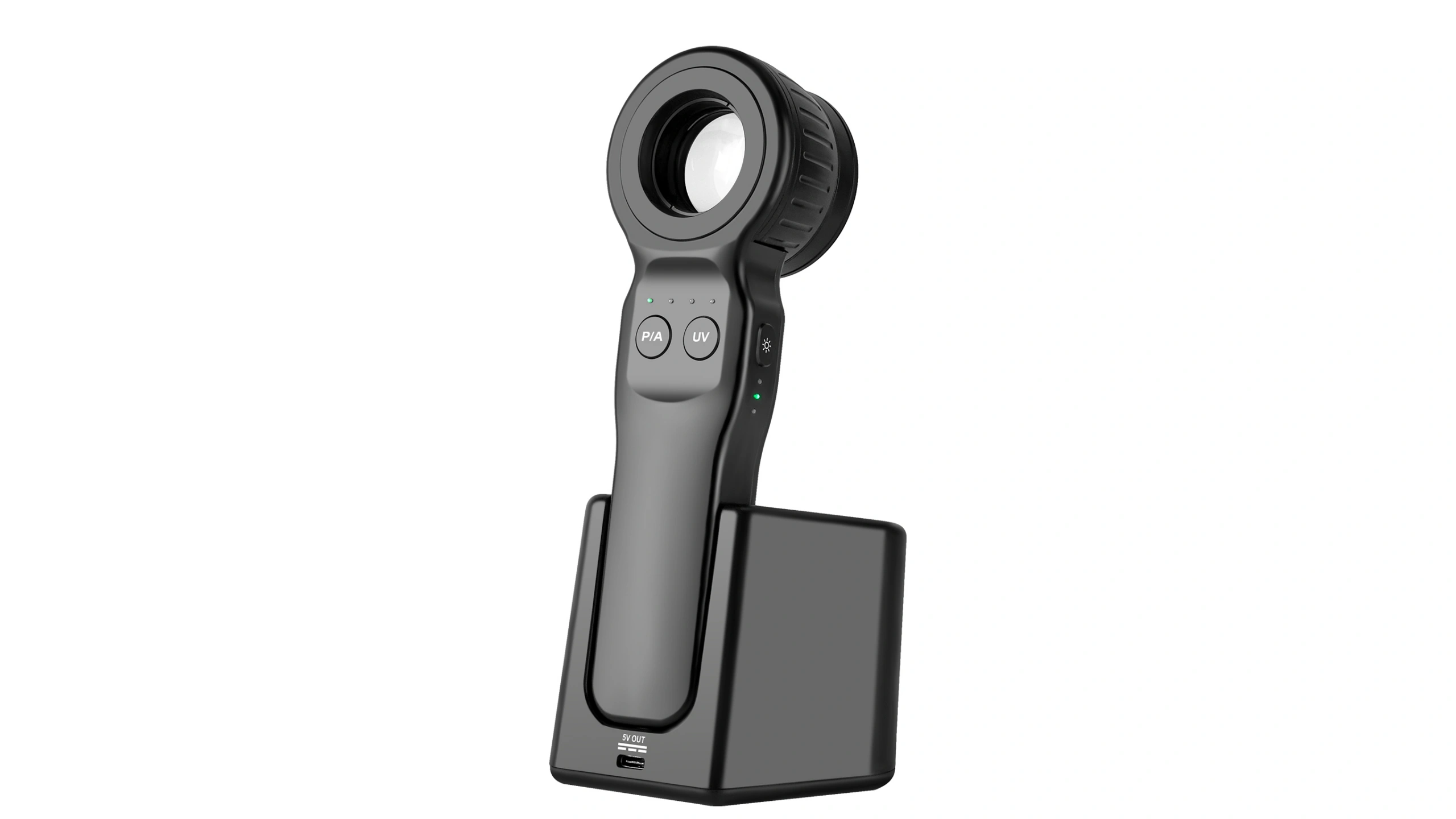
These practical details can have a significant influence on how comfortably a dermatoscope fits into everyday clinical practice.
Frequently Asked Questions
Does polarized dermoscopy require skin contact?
No. Polarized dermoscopy can be performed without direct skin contact because cross-polarization helps reduce surface reflection. However, polarized dermoscopy can also be used in contact mode when appropriate.
When is non-contact dermoscopy useful?
Non-contact polarized dermoscopy can be useful when the clinician wants to avoid pressure on the skin, preserve the appearance of superficial vessels, rapidly scan an area or examine a location where direct contact is undesirable.
How can clinics reduce cross-contamination during dermoscopy?
Clinics can combine appropriate cleaning and disinfection, removable contact accessories, compatible disposable protective covers and non-contact examination where suitable. Any reusable component that touches the patient should be handled according to the clinic’s infection-control policy.
Is non-contact dermoscopy always better than contact dermoscopy?
No. The two techniques serve different purposes. Contact dermoscopy can provide stable positioning and is commonly used with conventional non-polarized observation, while non-contact polarized imaging avoids direct compression and can be useful when vascular appearance or direct-contact considerations are important.
Should dermoscopic images be recorded for every examination?
Not necessarily. Image documentation depends on the purpose of the examination and the clinic’s own workflow. When images are needed for follow-up, comparison, consultation or patient records, capturing them during the dermoscopic examination and labeling them consistently can make later review easier.