






Article
How Can Dermoscopy Help Identify Basal Cell Carcinoma?
Basal cell carcinoma (BCC) is the most common type of skin cancer worldwide. It arises from the basal cells of the epidermis and is strongly associated with long-term ultraviolet (UV) exposure. Although BCC rarely metastasizes, it can grow locally and cause significant tissue destruction if left untreated. Early identification is therefore important to prevent deeper…
Basal cell carcinoma (BCC) is the most common type of skin cancer worldwide. It arises from the basal cells of the epidermis and is strongly associated with long-term ultraviolet (UV) exposure. Although BCC rarely metastasizes, it can grow locally and cause significant tissue destruction if left untreated. Early identification is therefore important to prevent deeper invasion and more complex surgical treatment.
In routine dermatology practice, distinguishing BCC from inflammatory conditions such as dermatitis or eczema can sometimes be difficult with naked-eye examination alone. Dermoscopy provides magnification and controlled lighting that allow clinicians to observe vascular structures and pigment patterns beneath the skin surface.
What Dermoscopic Structures Are Characteristic of Basal Cell Carcinoma?
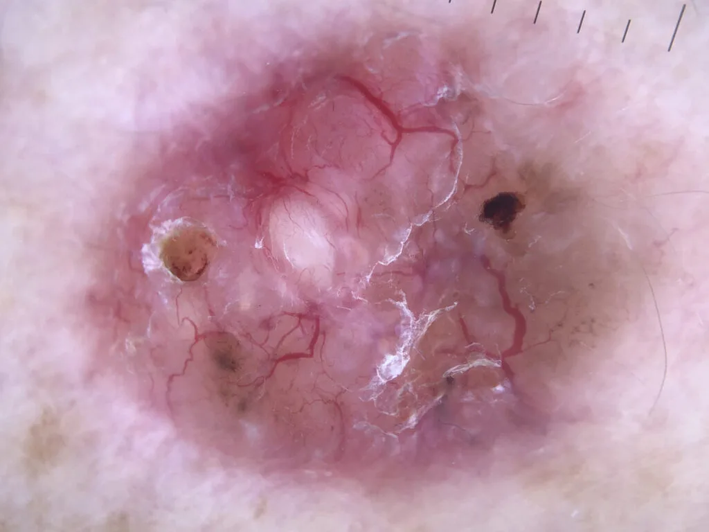
When evaluating basal cell carcinoma dermoscopy, dermatologists look for several characteristic structures that are rarely seen in benign lesions. One of the most important features is the presence of arborizing vessels. These vessels appear as large branching blood vessels that divide repeatedly into smaller branches, resembling the structure of a tree. Arborizing vessels are often bright red and clearly visible under dermoscopy, reflecting the abnormal vascular growth associated with BCC.
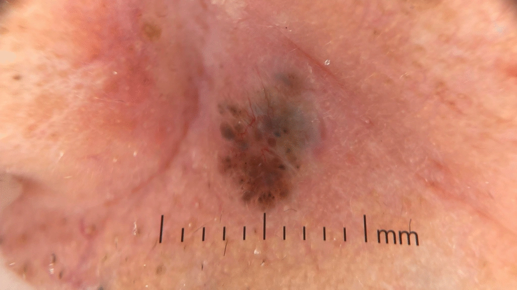
Another typical feature is the leaf-like area. These structures appear as brown or gray shapes with multiple lobes that resemble the outline of a leaf. Leaf-like areas usually represent pigmented tumor nests located within the superficial layers of the skin. Their presence strongly suggests basal cell carcinoma, particularly in pigmented lesions.
A third important dermoscopic pattern is the spoke-wheel area. This structure consists of radial projections that extend from a central darker hub, similar to the spokes of a wheel. Spoke-wheel structures are often observed in early BCC lesions and represent tumor nests arranged around a central axis. When arborizing vessels, leaf-like areas, or spoke-wheel patterns are present together, the probability of BCC is significantly increased in dermoscopic examination.
How Do Pigmented and Superficial BCC Appear Different Under Dermoscopy?
Different subtypes of BCC show distinct dermoscopic patterns that can help guide treatment decisions. Pigmented basal cell carcinoma dermoscopy often reveals multiple pigmented structures, including leaf-like areas, blue-gray globules, and spoke-wheel patterns. These structures reflect melanin accumulation within tumor nests and can make the lesion appear darker clinically.
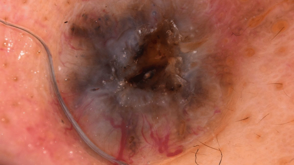
In contrast, superficial basal cell carcinoma dermoscopy typically shows a different pattern. Superficial BCC often presents with fine short vessels, small erosions, and multiple thin red vascular structures distributed across a slightly scaly background. These lesions are usually more superficial and may resemble inflammatory skin diseases at first glance.
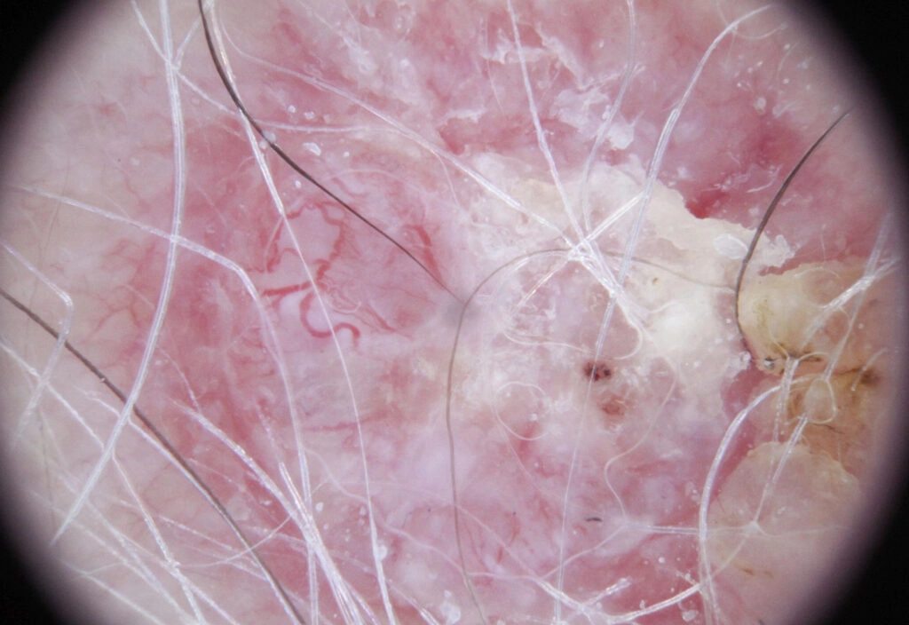
Recognizing these differences is important for treatment planning. Superficial BCC may be treated with topical therapies or photodynamic therapy in selected cases, while nodular or pigmented forms often require surgical removal. Dermoscopy therefore plays a key role not only in diagnosis but also in determining the most appropriate treatment approach.
Why Is Polarized Dermoscopy Useful for Observing BCC Structures?
Polarized dermoscopy offers an important advantage when examining basal cell carcinoma. By reducing surface reflection and glare, polarized light allows clinicians to visualize deeper skin structures more clearly. This is particularly helpful for observing vascular patterns and fibrotic areas that may not be visible with standard lighting.
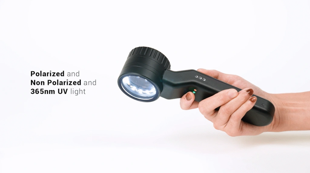
Each IBOOLO dermatoscope incorporates both polarized and non-polarized illumination modes. The polarized mode enables clearer visualization of arborizing vessels and other dermal structures characteristic of BCC. As a non-invasive imaging technique, dermoscopy allows clinicians to examine suspicious lesions repeatedly during follow-up without causing additional discomfort to patients. Consequently, dermoscopy has become an essential tool for the early detection and ongoing monitoring of BCC.
Can Standardized Dermoscopic Screening Reduce Unnecessary Biopsies?
Standardized dermoscopic examination protocols can improve diagnostic confidence and reduce unnecessary biopsies. By recognizing well-established basal cell dermoscopy patterns, clinicians can better distinguish suspicious tumors from benign lesions or inflammatory skin conditions. This targeted approach helps prioritize biopsy for lesions with high malignant potential while avoiding unnecessary procedures for benign conditions.
High-quality dermoscopic imaging is essential for reliable screening. Clear visualization of vascular patterns and structural features allows clinicians to make more accurate decisions during early assessment. Standardized dermoscopy therefore supports more efficient skin cancer screening while improving patient care.
Looking for a Reliable Dermoscopy Solution?
For dermatology clinics and medical distributors seeking high-quality dermoscopy solutions, advanced optical dermatoscopes can provide clear visualization of key diagnostic structures such as vascular patterns and pigment architecture.
Contact IBOOLO to request a quotation for professional dermoscopy devices and learn more about our clinical imaging solutions.
What Do You Get When You Choose an IBOOLO Dermatoscope?
(1) All of our dermatoscopes are hand-assembled by craftsmen with more than 20 years of experience. Before leaving the factory, each device goes through more than ten inspection procedures to ensure that every dermatoscope you receive meets strict quality standards.
(2) All of our products come with a two-year warranty. During this period, if any quality issues occur, we provide free replacement, repair, or return services.
(3) Finally, we offer fast shipping. If your order quantity is fewer than 10 units, we can usually arrange shipment within 24 hours. In addition, we provide DDP (Delivered Duty Paid) service for many countries, meaning you do not need to pay any import duties. The typical delivery time is around one week.