






Article
How Clear Do Skin Lesions Look Through the IBOOLO DE-3100?
IBOOLO introduced the DE-3100 in 2021 as our first professional handheld dermatoscope. Several years later, even after we have developed newer models, the DE-3100 remains one of the best-selling dermatoscopes in the IBOOLO product range. There are two main reasons. The first is value. The DE-3100 was designed to provide a professional dermoscopy workflow without requiring…
IBOOLO introduced the DE-3100 in 2021 as our first professional handheld dermatoscope. Several years later, even after we have developed newer models, the DE-3100 remains one of the best-selling dermatoscopes in the IBOOLO product range.
There are two main reasons. The first is value. The DE-3100 was designed to provide a professional dermoscopy workflow without requiring users to move immediately into a more complex or expensive system. The second reason is more important for everyday clinical use: image quality.
Specifications such as 10X magnification can tell part of the story, but real dermoscopic images are much more direct. In this article, we selected several representative skin and scalp conditions to show the structures that can be observed and recorded with the DE-3100.
How Clearly Can the DE-3100 Capture the Delta-Wing Jet Sign in Scabies?
One of the classic dermoscopic signs of scabies is the delta-wing jet with contrail sign.
The anterior part of the mite appears as a small brown or dark triangular structure, known as the delta-wing or delta-glider sign. Extending behind it is a thin, curved or S-shaped burrow, creating the appearance of a jet followed by a contrail.
The combination of the dark triangular mite and the whitish or grayish burrow is one of the most characteristic dermoscopic patterns associated with scabies.

What Does Basal Cell Carcinoma Look Like Under the DE-3100?
Basal cell carcinoma can show several recognizable dermoscopic structures in the same lesion.
One of the most typical findings is arborizing vessels: bright red vessels that branch repeatedly into progressively finer vessels, creating a tree-like appearance.
Other common features include erosion or ulceration, blue-gray ovoid nests, blue-gray globules and shiny white structures. In pigmented or nodular lesions, blue-gray nests may appear as relatively well-defined oval or rounded areas beneath the surface.
In a representative DE-3100 image, attention can therefore be focused on three particularly recognizable structures: central erosion or ulceration, surrounding arborizing vessels, and blue-gray ovoid areas.

What Can Be Seen in Discoid Lupus Erythematosus During Follow-Up?
The dermoscopic appearance of discoid lupus erythematosus (DLE) varies with lesion location and disease stage.
Common features include white scales, follicular keratotic plugs, perifollicular white halos, telangiectatic or arborizing vessels, speckled brown pigmentation and white structureless areas.
Scalp lesions may also show loss or alteration of follicular openings, perifollicular scaling and fibrotic white dots. In later-stage lesions, white structureless areas, telangiectasia and pigmentary changes can become more prominent.
During follow-up, these structures can be compared with previous dermoscopic images to observe changes in scaling, erythema, follicular appearance and pigmentation.

What Does Pityriasis Amiantacea Look Like Under the DE-3100?
The most characteristic feature of pityriasis amiantacea is thick, strongly adherent scale surrounding groups of hair shafts.
Under dermoscopy or trichoscopy, the scale may appear white, silver-white or yellowish, forming dense layers around individual hairs or bundles of hair. The hair shafts can appear to pass directly through or become enclosed by the adherent scale.
Additional findings may include diffuse yellowish scales, telangiectatic vessels and other vascular patterns. The vascular appearance may vary depending on the underlying scalp disorder associated with the pityriasis amiantacea.
The combination of hair shafts + tightly adherent scale + visible scalp background creates a very distinctive dermoscopic appearance.

What Optical and Lighting Features Does the DE-3100 Provide?
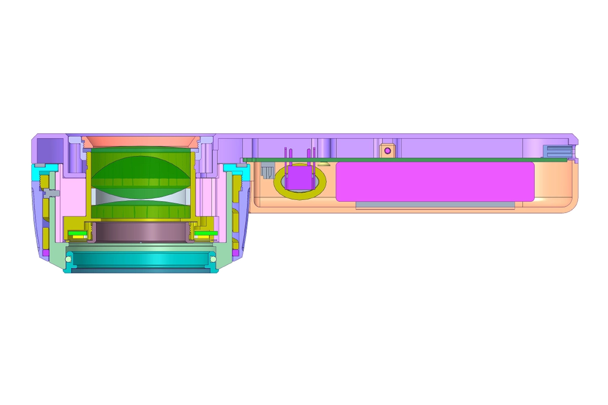
Behind these images is a relatively straightforward optical configuration.
The IBOOLO DE-3100 provides 10X optical magnification, giving users a close view of structures such as vessels, pigmentation, scale, follicular openings and other fine surface or subsurface details. It combines polarized, non-polarized and amber illumination to provide five lighting modes:
- Polarized Light
- Non-Polarized Light
- Amber Light
- White-Amber Mixed Polarized Light
- White-Amber Mixed Non-Polarized Light
Polarized illumination helps reduce strong surface reflection and makes subsurface colors and structures easier to observe. Non-polarized observation provides complementary information about more superficial features. Amber and white-amber mixed modes offer additional options for comparing color and contrast.
The DE-3100 uses a 1000 mAh rechargeable battery and is designed for portable handheld use.
Can the DE-3100 Be Used With and Without a Smartphone?
Yes. The DE-3100 supports both workflows.
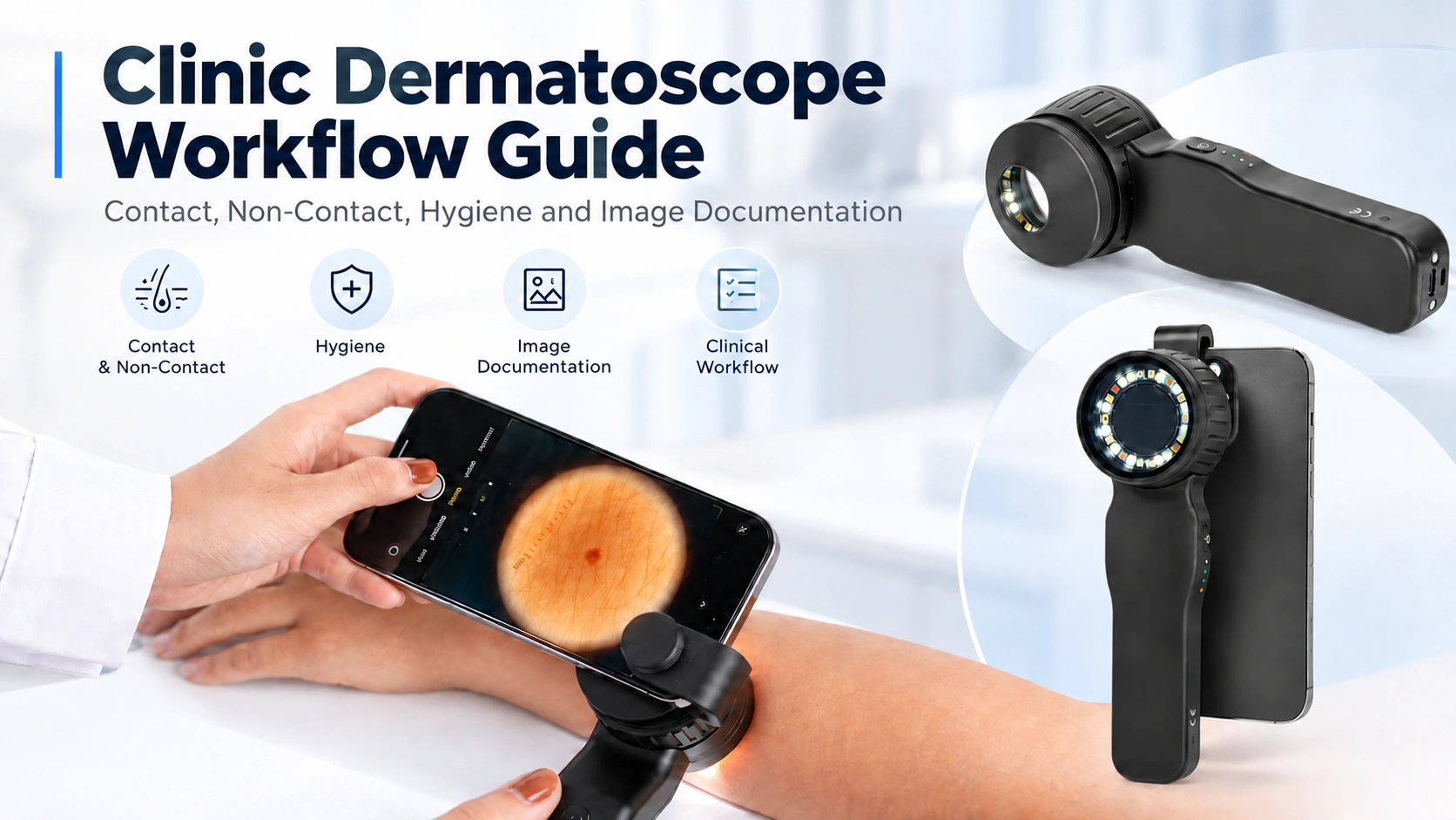
For direct examination, users can look through the dermatoscope without connecting a smartphone. This keeps the examination quick and straightforward.

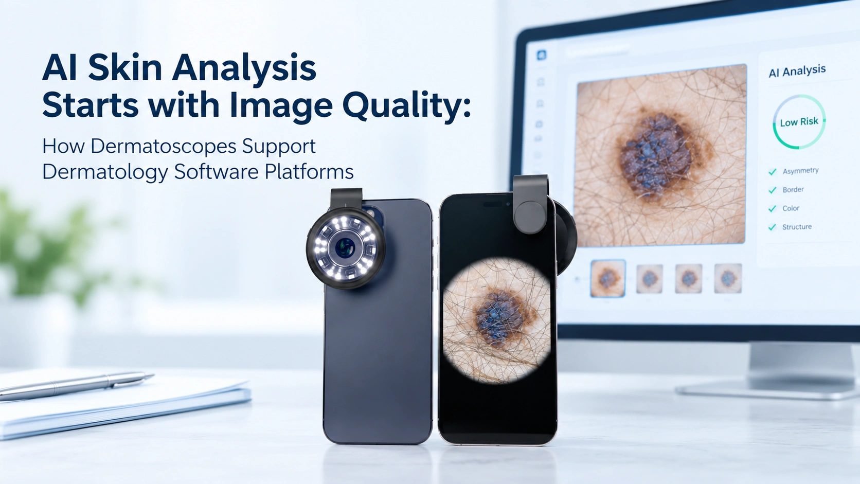
When images need to be captured, the DE-3100 can be connected to a smartphone through a compatible adapter. The same dermoscopic view can then be photographed for documentation, comparison, follow-up or professional communication.

This means users do not need to choose between a traditional handheld dermatoscope and a smartphone dermatoscope—the DE-3100 supports both.
Why Has the DE-3100 Remained One of IBOOLO’s Best-Selling Dermatoscopes?
The DE-3100 has been available since 2021, but its core configuration remains practical for everyday professional dermoscopy.
It combines:
- 10X optical magnification
- Five lighting modes
- Polarized and non-polarized observation
- 1000 mAh rechargeable battery
- Direct naked-eye observation
- Smartphone image capture
- Two-year IBOOLO warranty
Newer IBOOLO models provide additional optical modes and more advanced configurations, but the DE-3100 continues to offer the functions many users need in a compact professional handheld dermatoscope.
And when the question is simply How clear is the DE-3100?, specifications are only part of the answer.
The scabies burrow, arborizing vessels, blue-gray structures, follicular changes and adherent scalp scales shown in real dermoscopic images provide a much more direct demonstration.