






Article
How Do Dermatologists Identify Melanoma Under Dermoscopy?
Melanoma is one of the most dangerous types of skin cancer. Although it is less common than other skin cancers, it causes the majority of skin-cancer related deaths. The main reason is its strong ability to spread to other organs if it is not detected early. When melanoma is diagnosed at an early stage, simple…
Melanoma is one of the most dangerous types of skin cancer. Although it is less common than other skin cancers, it causes the majority of skin-cancer related deaths. The main reason is its strong ability to spread to other organs if it is not detected early. When melanoma is diagnosed at an early stage, simple surgical removal is usually effective. However, once the tumor grows deeper into the skin or spreads through the lymphatic system, treatment becomes more complex and survival rates decrease.
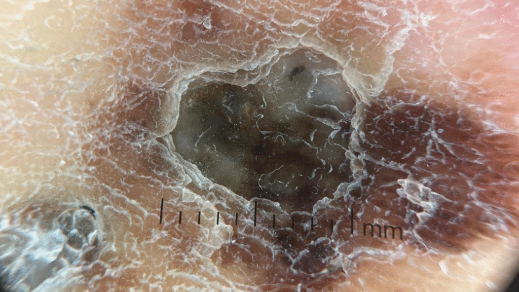
Dermoscopy has become an important tool for early detection. By using magnification and controlled lighting, doctors can observe skin structures that cannot be seen with the naked eye. This allows clinicians to detect early changes in melanocytic lesions and identify suspicious patterns before the disease progresses.
What Are the Common Dermoscopic Features of Melanoma?
When examining melanoma under dermoscopy, dermatologists look for several key visual patterns. One important feature is the atypical pigment network. In benign moles, the pigment network usually appears even and regular. In melanoma, the network lines often become thicker, uneven, or irregularly distributed across the lesion. This irregular pattern is one of the first warning signs that the lesion may be malignant.
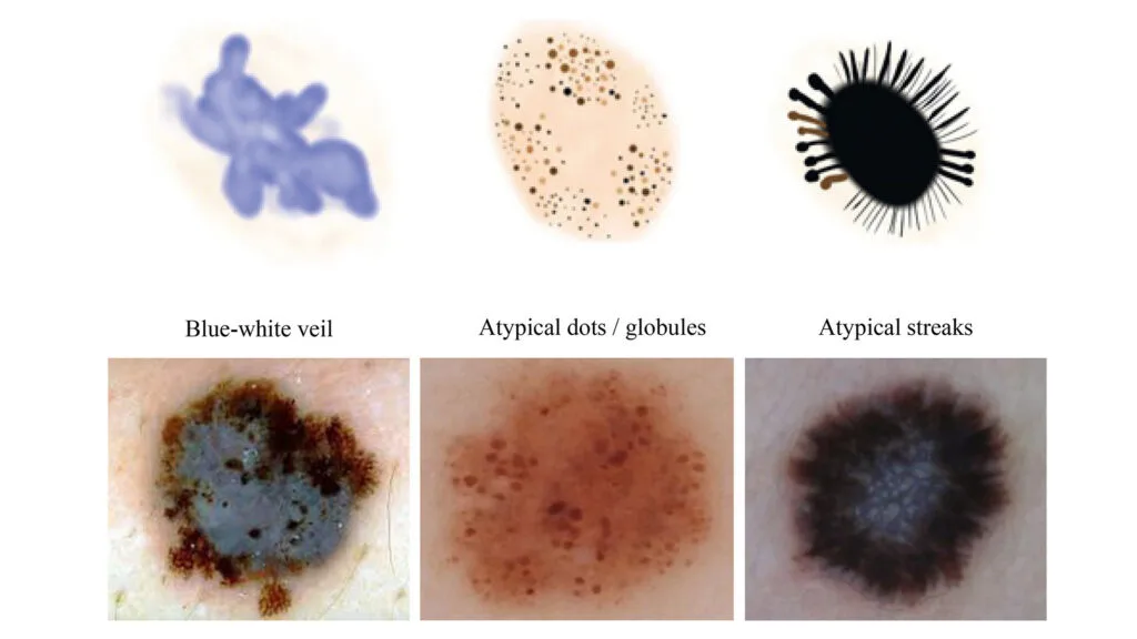
Another common feature is atypical streaks around the edge of the lesion. These streaks appear as lines or projections that extend outward from the border. They reflect uneven growth of melanocytes and often indicate that the lesion is expanding in an uncontrolled way.
A third important sign is the blue-white veil. This structure appears as an area of blue pigmentation covered by a cloudy white layer. It is often associated with dense melanin and changes in the upper skin layers. When the blue-white veil appears together with other irregular patterns, it strongly increases the suspicion of melanoma.
How Do Dermoscopy Images Help Distinguish Benign Moles From Melanoma?
In clinical dermatology, distinguishing benign melanocytic nevi from malignant melanoma cannot rely on naked-eye observation alone. Clinicians need the ability to look beneath the skin surface and evaluate the architectural arrangement of pigment structures and vascular patterns located within the underlying layers.
Dermoscopy bridges this gap by using polarized light and achromatic lenses to neutralize surface reflections, allowing a physician to analyze specific diagnostic criteria.
(1) Symmetry vs. Architectural Chaos
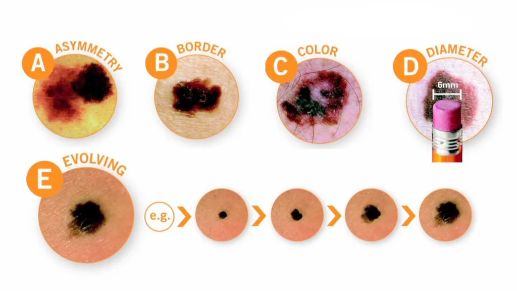
The first indicator is the overall distribution of structures. Benign moles generally follow a predictable, symmetric pattern, whereas melanoma is characterized by a chaos of colors and shapes.
• Benign Moles: Usually display a single, uniform pattern and a consistent color. The lesion is typically symmetric across two axes.
• Melanoma: Often displays “asymmetric distribution of structures.” One half of the lesion might show a thick network, while the other shows a structureless area or dots.
(2) The Pigment Network: Honeycomb vs. Atypical
The pigment network represents the dermo-epidermal junction. How these lines appear is a primary factor in the diagnostic decision.
• Benign Mole: The network looks like a delicate, light-brown honeycomb with thin lines and regular “holes.” These lines gradually thin out toward the periphery of the mole.
• Melanoma: The lines are thick, dark, and unevenly distributed. Crucially, in melanoma, the network may end abruptly at the edge rather than fading out, signaling invasive growth.
(3) Presence of Specialized Malignant Markers
Dermoscopy allows clinicians to see specific markers that are virtually invisible to the naked eye. The presence of any of these significantly increases the suspicion of malignancy: The Blue-White Veil, Irregular Streaks, and Regression Structures.
Why Is High-Quality Dermoscopic Imaging Important for Melanoma Detection?
Many important melanoma signs are small and subtle. Irregular pigment networks, streaks, or blue-white structures may be difficult to see if the image quality is poor. Clear optics and stable lighting allow these features to be seen more clearly and consistently.
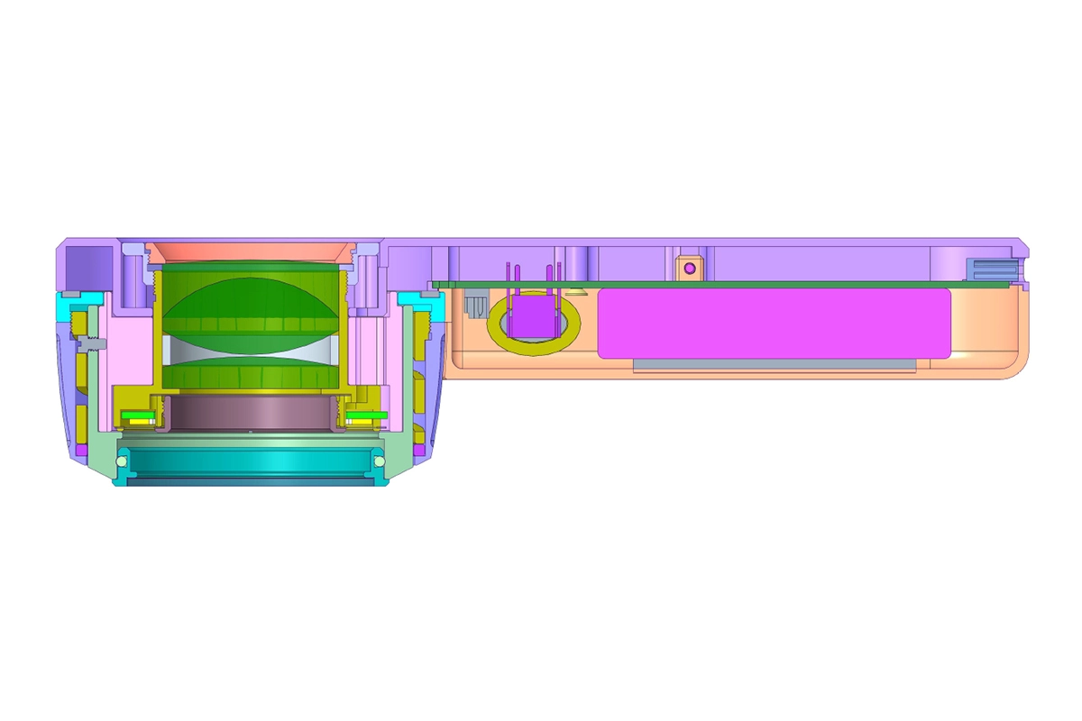
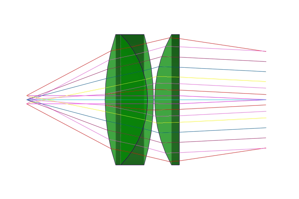
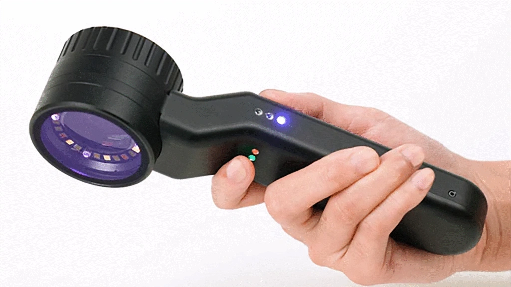
The IBOOLO DE-4100 PRO uses a high-precision optical lens system that minimizes distortion to a nearly negligible level. It provides 10X ultra-clear magnification, allowing vascular structures, pigment colors, and other fine skin details to be observed with high clarity.
The DE-4100 Pro features four illumination modes: polarized light, amber polarized light, non-polarized light, and UV light. Polarized light helps eliminate surface glare, allowing users to clearly observe structures within the dermal layer. Amber polarized light is designed to enhance the visualization of lesion borders and is particularly suitable for dermoscopic examination in individuals with darker skin tones. Non-polarized light is mainly used to observe surface texture and fine details of the skin, focusing on structures within the epidermis. The 365 nm UV light can assist in detecting fungal skin lesions and certain depigmentation disorders.
In addition, the device offers three adjustable brightness levels, allowing it to adapt to different ambient lighting conditions.
How Can Early Dermoscopic Diagnosis Improve Patient Outcomes?
Early diagnosis greatly improves the chances of successful treatment. When melanoma is detected before it grows deeply into the skin, surgical removal is usually sufficient. In many cases, patients can be treated with a simple outpatient procedure.
If melanoma is discovered at a later stage, the tumor may already have spread to lymph nodes or other organs. At that point, treatment may require additional surgery, immunotherapy, or systemic therapy. These treatments are more complex and carry higher risks.
By allowing doctors to recognize suspicious dermoscopic features of melanoma at an early stage, dermoscopy helps guide timely biopsy or removal of the lesion. This early intervention significantly improves survival rates.