








Article
The Most Common Benign Lesion: How to Identify Seborrheic Keratosis via Dermoscopy?
Seborrheic keratosis (SK) represents one of the most frequent reasons for dermatological consultations globally. Often referred to as senile warts or barnacles of aging, these benign epidermal tumors can cause significant psychological distress for patients who mistake them for malignant melanoma or pigmented basal cell carcinoma. Establishing a definitive diagnosis is the primary goal of…
Seborrheic keratosis (SK) represents one of the most frequent reasons for dermatological consultations globally. Often referred to as senile warts or barnacles of aging, these benign epidermal tumors can cause significant psychological distress for patients who mistake them for malignant melanoma or pigmented basal cell carcinoma. Establishing a definitive diagnosis is the primary goal of the clinical encounter, as it immediately alleviates patient anxiety and prevents unnecessary medical interventions.
While visual inspection is the first step, the integration of dermoscopy has revolutionized the diagnostic accuracy for SK, allowing clinicians to visualize subsurface morphological structures invisible to the naked eye. By utilizing advanced imaging tools like those provided by IBOOLO, practitioners can bridge the gap between clinical suspicion and diagnostic certainty.
What Are the Pathognomonic Dermoscopic Features of Seborrheic Keratosis?
The diagnostic reliability of dermoscopy in identifying seborrheic keratosis relies on the presence of specific morphological criteria that reflect the underlying histopathology of the lesion. The most consistent indicators of SK are comedo-like openings and milia-like cysts. Comedo-like openings, often described as plugs, appear as dark, circular, or irregular areas of keratin debris localized within follicular openings. These correspond to the keratin-filled invaginations of the epidermis seen in pathology.
Milia-like cysts are another hallmark feature, appearing as small, white-to-yellowish, round structures. These represent intraepidermal keratin cysts. When using an IBOOLO DE-4100 dermatoscope, these structures become sharply defined due to high-quality optics and polarized lighting. Furthermore, seborrheic keratosis often exhibits fissures and ridges, which create a brain-like or cerebriform appearance. This pattern is particularly common in acanthotic variants of SK. Another key feature is the moth-eaten border, where the edges of the lesion are not smooth but appear scalloped or indented. The presence of these benign indicators allows for a high degree of confidence in excluding malignancy.
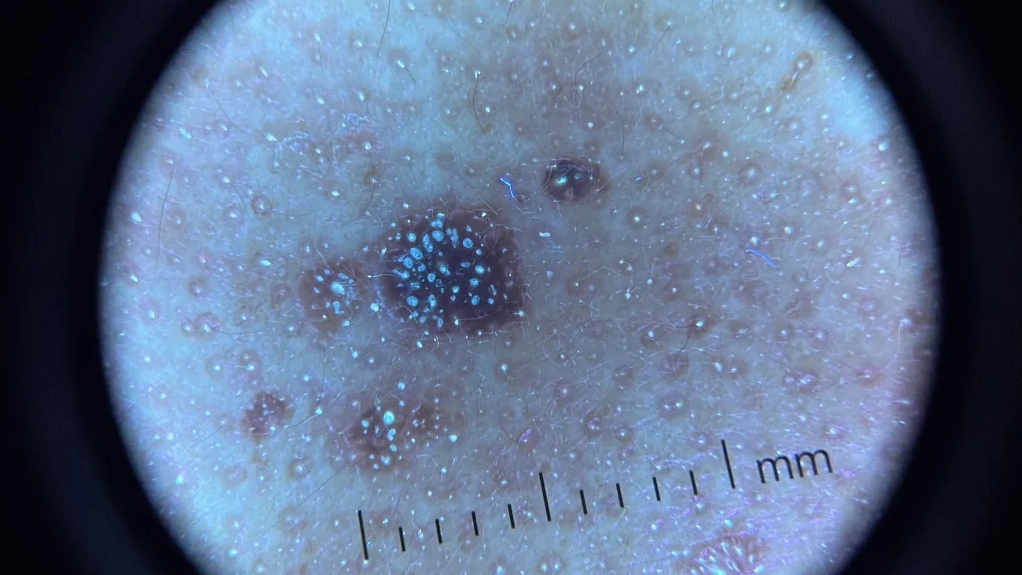
How Does Early Seborrheic Keratosis Dermoscopy Differ from Classic Lesions?
Dermoscopy of early seborrhoeic keratosis can be more challenging because the characteristic features may not yet be fully developed. Early lesions often lack prominent fissures or multiple cysts. Instead, they may show subtle pigmentation with a network-like pattern or light brown, structureless areas.
In the early stages, clinicians may observe only a few milia-like cysts or faint comedone-like openings. The surface of the lesion is usually flatter, and the stuck-on appearance may not be obvious. This can lead to confusion with melanocytic nevi or even early-stage melanoma. However, careful dermoscopic evaluation often reveals clues. The absence of a true pigment network and the presence of even a few keratin-related structures support the diagnosis of early seborrheic keratosis.
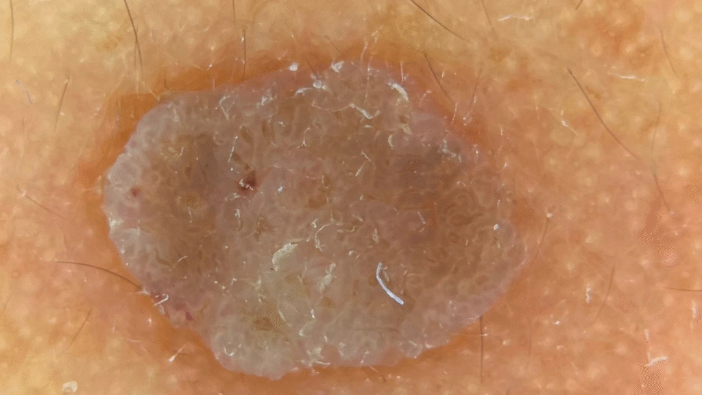
Seborrheic Keratosis under IBOOLO DE-3100 PRO Polarized Effect
What Changes Occur in Irritated Seborrheic Keratosis Dermoscopy?
When a seborrheic keratosis becomes inflamed due to friction, trauma, or immunological response, its dermoscopic appearance undergoes a dramatic shift. This state, known as irritated seborrheic keratosis, can be particularly alarming because the inflammation often masks typical benign features and introduces vascular patterns that mimic malignancy.
In irritated seborrheic keratosis dermoscopy, the most prominent change is the appearance of glomerular or looped vessels. These vessels are usually distributed in a clustered or hairpin arrangement and are often surrounded by a white, keratotic halo. The lesion may also exhibit squamous pearls or large, circular areas of keratinization. Unlike the chaotic neoangiogenesis seen in melanoma, the vascularity in irritated SK tends to be more uniform.
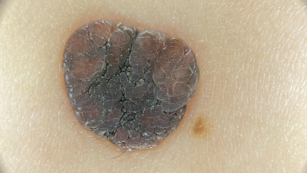
Seborrheic Keratosis under IBOOLO DE-3100 PRO Non-polarized Effect
How Is Seborrheic Keratosis Treated Effectively?
Once a diagnosis of seborrheic keratosis is confirmed via dermoscopy, the decision to treat is usually based on symptomatic relief or cosmetic preference. Because SK is strictly epidermal, treatments are focused on superficial destruction. Cryotherapy with liquid nitrogen remains the most common modality, causing the lesion to blister and slough off.
Curettage and electrodessication are also highly effective, allowing for the mechanical removal of the stuck-on tumor. For thinner lesions, topical therapies may be utilized. Regardless of the chosen method, the pre-treatment use of dermoscopy ensures that the clinician is not inadvertently treating a masked malignancy. The clarity provided by IBOOLO dermatoscope allows for precise mapping of the lesion’s borders, ensuring that the treatment is localized and minimizes damage to the surrounding healthy tissue.
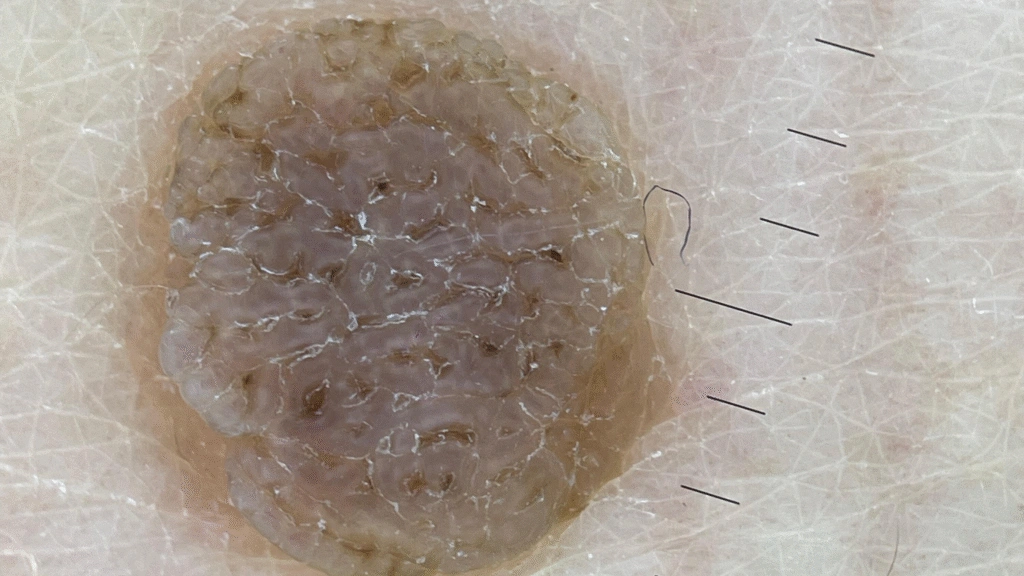
Seborrheic Keratosis under IBOOLO DE-500
Does Precision in Identifying Benign Features Improve Outcomes?
Efficiency in dermatology is derived from the precise mastery of benign features. When a practitioner can quickly identify the cerebriform patterns and milia-like cysts of seborrheic keratosis, the entire consultation becomes more streamlined. Advanced diagnostic tools are the foundation of this efficiency. By documenting lesions with IBOOLO’s high-definition dermoscopy solutions, clinicians can provide peace of mind through clear, visual evidence. Ultimately, the goal is to provide a diagnosis that is as accurate as it is fast.
Are you looking to enhance your diagnostic accuracy and clinical workflow? Contact us today to request a product demonstration of IBOOLO dermoscopy solutions.