







Article
What Does Human Skin Really Look Like Under a Dermatoscope?
Most people judge a skin lesion with the naked eye. We notice its color, size, shape, or whether it has changed over time. However, what we see is only the surface. A dermatoscope reveals structures that are invisible without magnification and specialized lighting. The difference between naked-eye observation and dermoscopy is similar to the difference…
Most people judge a skin lesion with the naked eye. We notice its color, size, shape, or whether it has changed over time. However, what we see is only the surface. A dermatoscope reveals structures that are invisible without magnification and specialized lighting. The difference between naked-eye observation and dermoscopy is similar to the difference between looking at an onion and looking at onion cells under a microscope. One shows the whole object, while the other reveals the fine structures that explain how it is built.
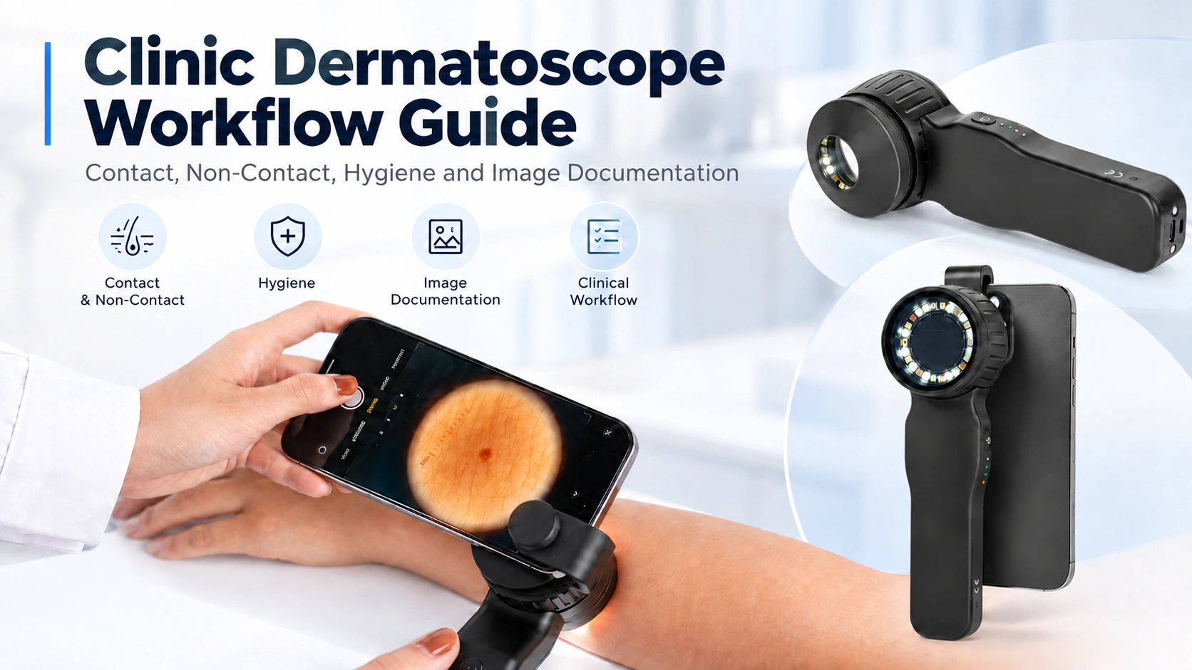
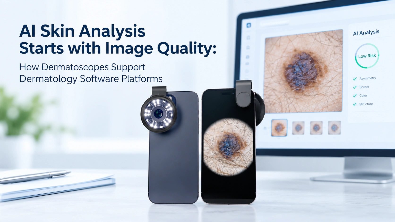
A dermatoscope combines optical magnification with polarized or non-polarized light to reduce surface reflection from the skin. This allows clinicians to visualize pigment distribution, blood vessels, keratin, and other microscopic structures within the epidermis and superficial dermis.
At IBOOLO, we believe that understanding what a dermatoscope actually reveals helps users appreciate why this instrument plays such an important role in skin assessment.
What Does a Common Benign Mole Look Like Under a Dermatoscope?
Most pigmented moles are benign melanocytic nevi. Under dermoscopy, these lesions often display organized and symmetrical patterns that reflect normal melanocyte growth.
A common junctional or compound melanocytic nevus frequently shows a regular pigment network. The network consists of evenly distributed brown lines surrounding lighter holes. The color is generally uniform, and the network maintains a consistent thickness across the lesion.
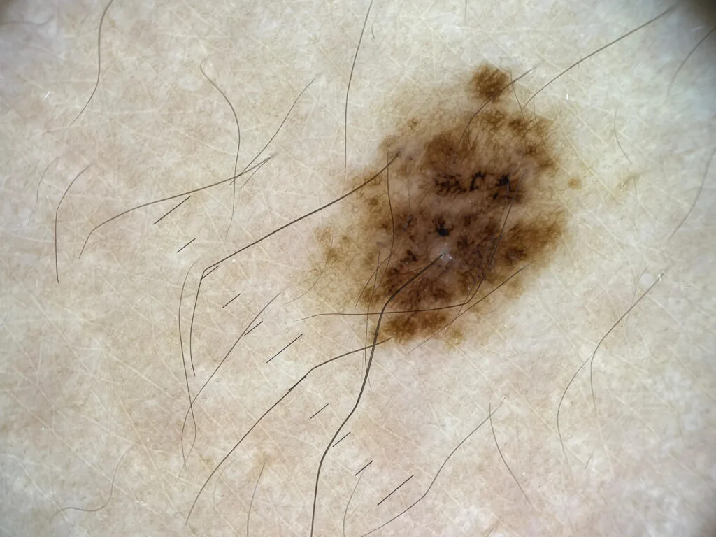
An intradermal nevus usually appears differently. Because the melanocytes are located deeper within the dermis, the pigment network often disappears. Instead, the lesion commonly demonstrates a homogeneous light brown or skin-colored appearance with comma-shaped vessels. Small white globules or terminal hairs may also be visible. The overall architecture remains symmetrical, with smooth borders and consistent coloration.

Dermatologists do not rely on a single dermoscopic feature. Instead, they evaluate the lesion as a whole while also considering the patient’s age, history, skin type, and lesion evolution.
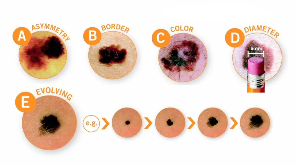
The well-known ABCDE rule remains an important first screening method for pigmented lesions. A represents asymmetry. B refers to irregular borders. C stands for multiple or uneven colors. D indicates a diameter greater than approximately 6 mm, although smaller melanomas can occur. E refers to evolution, meaning any change in size, color, shape, or symptoms over time. While the ABCDE rule is useful for visual self-examination, dermoscopy provides additional structural information that improves diagnostic confidence.
What Does Melanoma Look Like Under a Dermatoscope?
Melanoma is responsible for most skin cancer deaths despite representing only a small proportion of all skin cancers. Early detection is therefore essential.
Unlike benign nevi, melanoma usually demonstrates architectural disorder rather than symmetry. Multiple colors may be present within the same lesion, including light brown, dark brown, black, gray, blue, white, or red. Pigment networks become irregular, with variations in thickness and spacing. Atypical dots and globules may appear unevenly distributed. Streaks, pseudopods, regression structures, blue-white veils, and polymorphous vessels can also be observed depending on the melanoma subtype.
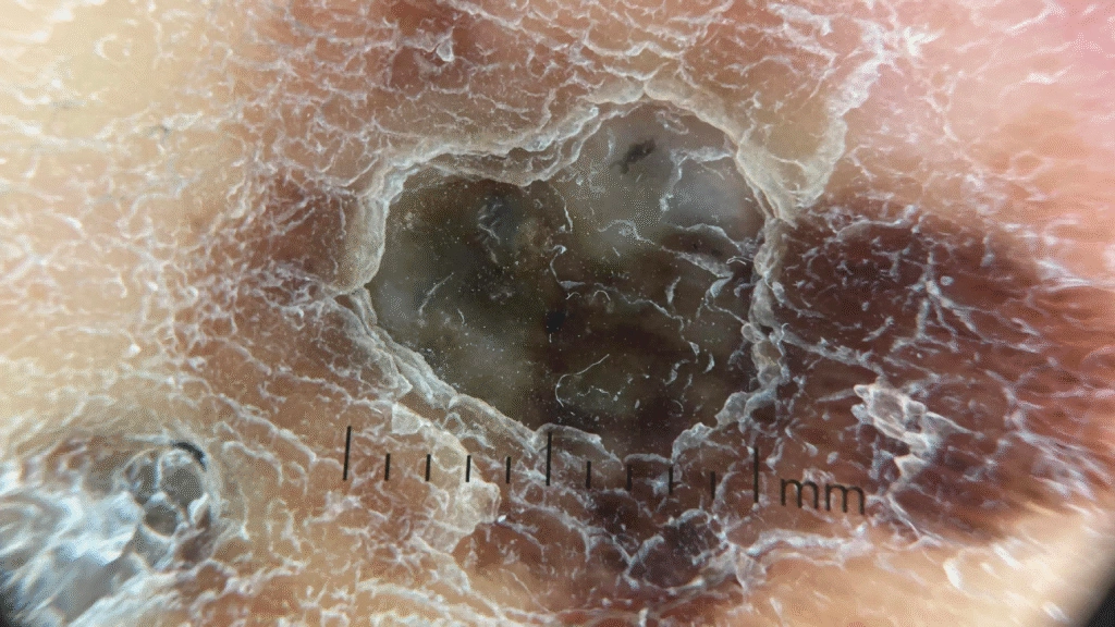
No single dermoscopic sign confirms melanoma by itself. Diagnosis depends on recognizing combinations of suspicious structures together with clinical history and lesion evolution. The value of dermoscopy lies in its ability to reveal these subtle abnormalities before they become obvious to the naked eye. Earlier recognition may lead to biopsy at a stage when melanoma remains thin and highly curable.
How Does Basal Cell Carcinoma Appear Under Dermoscopy?
Basal cell carcinoma (BCC) is the most common skin cancer worldwide. Unlike melanoma, BCC rarely spreads to distant organs, but delayed diagnosis may lead to progressive local tissue destruction.
Dermoscopy has greatly improved the recognition of BCC because the tumor often displays characteristic vascular and pigmented structures. One of the most recognizable findings is arborizing vessels, which are large branching blood vessels resembling tree branches. Blue-gray ovoid nests are another classic feature, representing aggregates of tumor cells located within the dermis. Other common findings include leaf-like areas, spoke-wheel structures, shiny white structures, and multiple small erosions.
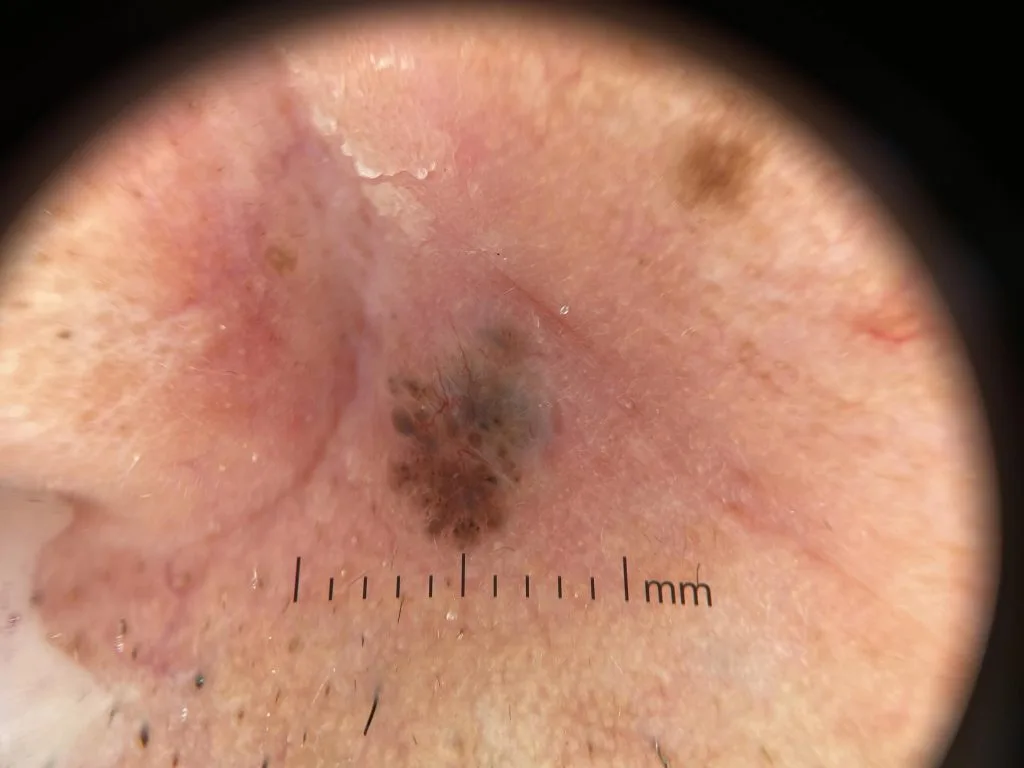
These dermoscopic features help distinguish BCC from benign lesions that may appear similar to the naked eye. Instead of relying only on lesion color or shape, clinicians can identify structural patterns that strongly suggest malignancy.
With true optical 10X magnification, the IBOOLO DE-4100 allows these characteristic vascular and pigmented structures to become clearly visible during examination.
Ready to see why IBOOLO is the #1 value pick for 2026?
*Includes manufacturer-direct support and global DDP shipping options.
How Does Dermoscopy Improve Everyday Clinical Practice?
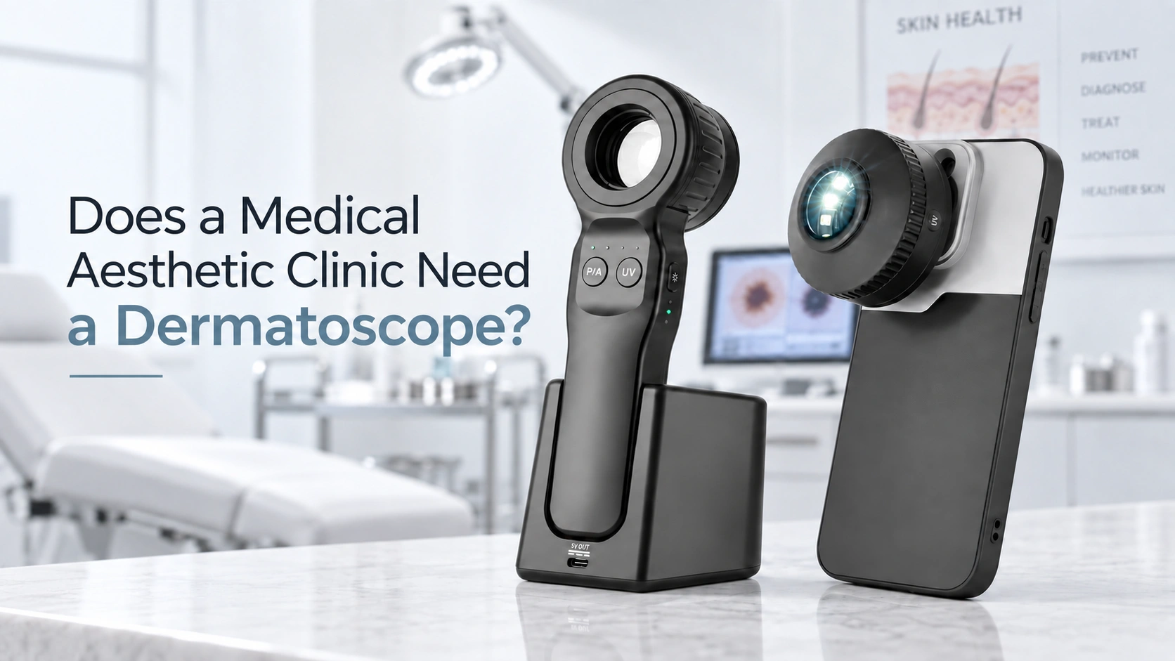
Dermoscopy is no longer limited to dermatology specialists. It has become an increasingly valuable tool for general practitioners, family physicians, emergency physicians, and other healthcare providers who regularly evaluate skin lesions.
Clinical studies have demonstrated that dermoscopy improves diagnostic accuracy for both pigmented and non-pigmented skin tumors when clinicians receive appropriate training. It can reduce unnecessary biopsies of benign lesions while increasing the early detection of malignant disease. Dermoscopy also assists in monitoring lesions over time, documenting changes with digital imaging, evaluating inflammatory skin disorders, identifying certain infectious diseases, and assessing hair and scalp conditions.
For primary care physicians, dermoscopy provides an additional level of confidence when deciding whether a lesion can be monitored, requires referral, or should undergo biopsy. This contributes to more efficient patient management and earlier identification of skin cancers.
Secure Your 2026 Diagnostic Edge
- Ready to Ship (In Stock)
- Worldwide DDP Shipping (No hidden tax)
- 2-Year Extended Warranty Included
Special: Use IBOOLO-TOP10 for an extra 10% OFF!
Average Response Time: 4 Hours