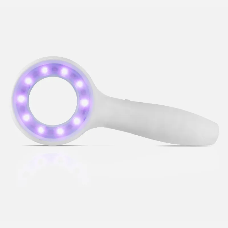
What Is Dermatoscopic Digital?

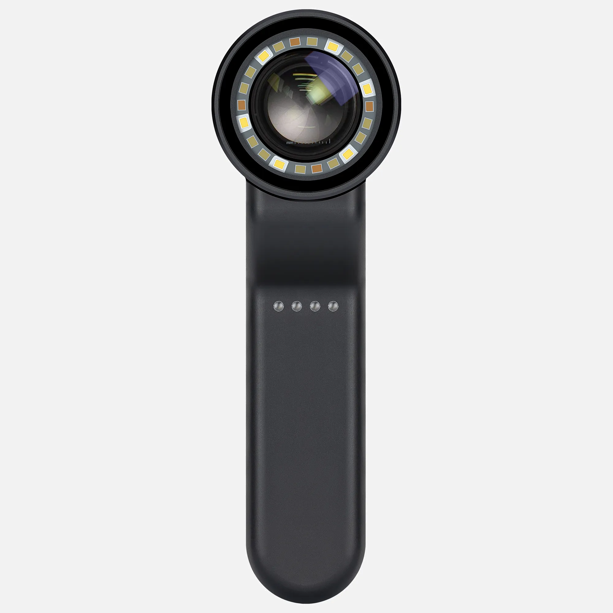
A digital dermatoscope, also known as a dermoscope or microscope, is a tool that provides high-resolution imaging to help detect skin conditions and cancer.

Digital dermatoscopy is a non-invasive diagnostic technique that can help evaluate skin aspects that are not visible to the naked eye. It is one of the most accurate methods for detecting changes in pigmented skin lesions, such as moles or nevi, which can lead to melanoma.

Digital dermatoscopy images are stored and compared to images obtained during a patient's next visit. Sequential digital dermoscopy imaging (SDDI) captures and stores images of suspicious lesions that are then monitored over time for changes. Studies have shown that SDDI allows for early detection of melanomas and leads to a decrease in the number of unnecessary excisions.

Dermatoscopes can be used for many purposes, including:

  • patient communication and education
  • Keeping and sharing patient records
  • Documenting changes over time
  • Clinical examinations
  • Cosmetics
  • Skin care
  • Medical schools

To perform a dermoscopy, a clinician will apply an ultrasound gel or oil onto the skin to improve the image clarity. Once the gel or oil is applied, the clinician will gently press the dermatoscope into the skin.

How Does Dermatoscopio Digital?

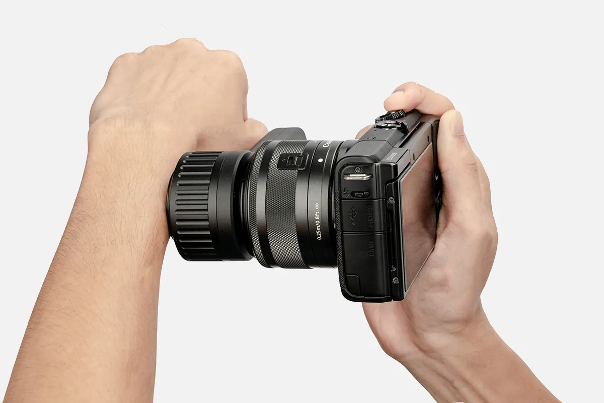
Digital dermatoscopy is a non-invasive, painless, and straightforward diagnostic technique that uses a camera to capture images of the skin. The images are then recorded and analyzed on the device, mobile phone, PC, or web-based software.

Here's how digital dermatoscopy works:

1. The patient stands in a designated spot for full-body imaging.

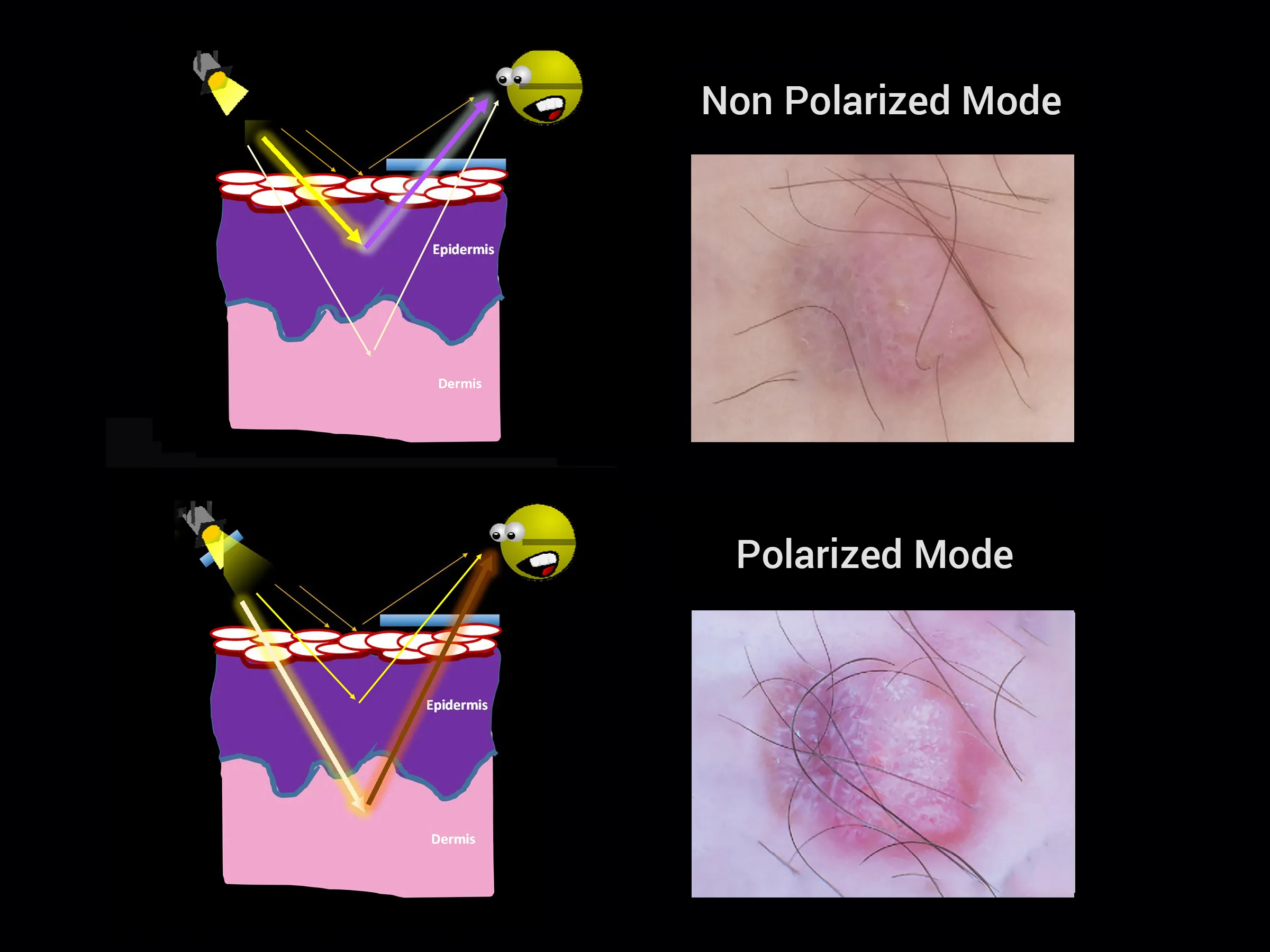
2. The camera allows light to pass through the skin and bounce back into the camera.

3. The camera sends the pictures to backend software, which compiles and generates a three-dimensional patient render.

4. The three-dimensional model is often viewable in applications provided by the device.

Digital dermatoscopy can help medical professionals:

    Capture high-quality images of their patient's skin

    Keep images organized in an online system

    Eliminate redundant paperwork

    Keep a detailed profile of their patients' lesion history

Forward images for the purposes of exchanging ideas with colleagues or seeking a second opinion

People May Ask

¿CuáL Es La Palabra Que Examina Tu Piel?

medical professional with a focus on diagnosing and treating skin issues. Se conoce como experto en dermatología además.

¿CuáL Es El Plane Solar?

Consiste en una técnica no invasive que permite visualizar estructuras profundas en la piel: registro fotográfico de alta definición asociado con la dermatoscopía. Particularmente se emplea en el análisis de lunares para identificar quiénes se sospecha de ser malignos y quiénes no.

¿CuáL Es El Proceso Del Examino Dermatologico?

La examinación cutánea is un componente esencial del examen físico estándar. All of the body's skin is meticulously examined as part of this examination. The assessment focuses on identifying abnormal signs in the skin, such as the cabelludo, orificios, uñas, and mucosal surfaces.

¿Qué Significa Un Mapa Lunar?

1. ¿Cuál es el mapeo lunar? A: La palabra "mapeo de lunares" más frecuentemente se denomina supervisión de un paciente con un riesgo elevado de melanoma. The dermatologist use a variety of tools.

How Are The Lunares Examined?

By examining your skin, the doctor can diagnose your lunar symptoms. The doctor examines the patient's skin from the top to the bottom during a dermatological examination. In the event that the doctor suspects a lunar specimen may be cancerous, the specimen will be removed and sent to a lab for microscopic examination (biopsia).

What Could Be Found in A Skin Biopsy?

Numerosas afecciones cutáneas son diagnosticadas mediante la biopsia de piel, entre ellas: enfermedades de la piel, tales como psoriasis, eccema, queratosis actínica ("precáncer") and verrugas. microbiological or bacterial skin infections.

¿CóMo Se Diagnosta Cancer Cutaneous?

Usted o su asesor médico verá si existen lunares, marcas de nacimiento u áreas con un color, tamaño, forma o textura anormales para realizar un examen de cáncer de piel. Should a portion of your skin appear abnormal, you may need to conduct tests to determine whether it is cancerous.

What Is The Name of The Examination Used to Identify Skin Cancer?

Some dermatologists use a technique called dermoscopy-also known as dermatoscopy, microscopy of epiluminescence [ELM], or microscopy of surface-in conjunction with the conventional medical examination to observe more clearly certain areas of the skin.

What Is The Duration of A Digital Dermatoscopy?

Takes between 30 and 40 minutes, though it always depends on the specific situation and the number of moons to be explored. It's not required to go with someone to take the test. There's no interaction with medications or pre-ingested food. There are no contraindications for digital dermatoscopy.

How Can One Perform A Digital Dermatoscopy?

What is the date of the digital dermatoscopy? This test is indicated for the general control of nevus, the prevention of melanoma, and the diagnosis of other skin lesions such as some benign or malignant tumors.

Dermatoscopio Digital Products

China high quality dermatoscopio digital

Skybasic 50X-1000X WiFi Portable Handheld Microscopes with Adjustable Stand, Wireless Digital Microscope, HD USB Microscope Camera Compatible with iPhone Android iPad Windows Mac Computer

China portable dermatoscopio digital suppliers & wholesalers

Suitable for children, this handheld digital microscope has an LCD screen and is suitable for adults. It has a 800x magnification and USB lab 32GB SD Card-equipped handheld digital microscope

China affordable dermatoscopio digital suppliers & wholesalers

The FOKH Skin Hair Analyser is a professional scale hair folliculle oil moisture magnifying detector skin pigment tester that is portable and rechargeable. It features a 5-in-50x/200x LCD digital microscope and is made in the United States.

China handheld dermatoscopio digital

The TOMLOV DM1S Wireless Digital Microscope is a fun and easy tool. 50X-1000X 1080P HD WiFi Compact Coin Microscope Camera Magnifier with Stand for iPhone, iPad, Android Phone, and PC - Trichome Mini Handheld USB Microscope

China handheld dermatoscopio digital suppliers & wholesalers

SUNRAYINNO DPM 1200 Handheld Digital Microscope: Easily examine small objects with this lightweight, compact device that features an articulating 4-inch display, a long run time of rechargeable lithium-ion batteries, and a compact design.

China Professional dermatoscopio digital

Compatible with Android phones, Mac computers, and Windows PCs, the MCJ USB digital microscope camera is a 50X-1600X portable handheld tiny microscope with an adjustable stand and an 8 LED high-definition USB microscope camera.

dermatoscopio digital suppliers & wholesalers

The Hiacinto Ear Wax Removal Tool Camera, a 7-inch IPS screen digital otoscope, and a 3.9mm HD ear scope with light, ear camera, and wax remover enables 32GB card support, photo capture, and video recording.

China affordable dermatoscopio digital

The True HD Macro 200x Zoom Imaging Opti-Tekscope Digital USB Microscope Camera (1600 x 1200) Advanced CMOS Sensor with 8 LEDs for video (Windows, MAC, Linux OS)

China high quality dermatoscopio digital suppliers & wholesalers

Children s Adult Electron Microscope - Black, GUVOP 50X-1000X Magnification WiFi Portable Handheld USB Microscope with 8 LEDs and Stand, Compatible with iPhone, Android

China customized dermatoscopio digital suppliers & wholesalers

Portable handheld microscopes with photo storage features, having a zoom range of 50X to 1600X USB digital microscopes USB Type C Computer Phone Electronic Digital Microscope

Top Reviews

Customer

Because I believe that 98% of reviews are LIES, I am not a huge fan of them. I'm going to tell you, this is fantastic! How much this aids in coin identification is beyond words. This makes it so much easier for me to detect problems, especially since my eyes are becoming worse. It allows you to record video and snap images. I have to deal with two shortcomings. The view finder does not allow you to zoom out far enough to see the entire coin. The first half must be photographed, followed by the opposite half. The second problem is that you can't link it to a computer to get a larger screen—at least not that I've worked out how to do so. If someone is seeking for ways to look at coins or anything similar, I would definitely suggest this to them.

Customer

It's easy to use, charged, and functions on my iPhone just as promised.

Nick

Both my phone (Android 14 with WiFi) and PC (Windows 11 with the free Microsoft Windows Camera program that I downloaded in accordance with the given instructions and connected with the included USB cable) function incredibly well. These are two photos I shot while using my computer. My $40 was more than well spent.

0

Have questions on gear or your order?

Our Gear Guides are here to help! Get personal advice from pro creatives

Name
Subject
Email address
How can we help?

Instant Answers

How Can Dermoscopy Images Be Captured?

Dermoscopy images can be captured and stored in different ways, such as: • Using a smartphone or tablet with dermoscopic adapter, which consisted in the package.• Using a digital camera

Dermoscopy images can be captured and stored in different ways, such as:

• Using a smartphone or tablet with dermoscopic adapter, which consisted in the package.
• Using a digital camera with dermoscopic adapter, there’s 49mm screw size camera adapter available to order now.

Compatible phone/tablet models:
All iPhone models, 95% Android phones, 90% tablet. For phone/tablet size in 5.25-14mm

Compatible camera models:
All camera with built 49mm filter screw, such as Canon EOS 70D, 80D, 90D; Canon EOS R7, R10, R50, R100; Canon M100, M200, M50, Mark II; Canon G7X Mark III, Sony ZV-1

How Can I Connect My Phone to My Dermatoscope?

There’s universal phone adapter for all our dermoscopes. Please check the installation procedure bellow or watch operation guide. Smartphone Connector (1) Place phone adapter screw in the center of smartphone’s

There’s universal phone adapter for all our dermoscopes. Please check the installation procedure bellow or watch operation guide.

Smartphone Connector

(1) Place phone adapter screw in the center of smartphone’s main camera.
(2) Screw magnet attachment on phone adapter.
(3) Put dermoscope’s back ring and magnet attachment together

Take The Best Images

You need to adjust the focus ring after the dermoscpe connected on smartphone to get the best images.

How Can I Clean My Dermoscopy after Usage?

Cleaning your dermoscopy after usage is important to prevent cross-contamination and infection. The cleaning method may vary depending on the type and model of your dermoscopy, so you should always

Cleaning your dermoscopy after usage is important to prevent cross-contamination and infection. The cleaning method may vary depending on the type and model of your dermoscopy, so you should always follow the manufacturer’s instructions. However, some general steps are:

• Turn off and disconnect your dermoscopy from any power source or device.

• Wipe off any visible dirt or debris from the dermoscopy with a soft cloth or tissue.

• Disinfect the dermoscopy with an alcohol-based wipe or spray, or a disinfectant solution recommended by the manufacturer. Make sure to cover all surfaces, especially the lens and contact plate.

• Let the dermoscopy air dry completely before storing it in a clean and dry place.

• Do not use abrasive or corrosive cleaners, solvents, or detergents that may damage the dermoscopy.

• Do not immerse the dermoscopy in water or any liquid, unless it is waterproof and designed for immersion.

You should clean your dermoscopy after each use, or at least once a day if you use it frequently. You should also check your dermoscopy regularly for any signs of damage or malfunction, and contact the manufacturer or service provider if needed.

Polarized VS Non-Polarized Dermoscopy

A dermoscopy is a device that allows the examination of skin lesions with magnificationand illumination. By revealing subsurface structures and patterns that are not visible tothe naked eye. It can

A dermoscopy is a device that allows the examination of skin lesions with magnificationand illumination. By revealing subsurface structures and patterns that are not visible tothe naked eye. It can improve the diagnose accuracy of skin lesions, such as melanoma,basal cell carcinoma, seborrheic keratosis, etc.

There are two main types of dermoscopy: Non polarized and polarized dermoscopy.We’ve fitted most of our dermoscopys with polarized and non-polarized light. They canbe used in multiple skin structures.

Non-polarized contact Mode

In non-polarized mode, the instrument can provide information about the superficialskin structures, such as milia-like cysts, comedo-like openings, and pigment in theepidemis.

The dermoscopy requires applying a liquid such as mineral oil or alcohol to the skin andplacing the lens in contact with the skin. This reduces surface reflection and enhancesthe view of subsurface structures.

Image with non-polarized light (DE-3100)

Polarized contact Mode

In polarized mode, the instrument allows for visualization for deeper skin structures,such as blood vessels, collagen, and pigment in the dermis.

The dermoscopy does not need to be in contact with the skin or use any liquid. Theirpolarized light can help to eliminate surface reflection and allow visualization ofvascular structures.

Image with polarized light (DE-3100)

Polarized non-contact Mode

The dermoscopy can also use polarized light to examine the skin without direct contact.

In polarized non-contact mode, the instrument allows for examination infected areasand lesions that are painful for the patient, or the difficult to contact pigmented lesions,such as nails and narrow areas.

The contact plate should be removed in this mode, and it does not require applying aliquid to the skin. As it doesn’t require pressure or fluid application on the skin, it canalso avoid cross-contamination and infection risk.

Image in polarized non-contact mode (DE-3100)

How Effectiveness Is Dermoscopy

Compared with visual inspection, the dermoscopy can be used to capture and store skin lesion photos, which play an important role in early skin cancer examination. The dermoscopy allows the

Compared with visual inspection, the dermoscopy can be used to capture and store skin lesion photos, which play an important role in early skin cancer examination.

The dermoscopy allows the examination of skin lesions with magnification and illumination. This can be greatly avoiding the factors that cause interference to visual detection. Such as lighting, skin color, hair and cosmetics.

Several studies have demonstrated that dermoscopy is useful in the identification of melanoma, when used by a trained professional.

It may improve the accuracy of clinical diagnosis by up to 35%
It may reduce the number of harmless lesions that are removed
In primary care, it may increase the referral of more worrisome lesions and reduce the referral of more trivial ones

A 2018 Cochrane meta-analysis published the accuracy of dermoscopy in the detection.

Table 1. Accuracy of dermoscopy in the detection of melanoma in adults
Detection Method Sensitivity, % Specificity, % Positive Likelihood Ratio NegativeLikelihood Ratio
Visual inspection alone (in person) 76 75 3.04 0.32
Dermoscopy with visual inspection (in person) 92 95 18 0.08
Image-based visual inspection alone (not in person) 47 42 0.81 1.3
Dermoscopy with image-based visual inspection (not in person) 81 82 4.5 0.23
ROC—receiver operating characteristic. *Estimated sensitivity calculated on the summary ROC curve at a fixed specificity of 80%.

As we can see, the dermoscope can improve the accuracy of diagnosis of skin lesions, especially melanoma.

Table 1. Accuracy of dermoscopy in the detection of melanoma in adults
Detection Method Sensitivity, % Specificity, % Positive Likelihood Ratio NegativeLikelihood Ratio
Visual inspection alone (in person) 79 77 3.4 0.27
Dermoscopy with visual inspection (in person) 93 99 93 0.07
Image-based visual inspection alone (not in person) 85 87 6.5 0.17
Dermoscopy with image-based visual inspection (not in person) 93 96 23 0.07
ROC—receiver operating characteristic. *Estimated sensitivity calculated on the summary ROC curve at a fixed specificity of 80%.

Characteristics of the dermatoscopic structure of the skin lesions include:

• Symmetry or asymmetry
• Homogeny/uniformity (sameness) or heterogeny (structural differences across the lesion)
• Distribution of pigment: brown lines, dots, clods and structureless areas
• Skin surface keratin: small white cysts, crypts, fissures
• Vascular morphology and pattern: regular or irregular
• Border of the lesion: fading, sharply cut off or radial streaks
• Presence of ulceration

There are specific dermoscopic patterns that aid in the diagnosis of the following pigmented skin lesions:

• Melanoma
• Moles (benign melanocytic naevus)
• Freckles (lentigos)
• Atypical naevi
• Blue naevi
• Seborrhoeic keratosis
• Pigmented basal cell carcinoma
• Haemangioma

No products in the cart.

No products in the cart.

Hot Search Terms