


DE-400 Dermatoscope – IBOOLO
IPhone Dermatoscope Attachments for Skin Photography
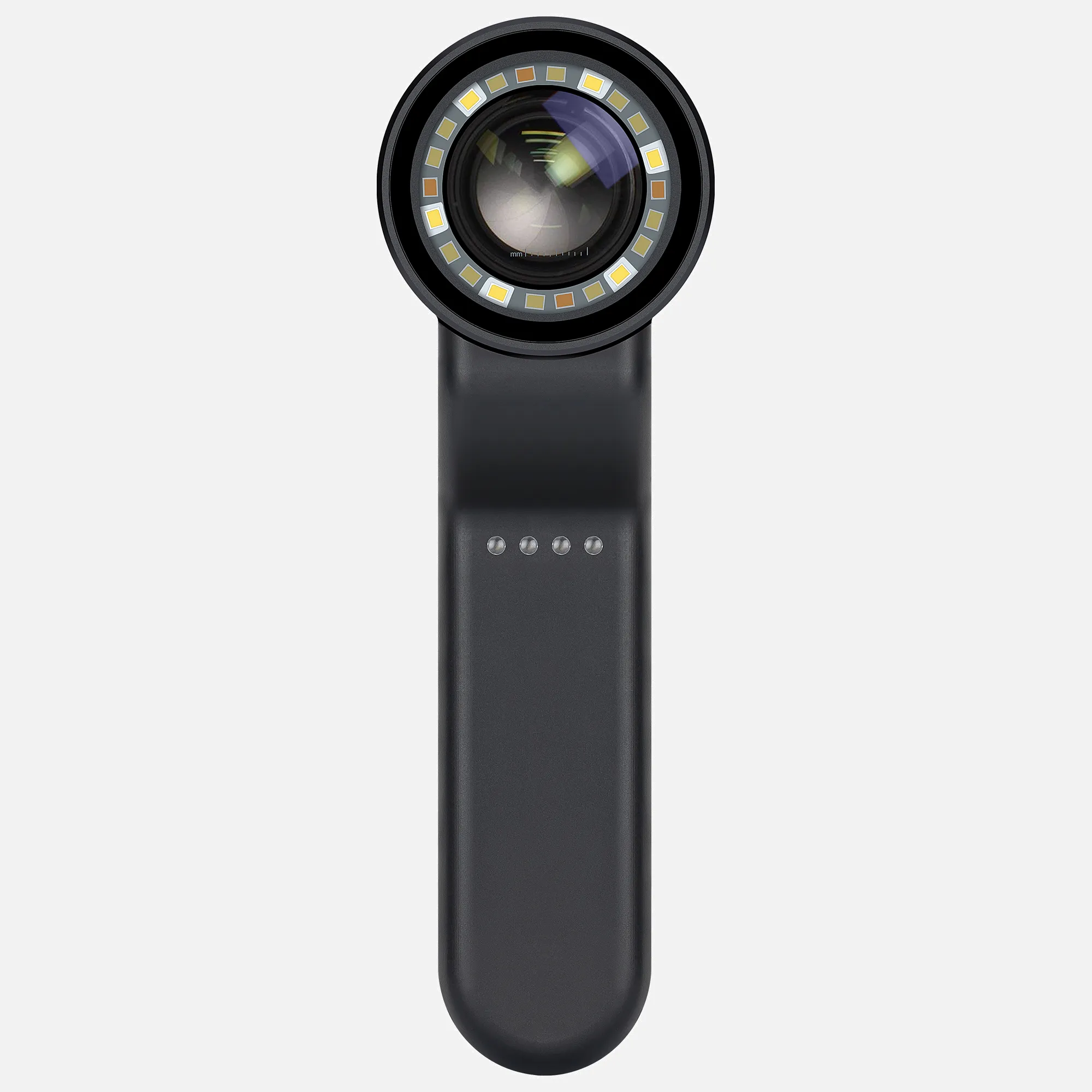
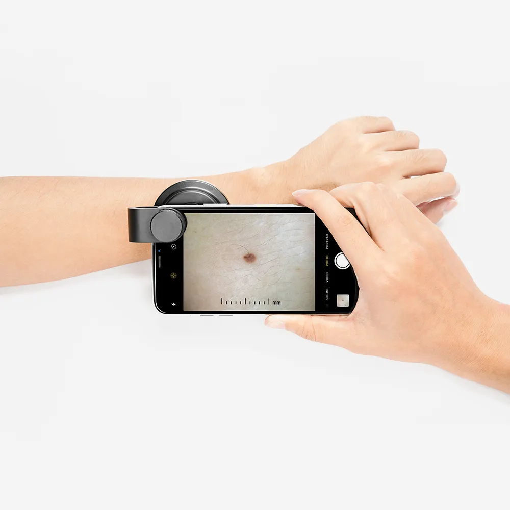
A dermatoscope is a specialized magnifying device that attaches to smartphones, including iPhones, to photograph skin lesions for tracking and diagnosis. Handheld models feature in-built transillumination lighting paired with 10x magnification optics to reveal subsurface skin characteristics invisible to the naked eye.
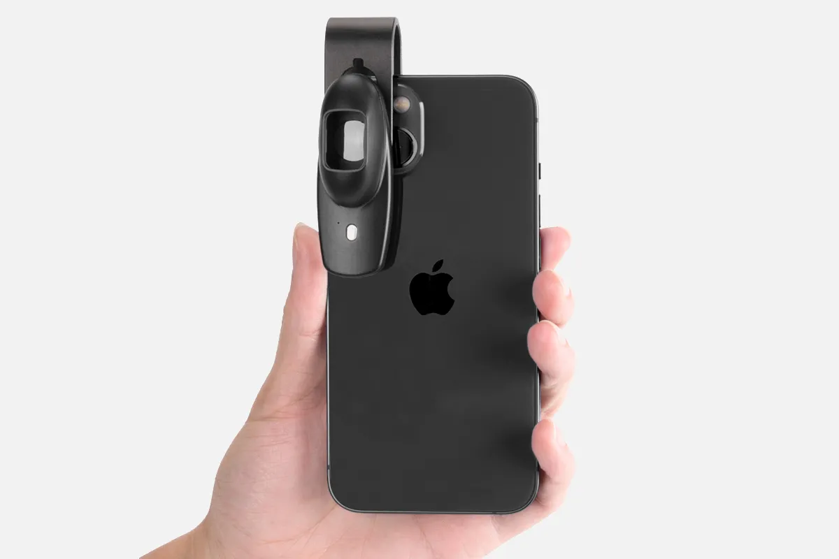
Specifically, iPhone dermatoscope adapters connect to the smartphone using its camera and display. Some models mount over the phone, while portable versions clip on. By illuminated lesions from varying angles, subtle visual features related to patterns, structure, blood vessels, and pigmentation become apparent. Quality images empower dermatologists to observe changes over time.
Additionally, iPhone-compatible dermatoscopes foster telemedicine by allowing easy transmission of clinical images to specialists for remote assessments. Some dermatoscopes integrate proprietary apps to measure lesions and reference analytics.
The iPhone's specialized dermatoscopy attachments leverage smartphone cameras to document magnified skin surface microscopy exams, enhancing clinical insights and care coordination. Select iPhone-compatible models offer advanced functionality like analytics to augment diagnostics.
Using An IPhone with A Dermatoscope Attachment
Here are key steps for conducting dermoscopy skin exams using an iPhone paired with a dermatoscopy attachment:
1. In iPhone settings, enable Macro photo control to override auto lens switching manually
2. Apply oil on the skin lesion to improve visualization
3. Attach the dermatoscopy securely onto the iPhone camera
4. Place the dermatoscopy flat on the skin, applying light pressure
5. Take multiple photos, refocusing on central areas of concern
6. Properly label stored photos for tracking
7. Review images closely, observing patterns and subtle visual characteristics
Different iPhone models may have unique camera settings to adjust. Some dermatoscopes connect using proprietary magnetic kits for stability, like the DermLite series.
Pairing an iPhone camera with a specialized dermatoscopy attachment allows capturing highly magnified clinical images to improve documentation of lesions over time. Combined with macro photography and proper technique, small visual clues apparent only under dermoscopy become visible.
People May Ask
There are certain melanoma variations that possess the potential to proliferate rapidly, posing a severe threat to life within a brief timeframe of six weeks. In the absence of prompt medical intervention, it has the capability to disseminate to various bodily regions. Nodular melanoma, a particularly hazardous subtype, exhibits a distinctive appearance from common melanomas and can develop in a matter of mere weeks.
Dermoscopy was shown to be more accurate than visual examination alone in both comparisons, according to the meta-analysis, with RDORs of (a) 4.7 (95% CI 3.0 to 7.5; P < 0.001) and (b) 5.6 (95% CI 3.7 to 8.5; P < 0.001).
Relative 5-year survival rates for skin cancers melanomaFive-year relative survival rate at SEER stageLocalized >99%74% regional35% awayThe total of all SEER phases is 94%.
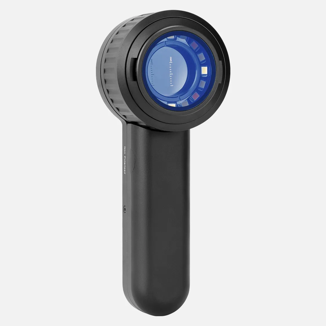
With the use of the DermLite product range, an expert eye can identify skin cancer and other skin diseases at an early stage. Each DermLite has an LED light source, a magnification lens, and the majority of them feature polarizing filters to reduce glare.
The LEDs on the DermLite DL3 should be pointed toward the lesion that needs to be checked when you hold it. Place the gadget |*|–25 mm away from the skin. The cross-polarized mode can be activated by pushing and holding the power button |A| on the right| side of the device for about a second.
Usually, the course of treatment for a malignant mole is the same as that for other malignancies. On the other hand, compared to malignancies of the internal organs, it will be simpler to reach the diseased tissue and perform surgery to remove the mole. Consequently, the majority of malignant moles are treated primarily with surgery.
The DL4W boasts a broad field of view, is more than 25% smaller than the previous generation, and has significantly brighter polarized and non-polarized white illumination when compared to the DL4 model. rapid processes. Snap connection is used by DL4W to provide processes that are incredibly quick and convenient.
The LEDs on the DermLite DL3 should be pointed toward the lesion that needs to be checked when you hold it. Place the gadget |*|–25 mm away from the skin. The cross-polarized mode can be activated by pushing and holding the power button |A| on the right| side of the device for about a second.
Use the included cable to connect the DermLite to both the Charging Port (CP) and any accessible USB port to charge it. The device has a roughly two-hour continuous operating time when completely charged. The battery, which is exclusive to 3Gen or an approved 3Gen dealer, may need to be replaced after years of use.
Identify Your Skin SpotsSkin cancer comes in three different forms: melanoma, basal cell carcinoma, and squamous cell carcinoma. The ability to recognize every kind of skin cancer is a skill taught to dermatologists. Make an appointment with the dermatologist over the phone if you find anything strange during your self-examination.
Dermatoscope Iphone Products

BeaverLAB Digital Microscope M1 WiFi 1000X HD Mini Coin Microscope Camera: a portable, handheld USB microscope that works with iPhone, Android, and iPad computers. Suitable for adults and children.

With a 4.3 LCD screen, two gooseneck lights, PC view, Windows compatibility, and compatibility with iPhones, the TOMLOV DM4 Pro Wireless Digital Microscope is a high-quality wireless microscope.

The 80-120X Clip-On LED Cell Phone Microscope is a compact, universal lens magnifier with LED and UV lights that can be used on smart phones.
Suitable with iPhone, Android, iPad, and PC, the Matatastudio M2C Wireless Digital Microscope is a handheld, portable, 1080p HD microscope with an adjustable stand for both indoor and outdoor use. It has a 50X–1000X magnification.

Pocket Phone Microscopes: Enjoy Microworld with this 200X Zoom Clip-On Microscope Magnifier with CPL Lens and LED Light, which is portable and fits iPhone, Samsung, Huawei, and Google. Available in black.
Quick Focus, Easy Operation, Chute Type Installation, Mini HD 400x Phone Microscope Lens with LED Light, DAUERHAFT 400x Microscope Lens for 14 Pro Max
A 400x magnifying kit, a mobile microscope lens for the iPhone 14 Pro Max, an easy-to-install smartphone microscope suitable for both adults and children, and an LED light
Microscope with 8 LEDs, compatible with iPhone & Android - Aopick Microscope Camera, Digital Coin Microscope for Adults Kids
Includes 6 ear pick scopes and an ear cleaning kit specifically for the iPhone. Microscope Camera - 1080P HD Otoscope with Light - Ear Cleaner with Camera - Ear Wax Removal
200x Microscope for Cell Phones: Compact Pocket Microphone with a Universal Clip and LED Light Attachment, Integrated CPL Lens Compatible with 99% of Android and iPhone Smartphones (Black)
Hot Products


News & Blog


Top Reviews
Evan b.
With the exception of being able to see every ridge and groove of a fingertip, this worked incredibly well when I hooked it up to my Windows PC and performed exactly like any other webcam. Although my demands kind of exclude that part of its pairing—I required it to examine etching paths on circuit boards—I haven't paired it with a mobile device yet.
Joe C
With its phone app, I had no trouble connecting this small, lightweight microscope, which offers excellent magnification. It may be pointed at the subject while still having enough room to operate thanks to the supplied stand. When I used it to service a watch, it was much easier to accomplish the task without having to take off my glasses as frequently as I would with a standard dissecting microscope. Even though I received this for free to review through the IBOOLO Vine program, the pricing is still quite affordable, and it works well for both still and video photography. I would gladly pay full retail for this. Additionally, if you'd want an even simpler viewing experience, it may be used with a tablet. It is fairly simple to use once you download the companion app, but it comes with very little instructions.
Marjorie
Very cool technology. I adore that it has a smart phone connection. On-the-go science!


