What Is Woods Lamp?

A Wood lamp is a tool that emits long-wave ultraviolet (UV) light. It can be used to help diagnose infections of the skin or scalp, such as bacterial or fungal infections. It can also identify the extent of pigmented or depigmented patches on the skin.

Certain cells appear fluorescent or show color When a Wood's lamp is used. If there is an infection in the area where the lamp is illuminating, the area will fluoresce.

Wood's lamps have a peak wavelength of 365nm, different from commercially available blacklight sources, which can have peak wavelengths of 375nm, 385nm, or 395nm. A longer peak UV wavelength produces more visible light, which results in less fluorescence.

Wood's lamps are called blacklight or ultraviolet light tests. They were developed by American physicist Robert Wood.

How Does Woods Lamp?

A Wood's lamp is a light that emits black light, which is ultraviolet light that is too dark for the human eye to see. The lamp emits long-wave UV radiation (UVR) through a high-pressure mercury arc and barium silicate and nickel oxide filter. This filter is opaque to all light except for a band between 320 and 400 nanometers, with a peak of 365 nanometers.

A Wood's lamp examination is a diagnostic test that involves exposing skin or hair to the lamp's black light. For example, a Wood's lamp can be used to detect the presence of a fungal scalp or skin infection. When viewed under a Wood's lamp, an infected area of the scalp may glow if it is infected with tinea, a type of ringworm fungus.

A Wood's lamp can also identify areas of vitiligo in light-skinned people. These areas will have sharper borders under black light and look bright blue-white or yellow-green.

Factors that can affect how a Wood's lamp works include:

  • Recent bathing or showering: Can decrease fluorescence
  • Medications, detergents, and fibers: Can cause inappropriate fluorescence
  • Substance production: Many substances produce false positives from a Wood's lamp, especially in pediatric dermatology

People May Ask

Which Illnesses Are Luminescent in The Dark?

Under Wood's light examination, a variety of skin conditions can fluoresce, including the following:> Infections caused by fungi, bacteria, head lice, and nits.ErythrasmaOther pigmentary illnesses, vitiligo, and porphyria cutanea tarda

Under Woods Lamp, What Color Is Ringworm?

Under this lamp, 50% of the strains of Microsporum canis, which is the most frequent cause of ringworm in cats, will cause the infected hairs to glow apple green.

What Other Name Would You Give A Wood Lamp?

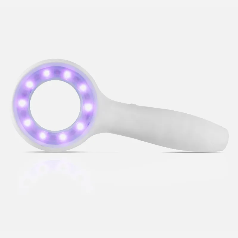
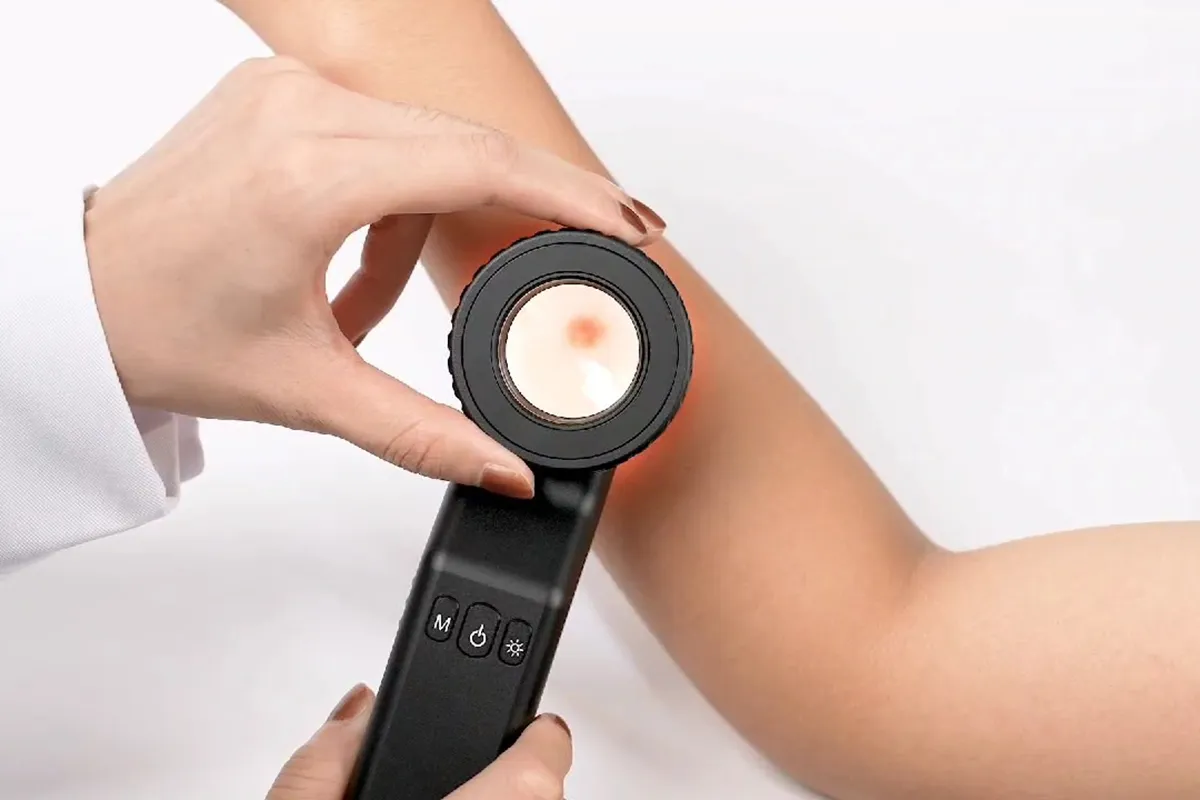
dark lightA lamp that generates mostly UV-A (long-wave) ultraviolet light and very little visible light is known as a blacklight, UV-A light, Wood's lamp, or ultraviolet light.

What Causes My Fingernails to Shine in The Dark?

This glow is the energy released when light is transformed by phosphor particles into visible light. Your teeth and fingernails are among the items that contain these particles.

Can Sun Damage Be Seen in A Woods Lamp?

The lamp will be shone by your practitioner a few inches from your skin when you enter a dimly lit room. When exposed to UV radiation, areas of sun damage and excess pigmentation will glow or fluoresce, whereas normal skin will appear somewhat blue. Acne can also be discovered by exposure to a Wood's lamp.

What Disorders of The Skin Does A Wood Lamp Reveal?

Frequently, the following skin conditions are diagnosed using Wood's lamp: disorders of pigmentation, such as vitiligo and melasma. The symptoms of Pityriasis Versicolor include an orange-colored rash on the back or chest.

Under The Woods Lamp, What Hue Is Healthy Skin?

Skin that is fluorescent, depigmented, or pigmented can be seen using the Wood's lamp test. Normal, healthy skin does not glow under a Wood's light; instead, it appears blue.

Under Wood's Lamp, What Glows White?

Under a Wood's lamp, in particular, the depigmentation of vitiligo looks remarkably white and well defined. Despite its deceptive name, Nevus depigmentosus is frequently hypopigmented, giving Wood's lamp merely a faint, off-white glow.

A Woods Lamp Is Just A Black Light, Right?

How is a Wood's lamp operated? Black light from a Wood's lamp is ultraviolet light that is invisible to the unaided eye. Also visible to the unaided eye is a tiny amount of light it releases in the violet spectrum. Certain cells appear differently when exposed to UV radiation on skin or other surfaces.

What Uses Does A Wood's Lamp Serve?

UV light from a Wood's lamp can be used as a diagnostic tool to identify bacterial or fungal infections of the skin or scalp. Where the Wood lamp is shining, if there is an infection present, the region will glow.

Woods Lamp Products

China Professional Woods Lamp

12-Way Dimmable Bedside Table Lamp with PLA Lamp Shade - UYKKE Bedside Lamp with Wood Base - Push Button Console Lamp for Bedroom Perfect for living rooms, children s rooms, offices, and college dorms (built-in LED light)

China smartphone Woods Lamp suppliers & wholesalers

Three-piece solid wood pendant light in a sophisticated matte black finish, ideal for kitchens, islands, dining rooms, living rooms, and bedrooms

China smartphone Woods Lamp

YAMEIWAN 28-Inch Gold Bedside Light Set of 2 for Bedroom - Contemporary Table Lamp with USB Charging Port for Living Room End Table - 2-Way Dimmable Bedside Table Lamp with Zipper Switch

China high quality Woods Lamp

LT2078-LWG Simple Designs Light Wood and Light Gray, Small Mid-Century Table Lamp

Woods Lamp suppliers & wholesalers

Six Clear Acrylic Blank Sheets and Six 3D Night LED Light Lamp Bases with Remote Control and USB Cable—a Set of 16 DIY Acrylic Lamp Bases in 4 Modes—for Bar and Shop Decoration (Square)

China Professional Woods Lamp suppliers & wholesalers

The ROTTOGOON Farmhouse Table Lamps Set of 2 features a rustic wood finish, two USB ports, and a 27-inch resin nightstand lamp with three-color temperature LED bulbs and a white fabric shade that may be used in the living room or bedroom.

China portable Woods Lamp

DSMJFU Wall Lights: Two-piece Modern Rotatable Wall Light Set, Rustic Wood Wall Light, Side Wall Light with Hammered Metal Shade, Living Room Hallway, Bedroom

China portable Woods Lamp suppliers & wholesalers

Small Table Lamp by DEWENWILS: Wood Tripod Nightstand Lamp with Linen Fabric Shade for Living Room, Nursery, Children s Room, College Dorm, Coffee Table, and Bookcase; 14.2-inch

China affordable Woods Lamp

Dark Bronze Four-Light Transitional Style Pendant Light in Wood/Metal Design by Cal Lighting FX-3536/4

China customized Woods Lamp suppliers & wholesalers

The Allesin Wooden Tripod Floor Lamp for Living Room is suitable for living rooms, bedrooms, and offices. It features continuous dimming, adjustable color temperature, and a stand.

Top Reviews

Gregory

USB is the only way to light this lamp. It lacks a battery compartment. It does provide a substantial amount of light. And will automatically light a room. The math formulas are nice. Any math enthusiast would love this. Fantastic item Two cheers for it!

Angela

Gorgeous contemporary lamp, I've had so many compliments on it. The light worked so well for the whole family during the 12-hour power outage. superb financial decision.

Huy Le

This was purchased for my WFH desk, which is now situated in an unlit corner of my living room. I will keep it on my desk and attempt to think of another method to bring light to my office even if, regrettably, the real light it emits is exactly as described and is much more appropriate as a bedtime lamp. Still, it looks great. (Switching it on and off is also humorous.)

0

Have questions on gear or your order?

Our Gear Guides are here to help! Get personal advice from pro creatives

Name
Subject
Email address
How can we help?

Instant Answers

How Can Dermoscopy Images Be Captured?

Dermoscopy images can be captured and stored in different ways, such as: • Using a smartphone or tablet with dermoscopic adapter, which consisted in the package.• Using a digital camera

Dermoscopy images can be captured and stored in different ways, such as:

• Using a smartphone or tablet with dermoscopic adapter, which consisted in the package.
• Using a digital camera with dermoscopic adapter, there’s 49mm screw size camera adapter available to order now.

Compatible phone/tablet models:
All iPhone models, 95% Android phones, 90% tablet. For phone/tablet size in 5.25-14mm

Compatible camera models:
All camera with built 49mm filter screw, such as Canon EOS 70D, 80D, 90D; Canon EOS R7, R10, R50, R100; Canon M100, M200, M50, Mark II; Canon G7X Mark III, Sony ZV-1

How Can I Connect My Phone to My Dermatoscope?

There’s universal phone adapter for all our dermoscopes. Please check the installation procedure bellow or watch operation guide. Smartphone Connector (1) Place phone adapter screw in the center of smartphone’s

There’s universal phone adapter for all our dermoscopes. Please check the installation procedure bellow or watch operation guide.

Smartphone Connector

(1) Place phone adapter screw in the center of smartphone’s main camera.
(2) Screw magnet attachment on phone adapter.
(3) Put dermoscope’s back ring and magnet attachment together

Take The Best Images

You need to adjust the focus ring after the dermoscpe connected on smartphone to get the best images.

How Can I Clean My Dermoscopy after Usage?

Cleaning your dermoscopy after usage is important to prevent cross-contamination and infection. The cleaning method may vary depending on the type and model of your dermoscopy, so you should always

Cleaning your dermoscopy after usage is important to prevent cross-contamination and infection. The cleaning method may vary depending on the type and model of your dermoscopy, so you should always follow the manufacturer’s instructions. However, some general steps are:

• Turn off and disconnect your dermoscopy from any power source or device.

• Wipe off any visible dirt or debris from the dermoscopy with a soft cloth or tissue.

• Disinfect the dermoscopy with an alcohol-based wipe or spray, or a disinfectant solution recommended by the manufacturer. Make sure to cover all surfaces, especially the lens and contact plate.

• Let the dermoscopy air dry completely before storing it in a clean and dry place.

• Do not use abrasive or corrosive cleaners, solvents, or detergents that may damage the dermoscopy.

• Do not immerse the dermoscopy in water or any liquid, unless it is waterproof and designed for immersion.

You should clean your dermoscopy after each use, or at least once a day if you use it frequently. You should also check your dermoscopy regularly for any signs of damage or malfunction, and contact the manufacturer or service provider if needed.

Polarized VS Non-Polarized Dermoscopy

A dermoscopy is a device that allows the examination of skin lesions with magnificationand illumination. By revealing subsurface structures and patterns that are not visible tothe naked eye. It can

A dermoscopy is a device that allows the examination of skin lesions with magnificationand illumination. By revealing subsurface structures and patterns that are not visible tothe naked eye. It can improve the diagnose accuracy of skin lesions, such as melanoma,basal cell carcinoma, seborrheic keratosis, etc.

There are two main types of dermoscopy: Non polarized and polarized dermoscopy.We’ve fitted most of our dermoscopys with polarized and non-polarized light. They canbe used in multiple skin structures.

Non-polarized contact Mode

In non-polarized mode, the instrument can provide information about the superficialskin structures, such as milia-like cysts, comedo-like openings, and pigment in theepidemis.

The dermoscopy requires applying a liquid such as mineral oil or alcohol to the skin andplacing the lens in contact with the skin. This reduces surface reflection and enhancesthe view of subsurface structures.

Image with non-polarized light (DE-3100)

Polarized contact Mode

In polarized mode, the instrument allows for visualization for deeper skin structures,such as blood vessels, collagen, and pigment in the dermis.

The dermoscopy does not need to be in contact with the skin or use any liquid. Theirpolarized light can help to eliminate surface reflection and allow visualization ofvascular structures.

Image with polarized light (DE-3100)

Polarized non-contact Mode

The dermoscopy can also use polarized light to examine the skin without direct contact.

In polarized non-contact mode, the instrument allows for examination infected areasand lesions that are painful for the patient, or the difficult to contact pigmented lesions,such as nails and narrow areas.

The contact plate should be removed in this mode, and it does not require applying aliquid to the skin. As it doesn’t require pressure or fluid application on the skin, it canalso avoid cross-contamination and infection risk.

Image in polarized non-contact mode (DE-3100)

How Effectiveness Is Dermoscopy

Compared with visual inspection, the dermoscopy can be used to capture and store skin lesion photos, which play an important role in early skin cancer examination. The dermoscopy allows the

Compared with visual inspection, the dermoscopy can be used to capture and store skin lesion photos, which play an important role in early skin cancer examination.

The dermoscopy allows the examination of skin lesions with magnification and illumination. This can be greatly avoiding the factors that cause interference to visual detection. Such as lighting, skin color, hair and cosmetics.

Several studies have demonstrated that dermoscopy is useful in the identification of melanoma, when used by a trained professional.

It may improve the accuracy of clinical diagnosis by up to 35%
It may reduce the number of harmless lesions that are removed
In primary care, it may increase the referral of more worrisome lesions and reduce the referral of more trivial ones

A 2018 Cochrane meta-analysis published the accuracy of dermoscopy in the detection.

Table 1. Accuracy of dermoscopy in the detection of melanoma in adults
Detection Method Sensitivity, % Specificity, % Positive Likelihood Ratio NegativeLikelihood Ratio
Visual inspection alone (in person) 76 75 3.04 0.32
Dermoscopy with visual inspection (in person) 92 95 18 0.08
Image-based visual inspection alone (not in person) 47 42 0.81 1.3
Dermoscopy with image-based visual inspection (not in person) 81 82 4.5 0.23
ROC—receiver operating characteristic. *Estimated sensitivity calculated on the summary ROC curve at a fixed specificity of 80%.

As we can see, the dermoscope can improve the accuracy of diagnosis of skin lesions, especially melanoma.

Table 1. Accuracy of dermoscopy in the detection of melanoma in adults
Detection Method Sensitivity, % Specificity, % Positive Likelihood Ratio NegativeLikelihood Ratio
Visual inspection alone (in person) 79 77 3.4 0.27
Dermoscopy with visual inspection (in person) 93 99 93 0.07
Image-based visual inspection alone (not in person) 85 87 6.5 0.17
Dermoscopy with image-based visual inspection (not in person) 93 96 23 0.07
ROC—receiver operating characteristic. *Estimated sensitivity calculated on the summary ROC curve at a fixed specificity of 80%.

Characteristics of the dermatoscopic structure of the skin lesions include:

• Symmetry or asymmetry
• Homogeny/uniformity (sameness) or heterogeny (structural differences across the lesion)
• Distribution of pigment: brown lines, dots, clods and structureless areas
• Skin surface keratin: small white cysts, crypts, fissures
• Vascular morphology and pattern: regular or irregular
• Border of the lesion: fading, sharply cut off or radial streaks
• Presence of ulceration

There are specific dermoscopic patterns that aid in the diagnosis of the following pigmented skin lesions:

• Melanoma
• Moles (benign melanocytic naevus)
• Freckles (lentigos)
• Atypical naevi
• Blue naevi
• Seborrhoeic keratosis
• Pigmented basal cell carcinoma
• Haemangioma

No products in the cart.

No products in the cart.

Hot Search Terms