


DE-400 Dermatoscope – IBOOLO
Dermoscopy Revelations: Advancing Skin Health with Precision
Explore the transformative world of dermoscopy, a pioneering technique in dermatology that revolutionizes the early detection and diagnosis of skin conditions.In the realm of dermatology, early detection is paramount. Dermoscopy, also known as chemiluminescence microscopy, stands as a sentinel in the fight against skin diseases, offering an unprecedented level of detail in the examination of skin lesions. This article takes you through the intricate labyrinth of dermoscopy, from its foundational principles to its role as a harbinger of hope in skin cancer detection.
The Science behind Dermoscopy
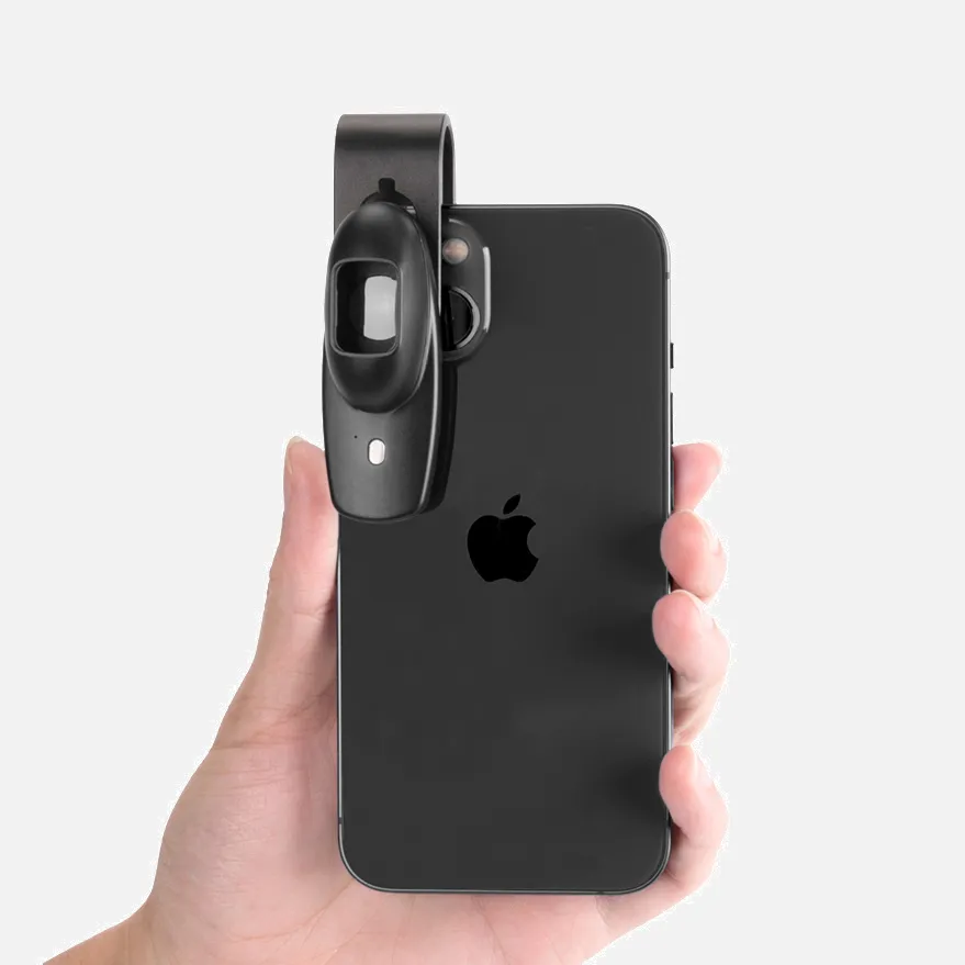
Dermoscopy examination employs a device that combines a high-powered magnification lens with polarized light to illuminate the surface and sub-surface structures of the skin. This non-invasive technique allows dermatologists to discern minute details that are invisible to the naked eye, paving the way for more accurate diagnoses.
A Historical Perspective
The journey of dermoscopy began with the advent of simple magnifying glasses in the early 20th century. Over the decades, technological advancements have culminated in the sophisticated digital dermoscopes of today, complete with imaging capabilities that facilitate the documentation and analysis of skin lesions.
Clinical Applications of Dermoscopy
Dermoscopic evaluation is a cornerstone in the diagnosis of pigmented skin lesions, including melanoma, the most aggressive form of skin cancer. Its high-resolution imaging aids in discerning the nuances of benign moles from malignant growths, thereby significantly improving early detection rates.
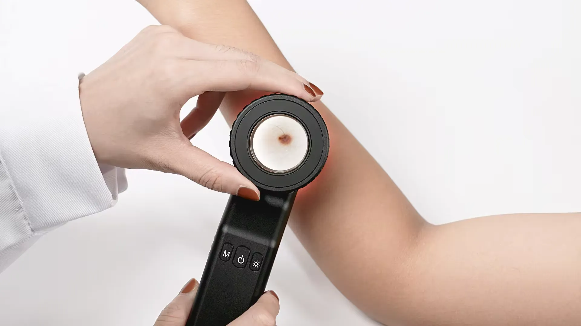
The Dermoscopy Examination Process
A typical dermoscopy examination begins with the preparation of the skin, followed by the application of the device to the area in question. The dermatologist then scrutinizes the lesion, looking for patterns and characteristics that may indicate a specific condition.
Interpreting Dermoscopic Images
Understanding the visual language of dermoscopy requires specialized training. Dermatologists are adept at identifying features such as pigment networks, streaks, and irregular borders, which can be indicative of various skin pathologies.
Innovations in Dermoscopy Technology
The integration of artificial intelligence into dermoscopy has opened new frontiers in automated image analysis. Machine learning algorithms are being developed to assist in the early detection of skin cancer, potentially reducing the rate of misdiagnosis.
Dermoscopy in Skin Cancer Screening
As a pivotal tool in skin cancer screening programs, dermoscopy has demonstrated its effectiveness in identifying atypical lesions that may warrant further investigation. It serves as an essential component of comprehensive skin examinations.
Comparative Analysis with Other Diagnostic Methods
While dermoscopy is a powerful diagnostic aid, it is not without its limitations. It is often used in conjunction with other diagnostic methods such as biopsy to provide a more complete assessment of skin lesions.
Educational Significance of Dermoscopy
The educational applications of dermoscopy are vast. It serves as a valuable teaching tool, enhancing the dermatological knowledge and diagnostic acumen of medical students and practitioners alike.
Despite its many advantages, dermoscopy also faces challenges such as the need for skilled interpretation and the quality of the device used. Ensuring standardized training and high-quality equipment is crucial for accurate diagnoses.
Precautions for Dermoscopy Examination
1. Patient preparation: patients should wear loose clothing to allow easy exposure of the area to be examined. Avoid using cosmetics or ointments that may affect the appearance of the skin before the examination.
2. Skin cleansing: The skin in the examination area should be thoroughly cleaned to remove dirt, oil, and dead skin cells, which helps to obtain clearer dermoscopic images.
3. Avoid direct sunlight: If the examination area is exposed to sunlight, it should be shielded first, as direct sunlight may affect the observation under the dermoscopy.
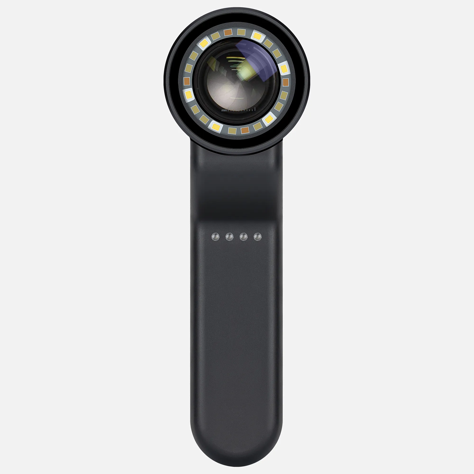
4. Use polarized light: Ensure that the polarized light function of the dermoscope is working properly to reduce surface reflection and improve image quality.
5. Appropriate magnification: Choose an appropriate magnification based on the size and location of the lesion for observation.
6. Avoid applying pressure: When using the dermoscope, avoid applying too much pressure to the examination area, as this may alter the appearance of the skin.
7. Recording and archiving: For important skin lesions, photographs should be taken and examination results should be recorded for subsequent comparison and analysis.
8.Professional operation: Ensure that dermoscopy examinations are performed by trained professionals to ensure the accuracy of the diagnosis.
9. Patient communication: During the examination, the doctor should maintain communication with the patient, explaining the steps being performed and any abnormalities observed.
10. Privacy protection: When conducting dermoscopy examinations, ensure that the patient's privacy is protected.
11. Follow-up: Based on the examination results, the doctor may recommend further examinations or regular follow-up visits for the patient.
12. Equipment maintenance: Regularly inspect and maintain the dermoscopy equipment to ensure it is in good working condition.
13. Avoid misinterpretation: Although dermoscopy improves diagnostic accuracy, it still has its limitations. Doctors should avoid making overly absolute interpretations of the observed results.
14. Provide educational information: provide patients with educational information about dermoscopy examinations to help them understand the examination process and possible outcomes.
Case Studies in Dermoscopic Diagnosis
Real-world applications of dermoscopy are best illustrated through case studies, demonstrating how this technology has led to the early detection and successful treatment of skin conditions.
The Future of Dermoscopy
As technology marches forward, the future of dermoscopy promises to be even more impressive. From improved imaging techniques to the integration of telemedicine, the scope of dermoscopy is set to expand, making skin health assessments more accessible and efficient. Dermoscopy technology stands as a testament to medical innovation, offering a clearer window into the skin's landscape. As we continue to refine and expand its capabilities, dermoscopy will undoubtedly remain a vital asset in the dermatologist's arsenal, leading us into a new era of skin health management.
What Does A Digital Marketing Agency Do?
A digital marketing agency is responsible for executing a wide range of tasks to ensure success in the digital marketing realm. Whether it's developing effective strategies, executing campaigns, or optimizing online presence, these agencies handle it all. Their extensive services cover various areas such as search engine optimization, social media management, content creation, email marketing, and pay-per-click advertising. They meticulously analyze market trends and consumer behavior to tailor their tactics for maximum impact. In essence, a digital marketing agency acts as a one-stop solution for businesses aiming to thrive in the digital landscape.
With its omni-channel, multi-channel, or single-channel approach, a digital marketing agency effectively connects with customers in the online realm. Through various channels like websites, blogs, email, and social media platforms, agencies engage with customers and ensure a comprehensive interaction experience.
People May Ask
A skin examination looks for birthmarks, moles, or other pigmented areas that don't seem normal in terms of size, shape, color, or texture. Your doctor may remove all or part of the abnormal skin during a biopsy, along with a tiny portion of surrounding normal tissue. Under a microscope, a pathologist examines the tissue to hunt for cancerous cells.
A benign tumor has edges that are smooth, regular, and distinct. In comparison to a benign tumor, a malignant tumor grows more quickly and has uneven borders. Moreover, a malignant tumor has the potential to spread to other bodily parts. Although a benign tumor can grow to be rather large, it won't travel to other parts of your body or infiltrate surrounding tissue.
The Use of Dermoscopy in BCC ManagementFor the assessment of residual tumor in BCC patients, dermoscopic examination following nonablative treatment has also shown promise. Remaining dermoscopic characteristics such as arborizing telangiectasias, ulceration, and pigmented formations are good markers of tumor persistence.
relative mass confinement.There is varying degrees of epidermal or follicular attachment.Large nuclear palisade-surrounded basaloid lobules.Because of the overproduction of mucin, loculules may be solid or exhibit central cyst development.Scleromyxoid stroma.A cleft forms in the stroma between the tumor lobules.Instead,Additional things...
How does a BCC look? Open sores, red spots, pink growths, shiny bumps, scars, or growths with slightly elevated, rolling edges and/or a center indentation are some of the appearances of BCCs. BCCs have the potential to leak, crust, itch, or bleed. The lesions usually appear on body parts that are exposed to the sun.
Using a dermascope gives a skin cancer specialist an additional advantage even if they can still detect malignant indications with their eyes. a color and structural clarity that would be invisible to the unaided eye or even a low-powered microscope. Color, particularly the distribution of pigments, can serve as a diagnostic tool in and of itself.
polarized light Non-polarized illuminationInstead,When light is polarized, the electric field only oscillates in one direction. Its electric field is oscillating in all directions.2. Light that is polarized is coherent by nature. Unpolarized light cannot be coherent in the natural world.
Dermoscopy is a non-invasive procedure that carries little risk of consequences. The rare chance of cross-infection between patients is the only problem, particularly when using contact dermoscopy.
Biopsy of the SkinYour dermatologist can typically identify psoriasis simply by looking at your skin. In the event that additional information is required to validate the diagnosis and exclude alternative etiologies of symptoms, such as cutaneous lupus or eczema, a skin biopsy might be conducted.
Your doctor will typically check your nails, scalp, and skin for symptoms of psoriasis in order to make the diagnosis. Inquiries concerning your health, medical history, and family history could also be made, including whether you: Feel sensations like skin that is burning or itching. had a recent illness or undergone a stressful event.
Dermoscopy Products

MEDCASE Radiance Otoscope with Light German Fiber Optic Otoscopes: Excellent for Professional Use - Ear Scope with LED Light and Speculum for Ear Examination and Diagnosis

Cancer (Astrology) - Discovering Love and Harmony in Every Relationship: Boxed Set of Cancer Horoscopes (Relationship Books for Dating Couples) Kindle攵子书
Cancer: A Special Gift for Cancer Patients And 121 Pages of Notepad/Journal with Matte Finish, 6 x 9 平装 – 2020年 1月 6日
Skin Cancer Diagnosis Made Simple: Over 220 full-color case discussions and a tutorial-based approach to skin cancer diagnosis for novices and experts alike. Kindle攵子书
The 30-Day Raw Vegan Plant-Based Detoxification & Regeneration Journal & Tracker: Healing Nevoid Basal Cell Carcinoma Syndrome A Reversing Conditions Journal and Tracker. Journal 2 - February 28, 2020 - Paperback
A Clear Guide to Medical Conditions: Diagnosis and Treatment of Basal Cell Carcinoma Kindle攵子书
The socio-economic significance of PGMs, PGM preconcentration and spectroscopy, chemistry and spectroscopy, adsorption and preconcentration, and PGM separation and determination are all covered in this section.

Ebook Enhanced Segmentation-Based Method Utilizing PSO and K-MEANS Algorithm for Basal Cell Carcinoma Identification from Skin Lesions
Diagnosis and Treatment of Common Disorders in Clinical Dermatology, Second Edition 2第二 版本
Dermoscopy, an Edition of Dermatologic Clinics (The Clinics: Dermatology Book 31) 1欬一 퉈本, Kindle电子乘
Related Products
Hot Products


News & Blog


Top Reviews
ampm247
My second tube of Sunspot Es is this one. Since my first tube's results were so good, I bought a second one after a friend convinced me not to use it for her issue. Regrettably, the FDA is refusing to let the firm disclose that its true benefit is treating minor skin malignancies. I mean business. My personal experience was with a basal cell carcinoma on my ankle that the doctor wanted to remove. Despite my hesitation, I tried this Sunspot. Es product. Over the course of two weeks, it performed flawlessly except from occasional stinging and itching. On his cheek, my spouse had a large area of keratoses. It looked terrible, and it bled every now and again. Reluctantly, he gave the Sunspot Es a try, and was astonished to discover how powerful it was. The spot took around a month to vanish since he neglected to apply a bandage. When applied correctly, it is an excellent product for treating kerotases, squamous cell, or basal cell carcinomas. I was aware that removing a malignancy using traditional techniques results in significant harm or destruction of the surrounding tissue, which
Dawn McKenna
I was pleasantly surprised to find this book contained much more than the dull, elementary content I had assumed based on its cover. This book has an easy-to-read, pleasant, and sympathetic tone, but its substance is jam-packed with reliable scientific data and practical advice. Rather than merely wearing sunscreen and waiting for my next skin check, I'm so happy to finally have information that I can truly utilize to improve myself. I now have a ton of good actions that I can do to strengthen my immunity and maybe stop future eruptions, in addition to the therapeutic alternatives that this book explained. For the first time, I've been informed of alternatives to avoiding the sun, and I find that information to be really valuable.
Cliente
Written for the BEST dermatoscopist, it's an excellent book with appropriate content, good photos, and helpful outlines. It's essential, in my opinion, for anybody with an interest in dermoscopy.


