


DE-400 Dermatoscope – IBOOLO
What Is A Dermatoscope
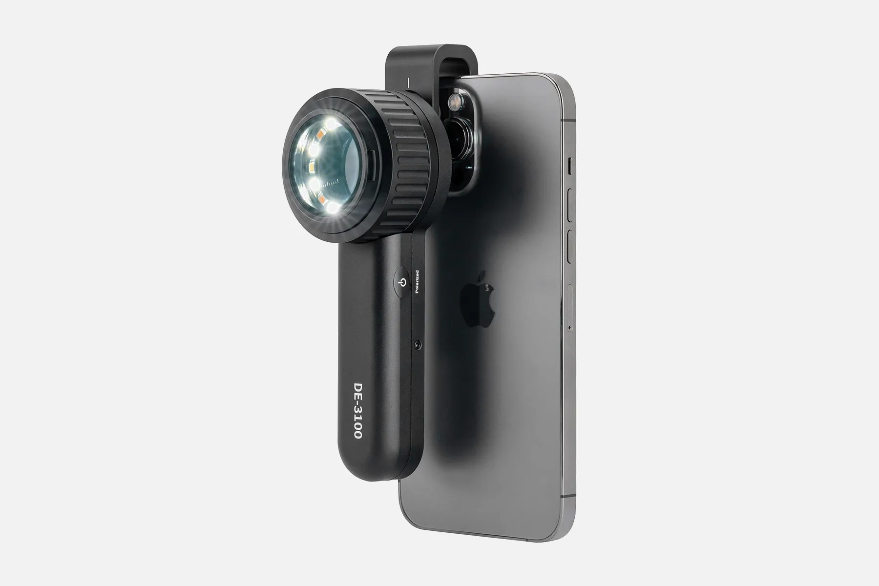
A dermatoscope is a handheld device that uses light and magnification to evaluate skin lesions. Dermoscopy is also known as dermatoscopy, chemiluminescence, or skin surface microscopy.
Dermoscopy allows doctors to visualize subsurface skin structures in the epidermis, dermo-epidermal junction, and papillary dermis. These structures are usually not visible to the naked eye.
Dermoscopy is mainly used to evaluate pigmented skin lesions. It has been shown to increase sensitivity for skin cancer detection, decrease the benign-to-malignant biopsy ratio, and allow for the diagnosis of thinner melanomas compared to naked eye exams.
How Does A Dermatoscope?
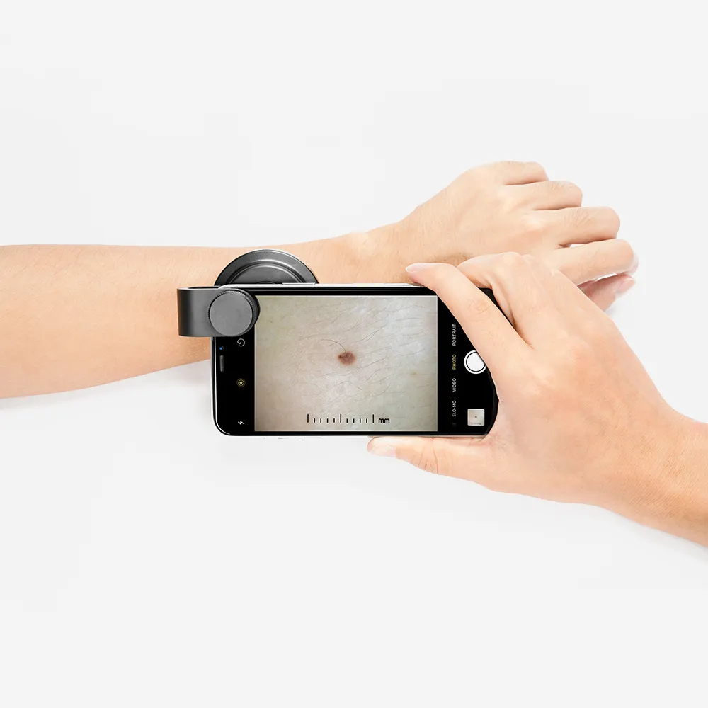
A dermatoscope is a hand-held device that uses light and magnification to examine skin lesions. It works by transilluminating a lesion to be studied at high magnification.
Here's how a dermatoscope works:
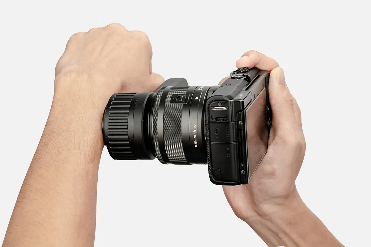
1. A clinician applies an ultrasound gel or oil, such as mineral oil, to the skin. The gel or oil improves the image clarity.
2. The clinician gently presses the dermatoscope into the skin.
3. The clinician holds the dermatoscope onto the skin to examine the area.
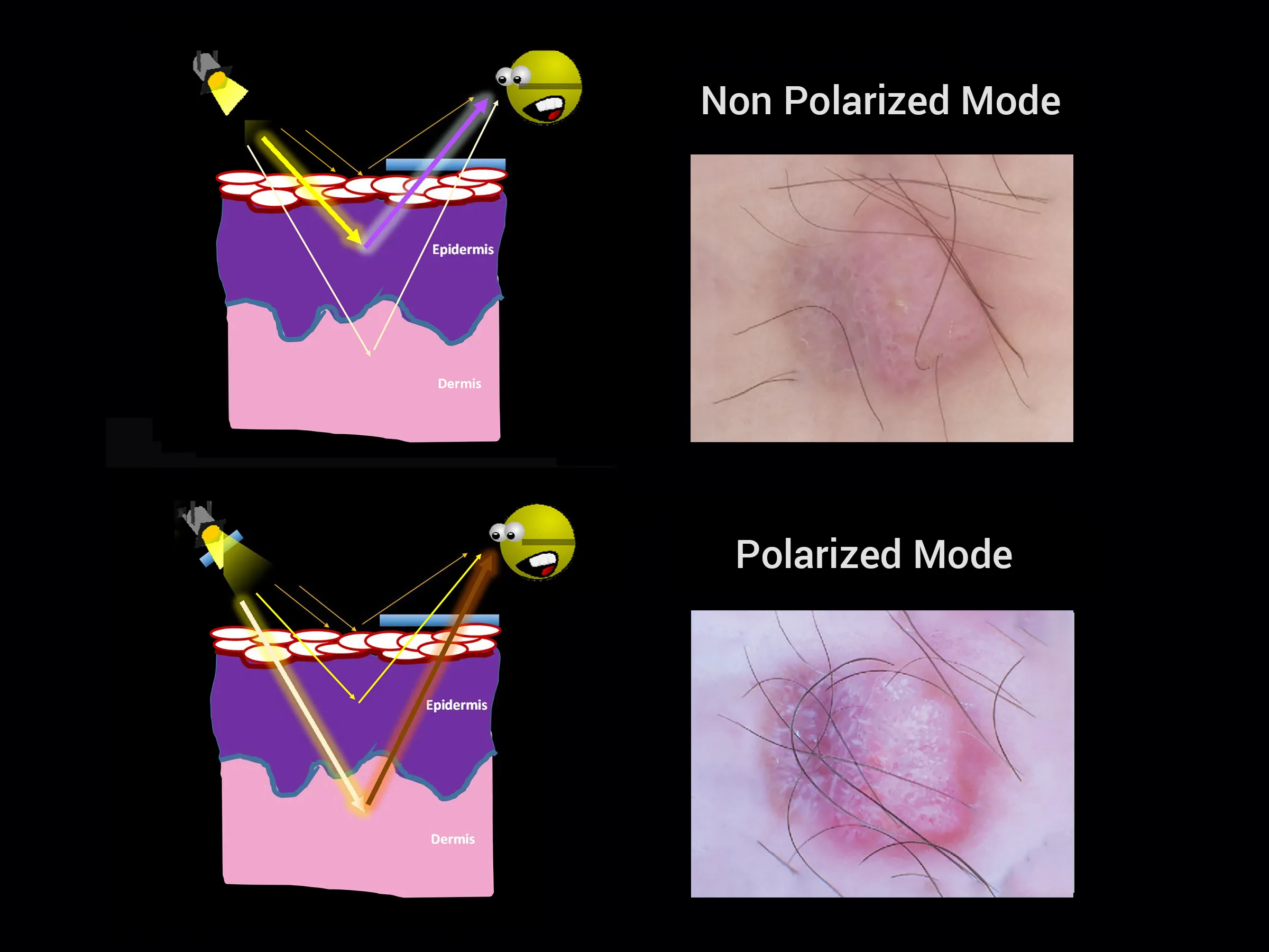
Dermoscopy is a noninvasive technique primarily used to examine cutaneous lesions. The principle of dermoscopy is based on the understanding that the stratum corneum reflects light, which reduces the ability to see structures under the skin. Applying a substance such as alcohol gel to the skin overcomes the refractive properties.
People May Ask
The pigmented lesion must lack pattern symmetry and color uniformity in addition to at least one of the following characteristics in order to be diagnosed as melanoma: numerous brown dots, pseudopods, radial streaming, scar-like depigmentation, peripheral block spots/globules, five to six colors, a blue-white veil,...Enhancing Diagnostic Quality by Using Dermoscopy to Identify Melanomahttps://www.ncbi.nlm.nih.gov › articles › PMC6375419
It is very helpful in separating melanoma from other pigmented lesions and in the early detection of malignant melanoma....
Using Dermoscopy to Identify Melanoma in Richmond, VAwww.mohsvirginia.comVisit https://mohsvirginia.com › Melanoma detection
The overall pooled sensitivity and specificity of dermoscopy were 95% (95% CI 85% to 99%) and 91.2% (95% CI 90.0% to 92.4%) for the diagnosis of BCC, respectively. The addition of dermoscopy to naked eye examination increased sensitivity from 67% to 85% (5 trials; 4455 lesions; P = ) when compared to the naked eye examination alone....
The efficacy of dermoscopy in the diagnosis of skin cancer - PMC - NCBIPMC7571636 can be found under articles at https://www.ncbi.nlm.nih.gov
Your physician applies a gel or oil to your skin. After that, they place the dermatoscope against your skin to take a close look at the affected area. Your skin is unaffected or damaged by this.Instead,Instead,Using a dermoscopy to examine your skin or mole - Cancer Research UKThe website cancerresearchuk.orgTests and scans are available at https://www.cancerresearchuk.org. LOL.
Cancer of the Basal CellDermoscopic parameters linked to BCCs include the lack of a pigment network and the presence of certain characteristics such as ulceration, arborizing vessels, enormous ovoid nests of blue-gray, multiple blue-gray globules, leaf-like areas, and spoke wheel areas.September 21, 2019Dermoscopy of Skin Cancers: Melanoma and Non-melanoma - PMChttps://www.ncbi.nlm.nih.gov › articles › PMC6712997
A doctor or individual can inspect and diagnose skin lesions and disorders, including melanoma, using a dermatoscope, a hand-held visual assistance equipment. Examining the nails, hair, and scalp can also be facilitated by it. A dermatologist's practice typically has a dermatoscope.18 Mar 2021...
What is visible through a dermatoscope? - Medical News Today
available at medicalnewstoday.com
https://www.medicalnewstoday.com › articles › dermatos...
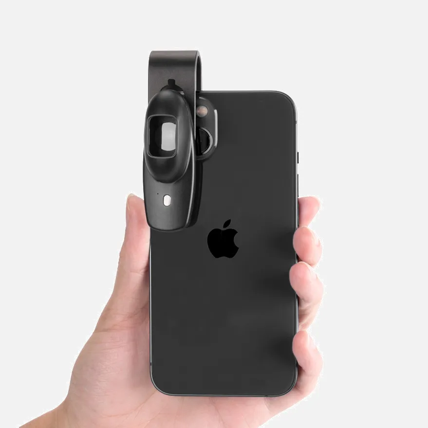
Dermatoscopes come in two primary varieties: handheld, portable, and stationary mounted. A magnifying optic with a magnification of at least ten times makes up a hand-held dermatoscope, together with a transilluminating light source....
The Wikipedia article on dermatologyWiki/Dermatiscopy https://en.wikipedia.org
Dermoscopy is a non-invasive diagnostic technique that offers several benefits to the physician, most notably early skin cancer diagnosis and improved melanoma detection accuracy....
The Diverse Applications of Dermoscopy in Melanoma Identification - MDPI ..
This gives dermatologists a strong, portable diagnostic instrument that's simple to use. When diagnosing skin cancer, dermatoscopes, also known as dermascopes, are the most common type of skin surface microscopy. It is possible to identify benign lesions without a biopsy and better understand those that need further attention....
The Skin Care Network - Dermoscopy for Skin Cancer Diagnosis
A handheld device known as a dermatoscope is used to perform dermoscopy. Subsurface skin structures in the epidermis, papillary dermis, and dermoepidermal junction-structures that are often invisible to the unaided eye-can be seen thanks to this method [2-4].September 9, 2023...
Synopsis of dermoscopy - UpToDateWolters Kluwer.Overview of the contents in https://www.uptodate.com/d...
What Is A Dermatoscope Products
Cordless handheld USB HD microscope with stand, Lepmerk digital microscope with 50-1000X magnification, and 8 LED endoscope camera Fit for iPhone, Android, iPad, Windows, and Mac Computer
A 7-inch LCD digital USB microscope with a 32GB TF card, a 12MP 1080P handheld video recorder with 1200x magnification, a PC view feature, a rechargeable battery, and a coin-filled light PCB soldering circuit board are all included.

Research-grade trinocular microscope compound lab with Siedentopf head, mechanical stage, ultra-precise focusing, and camera compatibility: Swift SW380T 40X-2500X

10x–220x Magnification Dino Lite USB Handheld Digital Microscope True Resolution: 0.3MP/1.3MP/5.0MP; Software for Windows, Mac, iOS, and Android Included; Supports PC, Tablet, and Mobile Devices

Magnification Nurugo Micro 400X The Tiniest Smartphone Microscope in the World for Photographic, Mechanics, Jewelry, and Cells - Comes with Brackets - Utilizing the Nurugo App to Share Media Is Simple
Eye Exam Polarized Skin Dermatology Light, xcvoc Esthetician UV Woods Lamp Skin Analyzer Portable 365nm Black Light Ringworm Detection Wood Lights for Pet Lice

With a 4.3-inch IPS screen, the BEAVERLAB Darwin M2 is a handheld digital microscope that is compatible with iPhone, Android computers, and 1600X pocket portable microscope camera for kids. It also has a 1080P HD coin microscope.
The coin microscope for adults with a 16MP camera sensor and a 10 inch HDMI LCD digital microscope by Dcorn is compatible with TVs, Macs, and Windows. It also comes with a coin guide and is a gift.
Andonstar Black Corlor 7-inch LCD 1080P Digital Microscope AD206 with 200X Magnification Zoom for Soldering Phone Repairs
UF-TOOLS 7-inch LCD digital microscope including a 12MP ultra-precise focusing camera, 1080P video microscopy, and a 64GB TF card with 1200x magnification Eight LED Lights for PC Watch Repair and Coin Circuit Board Soldering
Hot Products


News & Blog


Top Reviews
Libby
It is simple to use and performs admirably. I am pleased with my buy. The range of features appeals to me.
Wubble Gubble
I had to construct a homemade cure box, but it wasn't working out so well with the light I had because it felt too intense and overheated my prints. I picked this up to test if the increased coverage will make it function better. It stopped overcooking my prints and functioned flawlessly, distributing light more evenly. Since the battery lasts a long time and I haven't used it up yet, I merely intend to keep it plugged in whenever I need it. All things considered, there is more light than with my previous setup, but it is wider and more evenly distributed over my prints. My only task now is to construct a spinning plate because the built-in timer, which has two options—60 and 120 seconds—is a lifesaver and will enough for any project I can think of.
Tony
I love these large curing lights and I have another one of these that looks, feels, and performs the same as the lamp that I received from this brand where the only difference is the name that is stenciled on it. The light cures my resin quickly and is bright. When the power cord is plugged in, the light shines much brighter; however, when it is unplugged, the light still shines brightly enough to solve problems. I've used a variety of UV resins under this lamp and have not had any issues. All of the dragons and dinos in my photos were cured under this lamp without any problems. Simply press the button once to turn on the light for 60 seconds, or twice for 120 seconds, to adjust the brightness. I frequently multitask when doing UV resin work and I just double tap the button and walk away, come back later and flip things over and double tap the button again. I've never had to do more than 240 seconds with this light to cure a single pour of UV resin. I am happy with how this lamp performs. It is a couple do


