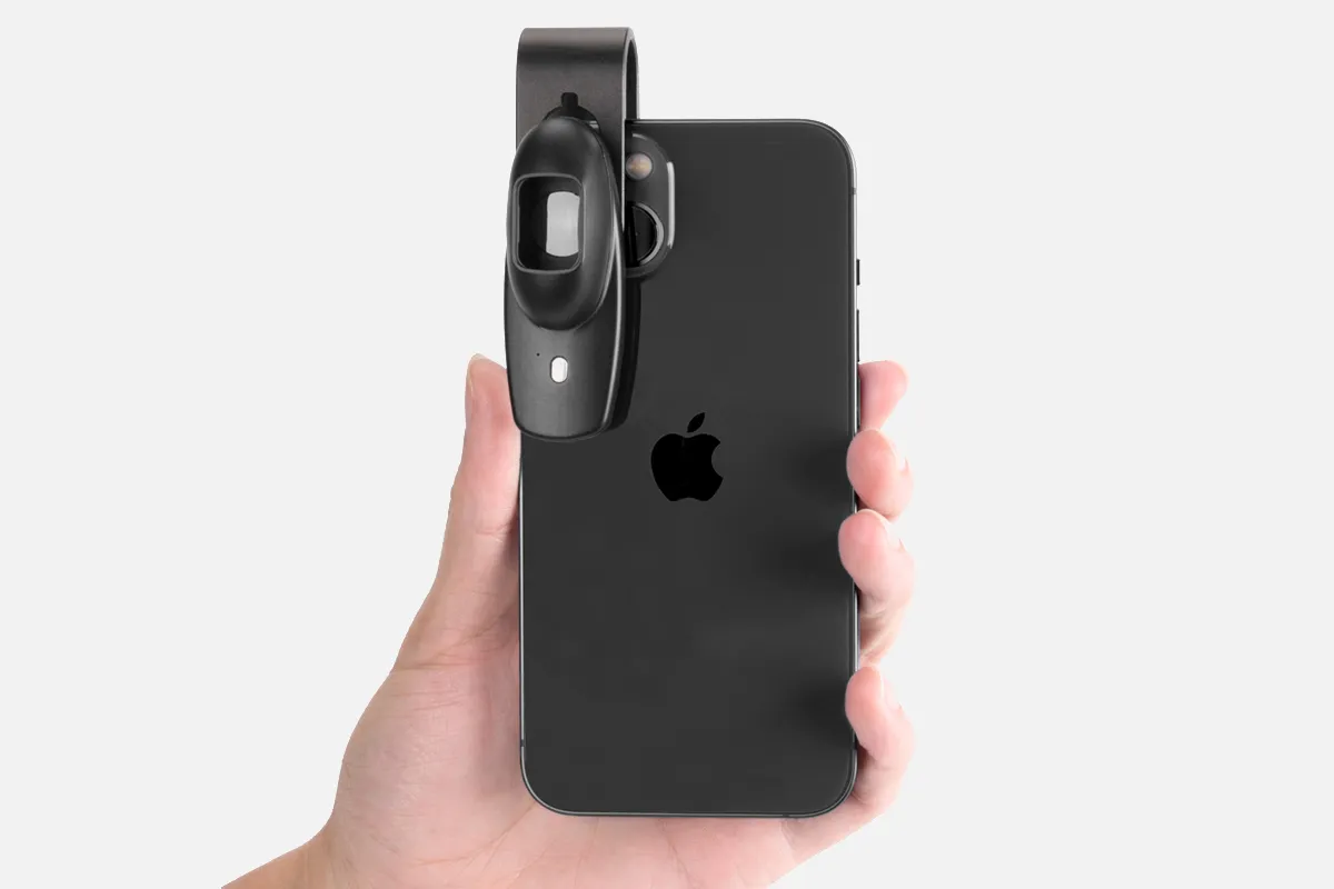
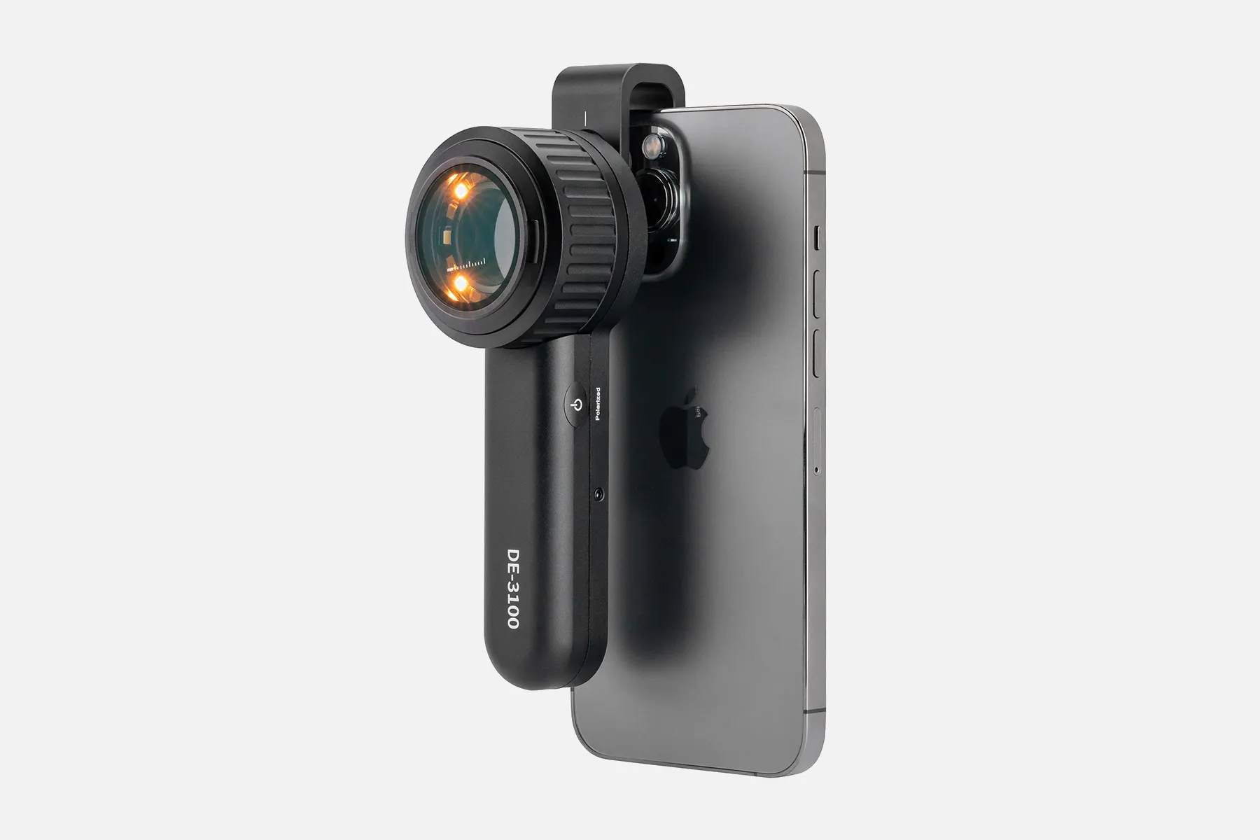
What Is A Mobile Phone Dermatoscope?

A mobile phone dermatoscope refers to a dermatoscope that interfaces with smartphones or tablets to capture clinical-grade dermoscopic images and video. A compatible accessory attaches over the mobile device's embedded camera to link it with the optics inside a standard dermatoscope.

By combining handheld dermoscopes with mobile computational power, mobile phone dermatoscopes aim to lower barriers to precision skin screening. The ability to conveniently document and share magnified subsurface lesions may assist telemedicine diagnosis when in-person clinician visits are limited.

Mobile configurations typically include adjustable lenses, specialized lighting modes, and secure mounting attachments to augment smartphone imaging with enhanced dermoscopic visualization capabilities. Captured photos/video can undergo software analysis as well.

While a licensed professional should diagnose medical conditions, mobile phone dermatoscopes expand access to observation tools for routine mole checks and baseline documentation – handy for rural regions or underserved groups where skin cancer risks may be high. Promoting early detection remains vital.

Mobile phone dermatoscopes enable broader populations to responsibly monitor skin health with greater anatomical precision than the naked eye allows. Further validation aims to support clinical care equity improvements through such technological applications.

How Does A Mobile Phone Dermatoscope?

Mobile phone dermatoscopes utilize a secure mounting device to align a smartphone’s embedded camera with the eyepiece of a conventional dermatoscope. This enables the capture of clinical-grade dermoscopic images and video.

The mobile phone device is first attached to the mount and paired with the accompanying app. The dermatoscope is then steadied over the skin area of interest using gentle pressure. Precise internal optics link the smartphone camera to visualize the enhanced magnified perspective under examination.

Intuitive mobile app controls allow smooth image and video capture workflows while maintaining full dermoscopic inspection. Advanced versions integrate specialized lighting modes, calibration tools, and analytics to quantify lesions over time. Documentation can be stored locally or transferred securely.

By leveraging smartphone capacities, mobile phone dermatoscopes provide streamlined operation for on-demand skin condition capturing, tracking, and coordination. Future intelligent algorithms may support automated diagnosis as well. Their convenience aims to promote responsible routine monitoring.

People May Ask

What Advantages Can A Dermoscopy Provide?

In order to diagnose melanoma at an earlier stage, when it may appear to be indistinguishable from a benign pigmented lesion, dermoscopy enables the detection of early melanomaspecific characteristics that are evident under the dermatoscope even when a melanoma is tiny in size (

Moles Are Still Removed by GPS?

Absolutely, but not generally for aesthetic purposes. Your first stop if you think a mole is cancerous should be your general practitioner (GP). In most cases, your GP can tell you right away whether a mole is benign (harmless), but if not, they will recommend you to a dermatologist for additional testing.Oct. 16, 2013...
Mole Removal: Can I use the NHS to have a mole removed?The website skinsurgeryclinic.co.ukInstead,How can I...? https://skinsurgeryclinic.co.uk › treatments-blog › can-i-...

Can Moles Be Checked Using GPS?

What transpires at your doctor's appointment. Your doctor will examine any odd skin or moles on your body. They could use a ruler or a marking scale to measure it....
Consulting a doctor | Skin cancer melanomaInstead,Visit cancerresearchuk.org.www.cancerresearchuk.org # receiving a diagnosis

How Are Dermatoscopes Made by Dermlite Used?

0:00 > 3:02It always appears polarized. In the middle. The pigment boost works similarly; all you have to do is tap thatFurther...Applying DermLite DL3N - YouTubewww.youtube.comhttp://m.youtube.com › Observe

What Varieties of Dermoscopy Exist?

Dermatoscopy can be performed in three major ways: Nonpolarized light, interaction [1] interaction with polarized light In [2] noncontact polarized light In [3]...
Skin examination - Wikipedia
wikipedia.org
Wiki/Dermatiscopy https://en.wikipedia.org

What Use Does A Dermatoscope Serve?

A handheld device known as a dermatoscope is used to perform dermoscopy. Subsurface skin structures in the epidermis, papillary dermis, and dermoepidermal junction-structures that are typically invisible to the unaided eye-can be seen thanks to this procedure [2-4].Aug. 9, 2023...
Synopsis of dermoscopy Current -The website uptodate.com can be accessed at https://www.uptodate.com/titles/overview-of-der...

Which Time Is Best for A Dermoscopy?

A basic skin exam is called a dermoscopy. Your doctor might do a dermatoscopy if you have a mole or pigmented skin lesion that is alarming. With a dermoscopy, your doctor can more precisely diagnose pigmented skin lesions, sometimes preventing the need for a skin sample or needless mole excision.20 May 2022...
Applications, Methods, and Outcomes of Dermoscopy - Verywell Health

What Dermoscopy Equipment Do General Practitioners Have?

Conclusions. Dermascopy is seen as having a significant role in enhancing skin examinations in primary care by both GPs who use it and those who do not. It was noted that a major obstacle to its broader adoption is the requirement for sufficient training in dermoscopy and dermatology more broadly.15 March 2022A qualitative study involving general practitioners on the use of dermoscopy in primary care

How Well Does A Dermatoscope Work?

Do dermatoscopes have accuracy? A 2018 Cochrane study found that when used by a qualified practitioner, dermatoscopes are more accurate than the human eye alone in the diagnosis of melanomas. This is important since it can save someone time and possibly avoid the needless operation.18 March 2021...
What is visible through a dermatoscope? - Medical News Today
available at medicalnewstoday.com
https://www.medicalnewstoday.com › articles › dermatos...

How Are Dermacopies Performed at Home?

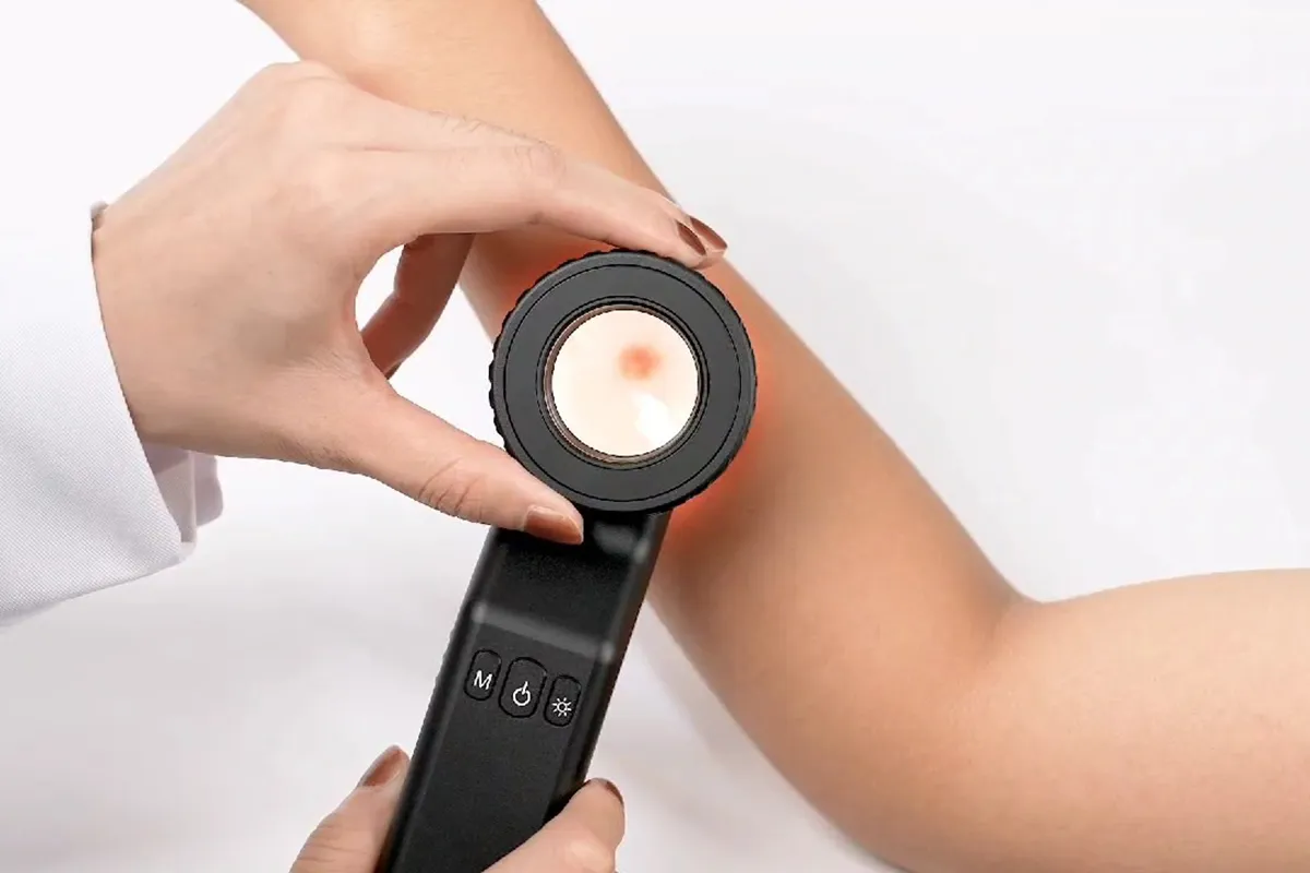
On the mole that bothers you, apply one drop of oil. Directly touch the skin with the dermatoscope, press down gently, and use your smartphone to take a picture. Touching the screen's center may be necessary to concentrate the mole's image. Take and preserve clear dermoscopic pictures....
At-Home Sklip® Dermoscopy - OHSUOhsu.eduSklipr-home: https://www.ohsu.edu/war-on-melanoma/n...

Mobile Phone Dermatoscope Products

China affordable mobile phone dermatoscope suppliers & manufacturers

The Pocket Microscope from UKCOCO Zoom Lens Micro Camera Hand Dermatoscope on-the-Go Wireless White Appendix with Miniature Camera LED Magnifying Glass Phone

mobile phone dermatoscope suppliers & manufacturers

Portable Digital Microscope, ViTiny Pro10, 10x - 200x

China handheld mobile phone dermatoscope

Microscope with built-in 8 LED lights, USB microscope industrial accessories, portable magnification, and appraisal for jewelry and phone repairs

China portable mobile phone dermatoscope suppliers & manufacturers

Portable Handheld Electronic Coin Magnifier, Teslong 10X to 200X Magnification Camera with Stand, Soldering Camera Ear Otoscope, and USB Digital Microscope

China Professional mobile phone dermatoscope suppliers & manufacturers

The little plus pocket microscope lighting, portable magnifying glass magnifier, magnifying lens glass for universal cell phone, and 60-100X clip-on microscope with LED light

China high quality mobile phone dermatoscope

SKYBASIC USB Digital Microscope Camera: A portable, handheld high-definition inspection camera with an adjustable stand, 50x–1600x magnification, and 8 LED lights Both iOS and Android device compatibility

China customized mobile phone dermatoscope

Three Gatuida phones for a 60x phone microscope and a handheld magnifying mirror Dermatoscope with a micro camera Microscope with Lights and a Clip-on Pocket Camera for White Cell Phone

China Professional mobile phone dermatoscope

Compact 200X Phone Mini Pocket Microscope with LED Light and Universal Clip, 99% Smartphone Compatible Portable Digital Microscope Camera Attachments, Microworld for Children and Adults (Black, 200X with CPL)

China wholesale mobile phone dermatoscope suppliers & manufacturers

Ideal for kids and adults, the Koolertron Mini Phone Microscope with LED Light, Universal Clip and CPL, 200X Portable USB Microscope Camera Accessory is compatible with iPhone, Android phones, and iPads.

China high quality mobile phone dermatoscope suppliers & manufacturers

OT-WiFi, or Opti-Tekscope, is a wireless HD digital microscope Camera: PC and Mac Compatible, Bright LED Illumination, 2 Megapixel Clarity, iOS/Android App Control, and Magnification Range of 50-1000x

Top Reviews

Jamal Uddin

This UV light is portable and easy to pack for travel because of its small, compact design. I use this when I go rock spotting. Although I haven't found anything worthwhile yet, I'm not giving up hope that I will eventually find something worthwhile. I appreciate that this device may be recharged, eliminating the need for batteries. simple to operate. Just one off the button. You can now move the light where you want it by pointing and shooting it.

Ali Albarack

To show you how wonderful this lens is considering its pricing, I've included a few shots to a game I like to play called "Guess What It Is!" You will examine the close-up picture in the first photograph and provide your best estimate as to what you believe it could be. The true nature of the object is shown in the second image! I wish you luck!

SHERYLE D.

My first one broke by accident, so I'm on my second. I adore that this pen camera captures crystal-clear images of coins and includes the adaptor required for more recent phones.

0

Have questions on gear or your order?

Our Gear Guides are here to help! Get personal advice from pro creatives

Name
Subject
Email address
How can we help?

Instant Answers

How Can Dermoscopy Images Be Captured?

Dermoscopy images can be captured and stored in different ways, such as: • Using a smartphone or tablet with dermoscopic adapter, which consisted in the package.• Using a digital camera

Dermoscopy images can be captured and stored in different ways, such as:

• Using a smartphone or tablet with dermoscopic adapter, which consisted in the package.
• Using a digital camera with dermoscopic adapter, there’s 49mm screw size camera adapter available to order now.

Compatible phone/tablet models:
All iPhone models, 95% Android phones, 90% tablet. For phone/tablet size in 5.25-14mm

Compatible camera models:
All camera with built 49mm filter screw, such as Canon EOS 70D, 80D, 90D; Canon EOS R7, R10, R50, R100; Canon M100, M200, M50, Mark II; Canon G7X Mark III, Sony ZV-1

How Can I Connect My Phone to My Dermatoscope?

There’s universal phone adapter for all our dermoscopes. Please check the installation procedure bellow or watch operation guide. Smartphone Connector (1) Place phone adapter screw in the center of smartphone’s

There’s universal phone adapter for all our dermoscopes. Please check the installation procedure bellow or watch operation guide.

Smartphone Connector

(1) Place phone adapter screw in the center of smartphone’s main camera.
(2) Screw magnet attachment on phone adapter.
(3) Put dermoscope’s back ring and magnet attachment together

Take The Best Images

You need to adjust the focus ring after the dermoscpe connected on smartphone to get the best images.

How Can I Clean My Dermoscopy after Usage?

Cleaning your dermoscopy after usage is important to prevent cross-contamination and infection. The cleaning method may vary depending on the type and model of your dermoscopy, so you should always

Cleaning your dermoscopy after usage is important to prevent cross-contamination and infection. The cleaning method may vary depending on the type and model of your dermoscopy, so you should always follow the manufacturer’s instructions. However, some general steps are:

• Turn off and disconnect your dermoscopy from any power source or device.

• Wipe off any visible dirt or debris from the dermoscopy with a soft cloth or tissue.

• Disinfect the dermoscopy with an alcohol-based wipe or spray, or a disinfectant solution recommended by the manufacturer. Make sure to cover all surfaces, especially the lens and contact plate.

• Let the dermoscopy air dry completely before storing it in a clean and dry place.

• Do not use abrasive or corrosive cleaners, solvents, or detergents that may damage the dermoscopy.

• Do not immerse the dermoscopy in water or any liquid, unless it is waterproof and designed for immersion.

You should clean your dermoscopy after each use, or at least once a day if you use it frequently. You should also check your dermoscopy regularly for any signs of damage or malfunction, and contact the manufacturer or service provider if needed.

Polarized VS Non-Polarized Dermoscopy

A dermoscopy is a device that allows the examination of skin lesions with magnificationand illumination. By revealing subsurface structures and patterns that are not visible tothe naked eye. It can

A dermoscopy is a device that allows the examination of skin lesions with magnificationand illumination. By revealing subsurface structures and patterns that are not visible tothe naked eye. It can improve the diagnose accuracy of skin lesions, such as melanoma,basal cell carcinoma, seborrheic keratosis, etc.

There are two main types of dermoscopy: Non polarized and polarized dermoscopy.We’ve fitted most of our dermoscopys with polarized and non-polarized light. They canbe used in multiple skin structures.

Non-polarized contact Mode

In non-polarized mode, the instrument can provide information about the superficialskin structures, such as milia-like cysts, comedo-like openings, and pigment in theepidemis.

The dermoscopy requires applying a liquid such as mineral oil or alcohol to the skin andplacing the lens in contact with the skin. This reduces surface reflection and enhancesthe view of subsurface structures.

Image with non-polarized light (DE-3100)

Polarized contact Mode

In polarized mode, the instrument allows for visualization for deeper skin structures,such as blood vessels, collagen, and pigment in the dermis.

The dermoscopy does not need to be in contact with the skin or use any liquid. Theirpolarized light can help to eliminate surface reflection and allow visualization ofvascular structures.

Image with polarized light (DE-3100)

Polarized non-contact Mode

The dermoscopy can also use polarized light to examine the skin without direct contact.

In polarized non-contact mode, the instrument allows for examination infected areasand lesions that are painful for the patient, or the difficult to contact pigmented lesions,such as nails and narrow areas.

The contact plate should be removed in this mode, and it does not require applying aliquid to the skin. As it doesn’t require pressure or fluid application on the skin, it canalso avoid cross-contamination and infection risk.

Image in polarized non-contact mode (DE-3100)

How Effectiveness Is Dermoscopy

Compared with visual inspection, the dermoscopy can be used to capture and store skin lesion photos, which play an important role in early skin cancer examination. The dermoscopy allows the

Compared with visual inspection, the dermoscopy can be used to capture and store skin lesion photos, which play an important role in early skin cancer examination.

The dermoscopy allows the examination of skin lesions with magnification and illumination. This can be greatly avoiding the factors that cause interference to visual detection. Such as lighting, skin color, hair and cosmetics.

Several studies have demonstrated that dermoscopy is useful in the identification of melanoma, when used by a trained professional.

It may improve the accuracy of clinical diagnosis by up to 35%
It may reduce the number of harmless lesions that are removed
In primary care, it may increase the referral of more worrisome lesions and reduce the referral of more trivial ones

A 2018 Cochrane meta-analysis published the accuracy of dermoscopy in the detection.

Table 1. Accuracy of dermoscopy in the detection of melanoma in adults
Detection Method Sensitivity, % Specificity, % Positive Likelihood Ratio NegativeLikelihood Ratio
Visual inspection alone (in person) 76 75 3.04 0.32
Dermoscopy with visual inspection (in person) 92 95 18 0.08
Image-based visual inspection alone (not in person) 47 42 0.81 1.3
Dermoscopy with image-based visual inspection (not in person) 81 82 4.5 0.23
ROC—receiver operating characteristic. *Estimated sensitivity calculated on the summary ROC curve at a fixed specificity of 80%.

As we can see, the dermoscope can improve the accuracy of diagnosis of skin lesions, especially melanoma.

Table 1. Accuracy of dermoscopy in the detection of melanoma in adults
Detection Method Sensitivity, % Specificity, % Positive Likelihood Ratio NegativeLikelihood Ratio
Visual inspection alone (in person) 79 77 3.4 0.27
Dermoscopy with visual inspection (in person) 93 99 93 0.07
Image-based visual inspection alone (not in person) 85 87 6.5 0.17
Dermoscopy with image-based visual inspection (not in person) 93 96 23 0.07
ROC—receiver operating characteristic. *Estimated sensitivity calculated on the summary ROC curve at a fixed specificity of 80%.

Characteristics of the dermatoscopic structure of the skin lesions include:

• Symmetry or asymmetry
• Homogeny/uniformity (sameness) or heterogeny (structural differences across the lesion)
• Distribution of pigment: brown lines, dots, clods and structureless areas
• Skin surface keratin: small white cysts, crypts, fissures
• Vascular morphology and pattern: regular or irregular
• Border of the lesion: fading, sharply cut off or radial streaks
• Presence of ulceration

There are specific dermoscopic patterns that aid in the diagnosis of the following pigmented skin lesions:

• Melanoma
• Moles (benign melanocytic naevus)
• Freckles (lentigos)
• Atypical naevi
• Blue naevi
• Seborrhoeic keratosis
• Pigmented basal cell carcinoma
• Haemangioma

No products in the cart.

No products in the cart.

Hot Search Terms