


DE-400 Dermatoscope – IBOOLO
Handyscope Portable Dermatoscope: Technical Analysis And Clinical Application Guide
Handyscope portable dermatoscope, created by IBOOLO, combines high-resolution imaging with intelligent analysis functions to aid in skin lesion detection. This article analyzes its working principles, usage methods, and value in early skin cancer detection and home self-examination.
Handyscope: Revolutionary Applications And Future Prospects of Portable Dermatoscopy
Handyscope is an innovative portable dermatoscope designed for dermatologists and home users, combining high-resolution imaging technology with the convenience of mobile devices to quickly and accurately examine skin lesions. Whether for early detection of skin cancer or monitoring chronic skin conditions, Handyscope demonstrates exceptional value. This article will delve into Handyscope's working principles, usage methods, technical features, and applications in medical and home settings, helping you fully understand this revolutionary device.
What Is Handyscope?
Basic Introduction And Definition of Handyscope
Handyscope is a portable dermatoscope designed specifically for dermatologists and medical professionals to quickly and accurately examine skin lesions. It combines high-resolution imaging technology with the convenience of mobile devices, allowing doctors to perform skin examinations anywhere. The core function of Handyscope is to help doctors observe microscopic structures of the skin surface through magnification and illumination technologies, thereby assisting in the diagnosis of skin diseases, especially early detection of skin cancer.
Differences Between Handyscope and Traditional Dermatoscopes
Compared to traditional dermatoscopes, Handyscope has the following significant differences:
Portability: Handyscope is compact and easy to carry, suitable for house calls or mobile medical scenarios.
Digital Integration: Handyscope can connect directly to smartphones or tablets, capturing and storing images in real time for subsequent analysis and tracking.
Ease of Operation: Traditional dermatoscopes typically require complex setup, while Handyscope is plug-and-play, suitable for quick examinations.
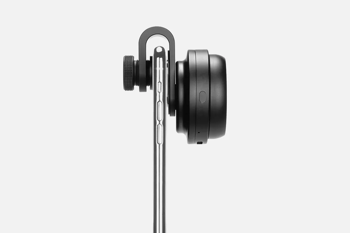
Product Structure And Components of Handyscope
Detailed Analysis of Handyscope's Core Components
Handyscope consists of the following core components:
Optical Lens: Provides high-magnification capabilities, ensuring clear images.
LED Illumination System: Provides uniform light source, reducing reflection and enhancing detail visibility.
Connection Interface: Supports USB or wireless connections, compatible with various mobile devices.
Housing Design: Lightweight and durable, ergonomically designed for easy handheld operation.
Mobile Device Connection Methods And Compatibility
Handyscope supports multiple connection methods, including:
USB Connection: Direct connection to smartphones or tablets via data cable.
Wireless Connection: Some models support Bluetooth or Wi-Fi connection, providing greater flexibility.
Compatibility: Supports iOS and Android systems, adapting to mainstream medical applications.
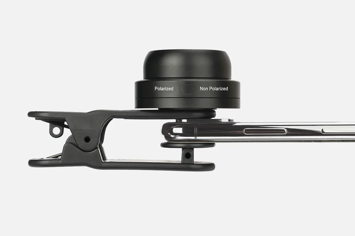
Working Principles of Handyscope
Optical Principles of Handyscope
Handyscope utilizes optical magnification and illumination technology to capture microscopic images of the skin surface through high-resolution lenses. Its LED illumination system employs polarized light technology to reduce reflection from the skin surface, allowing doctors to observe details such as pigmentation, blood vessels, and texture.
Image Capture And Processing Technology
Handyscope's built-in image processing technology can optimize image quality in real-time, including:
Auto Focus: Ensures image clarity.
Color Correction: Restores true skin colour.
Image Enhancement: Highlights details in lesion areas for easier diagnosis.
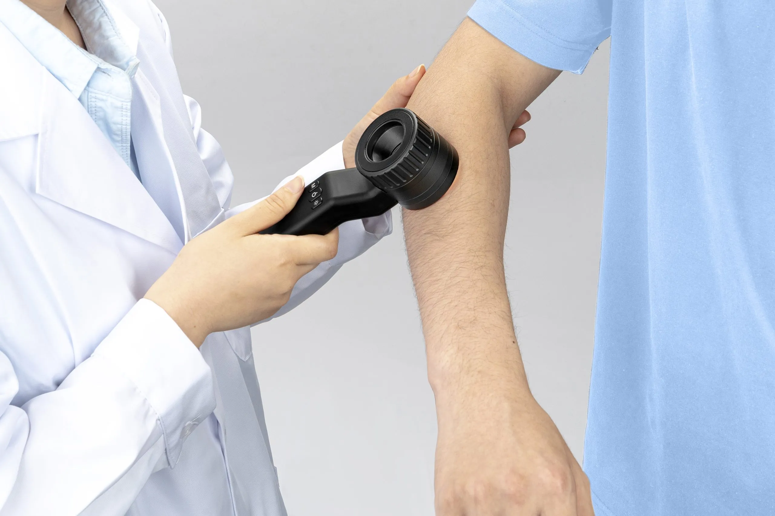
How to Correctly Use Handyscope
Detailed Steps for Using Handyscope
The steps for using Handyscope are as follows:
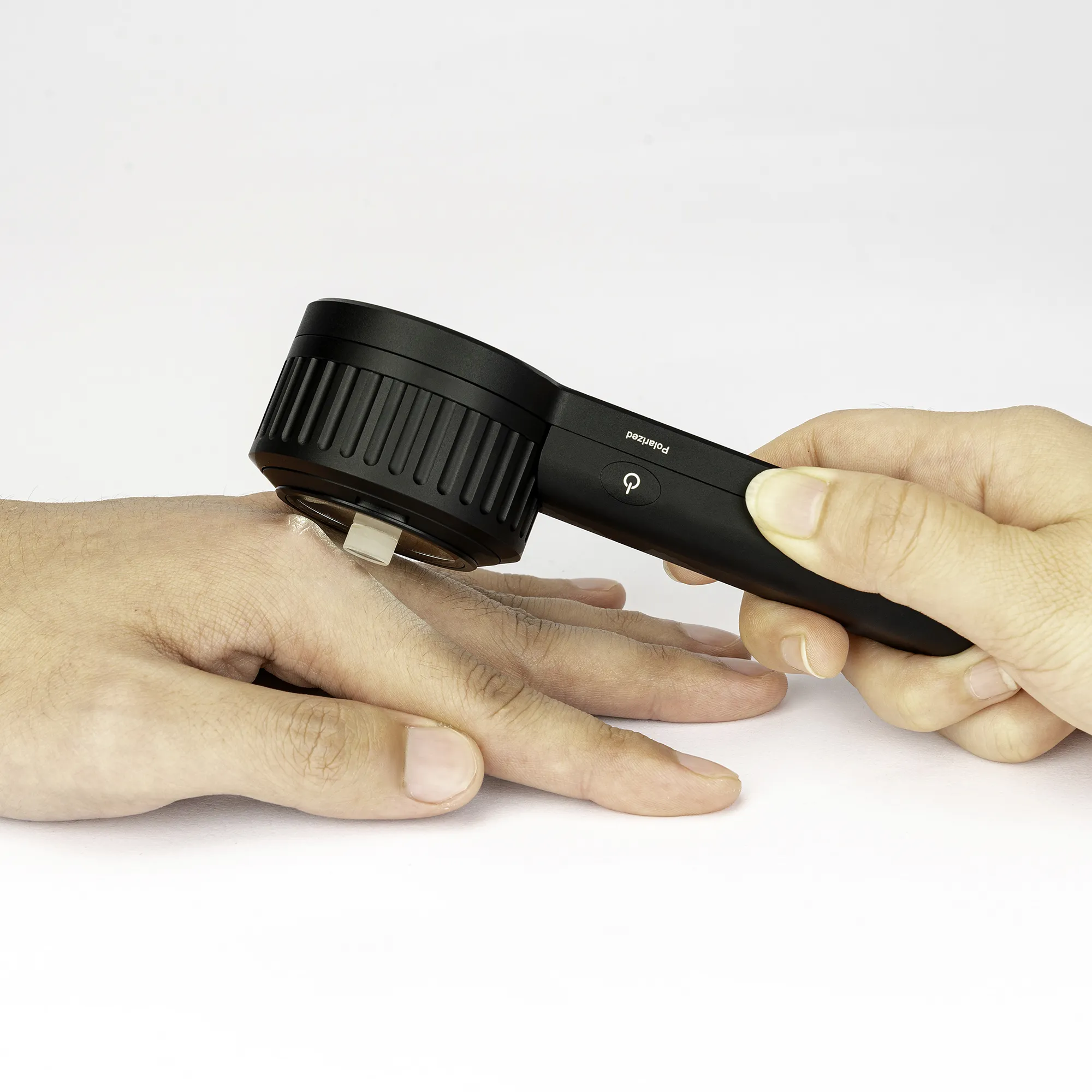
1. Preparation: Ensure the device is fully charged, connect to a mobile device, and open the relevant application.
2. Clean the Skin: Use an alcohol swab to clean the skin area to be examined.
3. Apply Coupling Agent: Apply a small amount of coupling agent to the skin to reduce reflection and improve image quality.
4. Position the Device: Gently place Handyscope on the skin, adjusting the angle to obtain the best view.
5. Capture Images: Press the shutter button to capture and save images to the device.
6. Analyze Images: Use tools in the application for further analysis of the images.
Common Operation Problems And Solutions
Blurry Images: Check if the lens is clean and ensure the device is placed stably.
Connection Failure: Restart the device and reconnect, ensuring drivers are installed.
Insufficient Light: Adjust LED brightness or change ambient lighting.
Application Scope of Handyscope
Application in Early Skin Cancer Detection
Handyscope performs excellently in early skin cancer detection, especially in identifying melanoma. Through high-resolution imaging, doctors can observe subtle changes in lesion areas, such as asymmetry, irregular edges, and uneven colouration, thereby improving diagnostic accuracy.
Other Skin Disease Diagnosis And Monitoring Applications
Besides skin cancer, Handyscope can also be used in the following areas:
Eczema and Psoriasis: Observe skin inflammation and scaling conditions.
Acne and Folliculitis: Assess the degree of lesions and treatment effects.
Pigmentary Skin Diseases: Monitor changes in pigmentation.
Advantages And Benefits of Using Handyscope
Advantages Compared to Traditional Examination Methods
Efficient and Convenient: No complex setup is required, examinations can be conducted anytime, anywhere.
High-Precision Imaging: Provides clear microscopic images, reducing misdiagnosis rates.
Digital Management: Supports image storage and sharing, facilitating tracking of disease development.
Practical Value for Patients And Doctors
For Patients: Reduces unnecessary biopsies, lowering psychological stress and economic burden.
For Doctors: Improves diagnostic efficiency, enhances patient trust, and improves quality of medical services.
7. Technical Features of Handyscope
Image Quality And Magnification
Handyscope is known for its excellent image quality, typically providing 10x to 20x optical magnification, capable of clearly capturing microscopic structures of the skin surface. Its high-resolution lens combined with advanced LED illumination technology ensures rich image details and true colours, helping doctors accurately identify lesion areas.
Introduction to Intelligent Analysis Functions
Handyscope is equipped with intelligent analysis functions that can automatically analyze images through built-in algorithms. For example:
Lesion Recognition: Automatically marks suspicious areas, helping doctors quickly locate problems.
Image Comparison: Supports historical image comparison, helping track changes in lesions.
Diagnostic Suggestions: Provides preliminary diagnostic suggestions based on the database, enhancing diagnostic efficiency.
Usage Precautions for Handyscope
Device Disinfection And Hygiene Maintenance
To ensure safe use, disinfection and hygiene maintenance of Handyscope are crucial:
Disinfection Method: Clean the lens and housing with 75% alcohol or medical disinfection wipes, avoiding corrosive cleaning agents.
Hygiene Maintenance: Clean the device promptly after each use to avoid cross-infection.
Common Errors during Examination
When using Handyscope, avoid the following common errors:
Insufficient Light: Failure to adjust LED brightness, resulting in blurry images.
Improper Use of Coupling Agent: Not applying or applying too much coupling agent, affecting image quality.
Unstable Device Placement: Unstable handheld operation causing image shaking, affecting observation effects.
Maintenance And Care of Handyscope
Daily Cleaning Methods for The Device
Daily cleaning is key to extending the lifespan of Handyscope:
1. Turn Off the Device: Ensure the device is turned off and disconnected before cleaning.
2. Clean the Lens: Use a specialized lens cloth to gently wipe, avoiding scratches.
3. Clean the Housing: Use alcohol swabs to wipe the housing, removing stains and bacteria.
4. Check the Connection Port: Ensure the USB or wireless connection port is free of dust or foreign objects.
Tips for Extending Device Lifespan
Avoid Dropping: Handle with care during use, avoiding impacts to the device.
Regular Charging: Keep the battery well-charged, avoiding over-discharge.
Storage Environment: Store the device in a dry, cool place, avoiding high temperature or humid environments.
Value of Handyscope in Home Use
Suitable Situations for Home Self-Examination
Handyscope has important value in home self-examination, particularly suitable for the following scenarios:
Skin Lesion Monitoring: Regularly check moles or spots on the skin, observing whether there are changes.
Skin Disease Management: Track the development of chronic skin diseases such as eczema and psoriasis.
Early Warning: Seek medical attention promptly for professional diagnosis after discovering suspicious lesions.
How to Use in Conjunction with Professional Medical Advice
When using Handyscope at home, follow these recommendations:
1. Learn Basic Operations: Master the device usage methods through manuals or video tutorials.
2. Record Examination Results: Save images from each examination for subsequent comparison.
3. Consult Professional Doctors: Promptly provide images and records to doctors for reference when abnormalities are found.
Integration of Handyscope with Artificial Intelligence
Intelligent Diagnostic Assistance Functions
The integration of Handyscope with artificial intelligence (AI) makes it excel in diagnostic assistance:
Lesion Classification: AI algorithms can automatically identify and classify common skin lesions, such as moles, melanoma, etc.
Risk Assessment: Assess the malignant risk of lesions based on image characteristics, providing preliminary diagnostic suggestions.
Learning Optimization: AI systems continuously improve diagnostic accuracy and reliability by learning new data.
Data Analysis And Lesion Tracking
The AI functions of Handyscope also support data analysis and lesion tracking:
Historical Data Comparison: Automatically compare multiple examination results, identifying trends in lesion changes.
Report Generation: Generate detailed examination reports, including images, analysis results, and recommendations.
Remote Consultation: Support uploading data to the cloud, facilitating remote consultation and collaboration among doctors.
As a portable dermatoscope, Handyscope is changing the way skin lesions are diagnosed and monitored through its high-resolution imaging, intelligent analysis functions, and convenient operation. This article has detailed the basic definition, core structure, working principles, and usage steps of Handyscope, and discussed its application value in early skin cancer detection, chronic skin disease management, and home self-examination. Additionally, the article has analyzed the differences between Handyscope and traditional dermatoscopes, as well as their intelligent diagnostic assistance functions when integrated with artificial intelligence. By understanding the technical features and advantages of Handyscope, doctors and patients can better utilize this tool to improve the efficiency and accuracy of skin health management.
People May Ask
Golf Performance Standards Based on Age
Age Categories Median Golf Scores for Males
50s Range 96 to 101
60s Range 98 to 103
70s Range 100 to 105
80s Range 102 to 107
Additional Data Points•
No, amblyopia does not resolve spontaneously, and children are not able to naturally outgrow it. In the absence of treatment, amblyopia can potentially lead to long-term vision complications, including the potential loss of sight in the affected eye.
Technically, there is no specific age restriction for amblyopia treatment. However, Dr. Borriello emphasizes that the optimal time for its management is during childhood.
Does the act of wearing glasses constitute a disability? The wearing of glasses, regardless of its corrective power, is not classified as a disability. Legally, visual impairment is evaluated based on the concept of "best corrected vision," which refers to an individual's optimal visual clarity achieved through the use of corrective lenses.
Exploring the Mechanics of a Golf Handicap? Here's a Simplified Approach to Calculate Yours.
To determine your golf handicap, consider averaging the top eight scores from your last 20 rounds.
Employ this formula to assess your handicap for various courses: Course Handicap = (Handicap Index multiplied by (SR divided by 113)) plus (CR minus Par).
Published on
In essence, it is crucial to prioritize the individual before their disability. Those with disabilities are, ultimately, human beings at heart. Assigning a label that solely identifies a person with a specific condition can be deemed disrespectful and reductive of their humanity.
2. Preferred and Undesirable Vocabulary
Avoid Employ
Terms like (the) handicapped, (the) disabled The phrase 'disabled (people)'
Expressions such as afflicted by, suffers from, victim of Prefer 'has [specific condition or disability]'
Phrases like confined to a wheelchair, wheelchair-bound Instead, use 'wheelchair user'
Additional 10 examples•
Examples of visual impairments encompass, yet are not comprehensive to: total loss of sight, impaired vision, abnormalities in ocular movement or motility, difficulty in utilizing both eyes synergistically, a misalignment in the alignment of the eyes known as strabismus, a condition where one eye fails to develop properly known as amblyopia, issues with focusing such as accommodative disorders, as well as visual sensory...
The Britannica Dictionary's interpretation of HANDICAP is as follows: [quantifiable] 1. occasionally deemed insensitive: a bodily or cognitive state that can potentially constrain an individual's capabilities, encompassing both physical and mental impairments.
1. A hindrance that significantly complicates the attainment of goals and achievements. 2. Occasionally deemed offensive: a limitation in one's physical abilities.
Handyscope Products

The AmScope SE400-Z is a professional binocular stereo microscope with a boom arm mount, LED illumination, 10X and 20X magnification, 1X objective lens, and 110V–120V power supply.
TOMLOV Coin Microscope 1000, Model DM4
Amoper 7-inch 1600X Digital Microscope with Silicone Repair Pad, 28 Light Coin Microscope, 12-MP Sensor Soldering Electronic Repair Microscope with IPS Touch Control Panel, 32GB—

Handheld, compact, mini, recorder, camera with six LED lights, Bee-Bot Flexi-Scope Digital USB Kids Microscope 10X-200X

dimer-glass calcite

An essential tool for inspectors, surveyors, engineers, and architects is the AdirPro Pocket Stereoscope, a lightweight wearable microscope with two lenses that change in distance from 55 to 75 mm.
Eisco Labs Electroscope Demonstration

攰字听诊器 - 直接传输到蓝牙耳机 - 无系绳听诊自由 - 放大听诊器 - 电子听诊器
With eight adjustable LED lights, this 4.3-inch handheld USB microscope is perfect for adults PCB soldering and children using it outside. It has a 50X–1000X magnification coin microscope video camera.

DM9 7 LCD Digital Microscope 1200X, 1080P Coin Microscope Magnifier, 12MP Adult Ultra-Precise Focusing Soldering Microscope, PC View, 32GB
Hot Products


News & Blog


Top Reviews
Kirt Keltner
It is intended for the purpose for which I purchased it—close-up visual examination of minute flaws that are invisible to the unaided eye. In the deburr section, everyone is now requesting to utilize it, and I do allow them to do so if it improves their work.
Brandon Friedrich
About nine months ago, I made this transaction. After setting it up, I hadn't used it, and the Keypad was gone. After I wrote an email to their company's assistance, I received a brand-new, fully functional unit. To return it, all I need is the shipping label that they will provide. Excellent piece of gear. I solder solderless semiconductors onto PC boards with it. The company offers excellent customer support. Kevin Rea Lancaster, CA, United States
Pat C
For my nine-year-old granddaughter, I purchased this. It is quite simple to use. I adore it as much as she does. We are examining a grain of salt as well as insects, stones, and flowers. It's an excellent present. I heartily endorse it.


