


DE-400 Dermatoscope – IBOOLO
What Is Dermatoscopio Dermlite?
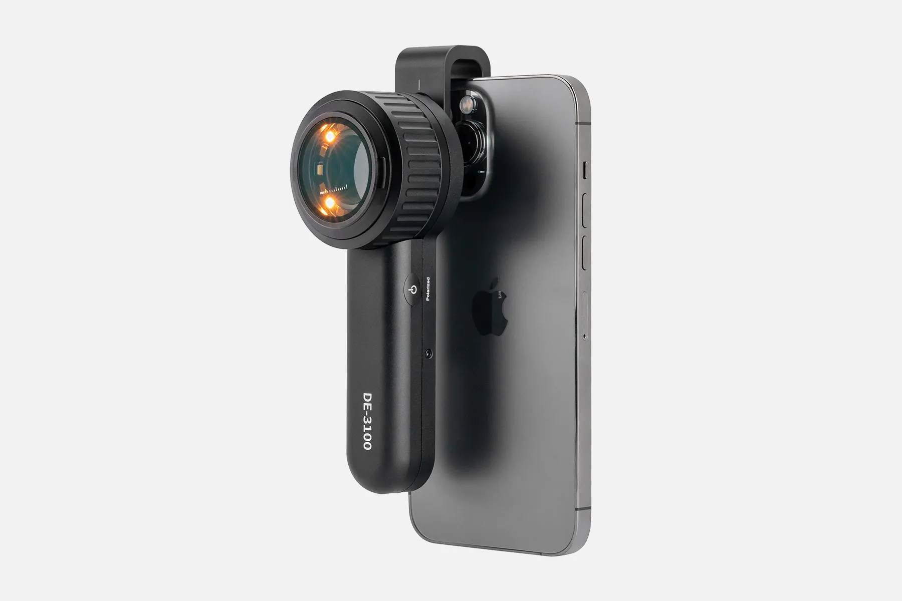
The DermLite DL100 from 3Gen is an advanced digital dermatoscope that utilizes innovative optics and software for skin evaluation. It features a no-oil, non-contact platform with 10x magnification and high-intensity LED lighting with cross-polarizers.
This dermatoscope enables detailed visualization of subsurface skin structures without requiring direct contact or immersion fluid. The specialized optical system produces color-corrected images to reveal information about skin lesions' color, size, shape, and morphology.
Embedded image processing software provides additional functionality like side-by-side comparisons for tracking changes over time. Dermatoscopy can be integrated with smartphones or PCs for image capture, storage, and analysis.
The 3Gen DermLite DL100 is a versatile, non-contact dermatoscope leveraging advanced optics and digital technology. It provides dermatologists with an enhanced platform for evaluating pigmented and non-pigmented skin abnormalities. Detailed dermoscopic images facilitate remote consultations and assist in the early detection of skin cancers.
How Does Dermatoscopio Dermlite?
The DermLite DL100 from 3Gen is an advanced digital dermatoscope that utilizes innovative optics and software for skin evaluation. It features a no-oil, non-contact platform with 10x magnification and high-intensity LED lighting with cross-polarizers.
This dermatoscope enables detailed visualization of subsurface skin structures without requiring direct contact or immersion fluid. The specialized optical system produces color-corrected images to reveal information about skin lesions' color, size, shape, and morphology.
Embedded image processing software provides additional functionality like side-by-side comparisons for tracking changes over time. Dermatoscopy can be integrated with smartphones or PCs for image capture, storage, and analysis.
The 3Gen DermLite DL100 is a versatile, non-contact dermatoscope leveraging advanced optics and digital technology. It provides dermatologists with an enhanced platform for evaluating pigmented and non-pigmented skin abnormalities. Detailed dermoscopic images facilitate remote consultations and assist in the early detection of skin cancers.
People May Ask
Identify Your Skin SpotsSkin cancer comes in three different forms: melanoma, basal cell carcinoma, and squamous cell carcinoma. The ability to recognize every kind of skin cancer is a skill taught to dermatologists. Make an appointment with the dermatologist over the phone if you find anything strange during your self-examination.
The diameter of a scabies mite is 0.2–0.5 mm. It burrows a track in the stratum corneum of the epidermis to deposit its eggs. With a dermatoscope that has an x 10 magnification, both mites and burrows are plainly visible.
Compared to not using dermoscopy, the diagnosis accuracy for melanoma was significantly greater (log odds ratio 4.0 [95% CI 3.0 to 5.1] versus 2.7 [1.9 to 3.4]; an improvement of 49%, p = 0.001). The level of experience of the examiners had a substantial impact on the accuracy of the dermoscopy diagnosis.
Do dermatoscopes have accuracy? A 2018 Cochrane study found that when used by a qualified practitioner, dermatoscopes are more accurate than the human eye alone in the diagnosis of melanomas. This is important since it can save someone time and possibly avoid the needless operation.
sanitization and cleaning...
Isopropyl alcohol (70% vol.) can be used to wipe out and sanitize the DermLite body. In the optical sections of the device, avoid using alcohol or disinfectants.
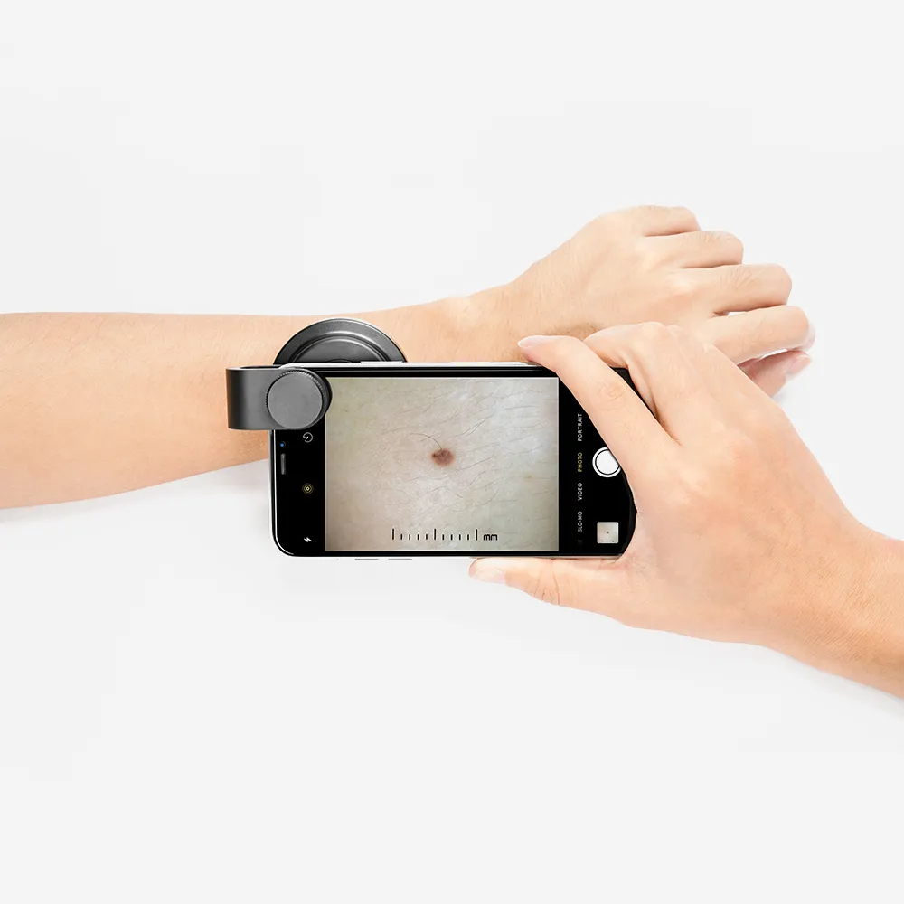
Similar to a magnifying glass, a dermatoscope is a portable device. It has the ability to magnify objects up to ten times. Your physician applies a gel or oil to your skin. After that, they place the dermatoscope against your skin to take a close look at the affected area.
With a dermatoscope, features can be evaluated down to the reticular dermis, and pictures can be taken for further analysis. Transillumination of a lesion for high magnification examination and visualization of minor details is the fundamental idea behind dermoscopy.
Handheld dermatoscope including changeable polarization, dermoscopic white and UV, torch LED, and 10x magnification. desktop charging base with auxiliary USB output and IceCap storage. 2 meter USB-C to USB-USB cable.
0:00 > 3:02It always appears polarized. In the middle. The pigment boost works similarly; all you have to do is tap that more
DermLite and Prompt Identification of Skin CancerWith the use of the DermLite product range, an expert eye can identify skin cancer and other skin diseases at an early stage. Each DermLite has an LED light source, a magnification lens, and the majority of them feature polarizing filters to reduce glare.
Dermatoscopio Dermlite Products
Featuring WF10x and WF20x eyepieces, 20X/40X/80X magnification, 2X and 4X objectives, upper and lower LED lighting, a reversible black/white stage plate, a pillar stand, and 120V or battery power, the AmScope SE306R-PZ-LED forward-mounted binocular stereo

A complete science kit that includes 10 microscope slide specimens, 5 blank slides, a 16-page user guide, and a phone adapter is the perfect choice for students studying science with a 40x–1000x microscope for adults.
GUVOP 50X-1000X Magnification WiFi Portable Handheld USB Microscopes with 8 LED & Stand: Wireless Digital Microscope Camera Compatible with iPhone, Android, Electronic Microscope for Kids and Adults - Blue

BEBANG Portable Microscope for Children Students Adult Microbiology Observation Laboratory Learning, Educational Set, 60x–120x Handheld Mini Microscope with LED Light and 5 Slide Samples
20x/50x/100x Magnification, 17mm Field of View, Pen Light Included in AmScope H2510 Handheld Stand Measuring Microscope
Arsir 7.3*4.4 Large Folding Lighted Magnifier with 48 LED Lights (3 Modes), Rectangular Handhold, Full-Page 5X Magnifying Glass for Reading Black Magnifying Lens Gifts for Seniors Who Read Books and Prints
Radio Frequency Face Machine for Residential Use High Frequency Face Massager with EMS, Light Therapy for Wrinkles Lifting and Anti-Aging Skin Tightening and Rejuvenation

Skin Tightening, Wrinkle Reduction, Hair Care: YourMate PhotoTherapy Device: High Frequency Facial Wand Machine with Argon Tubes for Face, Neck, and Hair

For kids and adults, a 3.9mm 720PHD WiFi ear scope with 6 LED lights that is compatible with both Android and iPhone is available with a wireless otoscope ear camera with dual view.

USSERA Medicube Age-R (Device alone)
Hot Products


News & Blog


Top Reviews
Lane Nelson
Works flawlessly on both Android and iPhone. Excellent for keeping an eye on your child's ears to determine whether the problem is ongoing or just transient. Helped me avoid a third round of antibiotics and surgical procedures recommended by a pushy specialist, after I showed a series of photos to his pediatrician, that showed the ears clear themselves. My sister used it to identify a tick that looked like a tiny black dot on a skin.
Barbara Sanchez
This ottoscope has performed admirably. Simple to operate and link to a phone. You can record videos and take photos, and you can store those as well. It also has a good battery life. I've only charged it once in the roughly five months I've had it.
Cindy M.
You "see" what this small lightweight endoscope picks up on its camera on your cell phone via an app. You don't actually look in the scope like your doctor does when examining you. This means you can easily look in your own ears and see a large image on your phone which is an added benefit. This unit came with a small card with two bar codes, one for downloading the app on your phone and a second to turn off ads on the app. Once the app is installed on your phone, you push the button to turn on the endoscope and find it via phone wifi settings to connect to it. Once you tell the app what kind of device it is, you are good to go. Clean a cone with alcohol, put it on the scope, and look in your ear (or wherever you need to look). You can capture photos with the touch of a button that you could easily send to your doctor if needed. The scope came fully charges, has a charger cord, little tools for use in your ear, and four starter alcohol swabs for cleaning before and after use. For the price, this will be a val


