


DE-3100 Dermatoscope – IBOOLO
Dermatoscope: Key Technology to Enhance Diagnostic Accuracy of Skin Lesions
What Is A Dermatoscope?
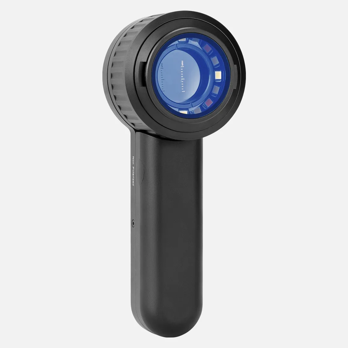
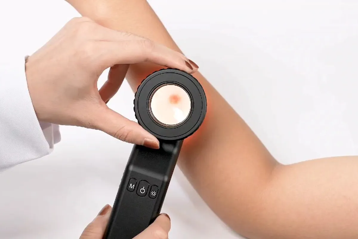
A dermatoscope, also known as a dermatoscope or chemiluminescence microscope, is an advanced non-invasive diagnostic aid technology. It is essentially a microscope that can magnify the skin from tens to hundreds of times, mainly used for observing and diagnosing pigmented skin disorders. Through cross-polarized light technology, the dermatoscope utilizes polarizing filters to eliminate diffuse reflection from the skin surface and selectively collects transmitted light, enabling an in-depth observation of the epidermal structure, pigmentation, blood vessels, and skin appendages.
What Can A Dermatoscope See?
A dermatoscope can observe details that are difficult to perceive with the naked eye, including but not limited to the following aspects:
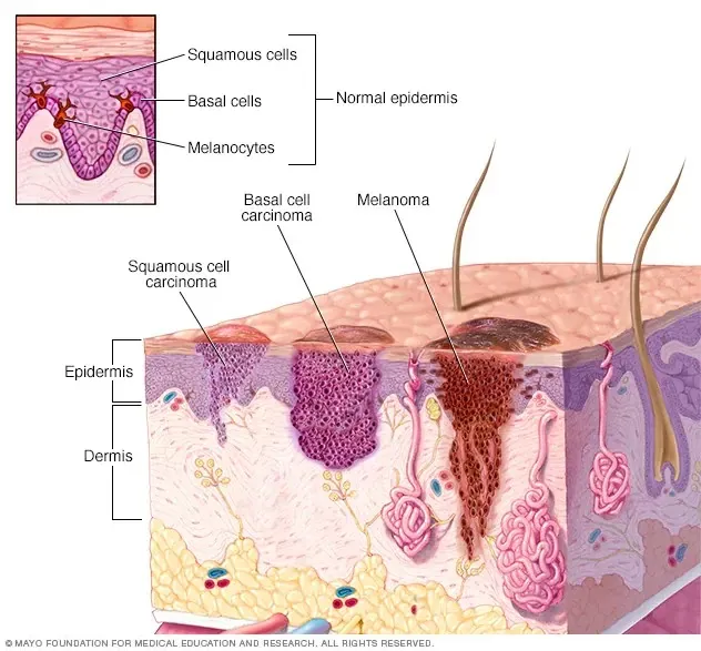
1. Epidermal structure: The dermatoscope can observe the epidermal layer in depth and analyze the microscopic structure of the skin.
2. Pigment distribution: Through the dermatoscope, doctors can see the pigment deposition and distribution patterns on the skin, which is crucial for diagnosing pigmented skin lesions.
3. Vascular morphology: The dermatoscope can reveal the vascular morphology and blood flow in the skin, helping to identify vascular-related lesions.
4. Skin appendages: Including hair follicles, sweat glands, and other skin appendage structures, the observation of which is very important for the diagnosis of certain skin lesions.
Uses of Dermatoscopes
Dermatoscopes have widespread applications in dermatological diagnosis. They can not only assist in the diagnosis of common moles, Spitz nevi, malignant melanoma, basal cell carcinoma, and other skin tumors but also help in differentiating pigmented skin tumors from vascular tumors, dermatofibroma, and others. Additionally, dermatoscopes can be used to observe pathological changes at the sub-microscopic level in the skin, providing important information for clinical diagnosis.
How Do Dermatoscopes Work?
The principle of dermatoscopes is mainly based on optical magnification and polarized light technology. They work through the following steps:
1. Magnification: dermatoscopes can magnify the skin surface from tens to hundreds of times, allowing doctors to observe minute details that are difficult to distinguish with the naked eye.
2. Polarized light technology: dermatoscopes use cross-polarized light technology, utilizing polarizing filters to eliminate scattered light from the skin surface, reducing glare and reflection for clearer observation.
3. Transmitted light collection: dermatoscopes selectively collect light transmitted through the skin, enabling doctors to observe deeper structures such as the epidermis, dermal-epidermal junction, and papillary dermis.
4. High-resolution imaging: dermatoscopes are equipped with high-resolution imaging systems that provide clear images, aiding doctors in precise diagnosis.
5. Non-invasive examination: dermatoscopes are non-invasive examination tools that do not require any cutting or trauma to the skin, making them safer and more comfortable for patients.
6. Image recording and analysis: Images obtained during dermatoscope examinations can be digitally recorded, facilitating archiving, comparison, and further analysis. This is useful for monitoring changes in lesions and evaluating treatment efficacy.
7. Free switching between observation levels: dermatoscopes allow doctors to freely switch between observing the epidermis, dermal-epidermal junction, and papillary dermis, obtaining skin information at different depths.
Can Dermatoscopes Detect Cancer?
Dermatoscopes are non-invasive diagnostic techniques that can significantly improve the accuracy of clinical diagnosis, especially in detecting skin cancer. By definition, a dermatoscope (also known as a chemiluminescence microscope) uses optical magnification principles, aided by polarization or immersion methods, to reflect the color and structural characteristics of the epidermis and papillary dermis. This technique can reveal surface structures and those beneath the skin's surface that are difficult for the naked eye to perceive, playing an important role in differentiating melanoma from other pigmented skin tumors.
Dermatoscopes have also become increasingly mature in the differential diagnosis of a series of non-melanocytic tumors, represented by basal cell carcinoma and seborrheic keratosis, with relatively high specificity and sensitivity in diagnosis. Furthermore, dermatoscope examinations have become an important tool for professional clinicians to assist in diagnosis and are increasingly being used in primary healthcare facilities.
Dermatoscope examinations can be used by general practitioners, helping to improve the accuracy of diagnosing basal cell carcinoma or cutaneous squamous cell carcinoma (BCC or cSCC). The dermatoscope is a handheld device that uses visible light and can be part of a clinical examination of suspicious skin lesions. Compared to naked-eye observation, dermatoscope examinations have higher accuracy in diagnosing melanoma.
Dermatoscopes can indeed serve as an effective tool for detecting skin cancer, including melanoma and non-melanoma skin cancer types. However, it is worth noting that dermatoscopes are an auxiliary diagnostic tool, and the final diagnosis may still require a combination of other examinations and histopathological evaluations.
Are Dermatoscopes Accurate?
As a non-invasive diagnostic aid, dermatoscopes have relatively high accuracy in dermatological diagnosis. According to the literature, dermatoscopes can increase diagnostic accuracy by 20% to 30%, thereby reducing unnecessary skin biopsies and surgeries. Furthermore, dermatoscope examinations have become a routine diagnostic tool for dermatologists in countries such as Europe and the United States, and are increasingly gaining attention from dermatologists worldwide.
The accuracy of dermatoscopes is also reflected in their ability to help doctors observe structures on and beneath the skin's surface that are invisible to the naked eye, playing an important role in distinguishing melanoma from other pigmented skin tumors. As mentioned, dermatoscope examinations can significantly improve the accuracy of clinical diagnosis. There have been numerous clinical studies on the diagnosis of basal cell carcinoma (BCC) using dermatoscopes in other countries, and a certain consensus has been reached.
Additionally, the article also confirms that compared to naked eye examination, dermatoscopes can improve the diagnostic accuracy of skin cancer (including melanoma). This indicates that dermatoscopes are a reliable diagnostic tool, particularly in enhancing the accuracy of skin lesion diagnosis.
Advantages of Dermatoscope Examinations
The advantages of dermatoscope examinations lie in their non-invasive nature, high resolution, and ease of use. They do not require surgery or invasive procedures, yet can provide high-definition images to aid doctors in accurate diagnosis. dermatoscope images can also be digitally photographed or recorded, facilitating archiving or continuous monitoring of lesion changes. Research has shown that dermatoscopes have a specificity of 98% in the diagnosis of malignant melanoma, even higher than clinical diagnosis, making them an indispensable tool for dermatologists.
People May Ask
In order to diagnose melanoma at an earlier stage, when it may appear to be indistinguishable from a benign pigmented lesion, dermoscopy enables the detection of early melanomaspecific characteristics that are evident under the dermatoscope even when a melanoma is tiny in size (
Absolutely, but not generally for aesthetic purposes. Your first stop if you think a mole is cancerous should be your general practitioner (GP). In most cases, your GP can tell you right away whether a mole is benign (harmless), but if not, they will recommend you to a dermatologist for additional testing.Oct. 16, 2013...
Mole Removal: Can I use the NHS to have a mole removed?The website skinsurgeryclinic.co.ukInstead,How can I...? https://skinsurgeryclinic.co.uk › treatments-blog › can-i-...
Do dermatoscopes have accuracy? A 2018 Cochrane study found that when used by a qualified practitioner, dermatoscopes are more accurate than the human eye alone in the diagnosis of melanomas. This is important since it can save someone time and possibly avoid the needless operation.18 March 2021...
What is visible through a dermatoscope? - Medical News Today
available at medicalnewstoday.com
https://www.medicalnewstoday.com › articles › dermatos...
A doctor or individual can inspect and diagnose skin lesions and disorders, including melanoma, using a dermatoscope, a hand-held visual assistance equipment. Examining the nails, hair, and scalp can also be facilitated by it. A dermatologist's practice typically has a dermatoscope.18 March 2021...
What is visible through a dermatoscope? - Medical News Today
available at medicalnewstoday.com
https://www.medicalnewstoday.com › articles › dermatos...
In contrast to non-polarized sunglasses, polarized lenses include a chemical coating that lowers glare. It may therefore be more challenging to see in bright light when using non-polarized sunglasses. In order to reduce glare, polarized glasses filter horizontal light waves while allowing vertical light waves to pass through the lens.April 8, 2022...
Sunglasses with or without polarization: What's the difference?Instead,www.myvision.orgPolarized vs non-polarized eyeglasses: https://myvision.org/...
You will not experience glare or any of its more uncomfortable or potentially harmful consequences if you use polarized sunglasses. Comparing your eyesight with non-polarized lenses, you should be able to see more contrast and clarity. The coating that polarized sunglasses have to block glare is absent from non-polarized sunglasses.26 May 2022The Difference Between Polarized and Non-Polarized Sunglasses | Warby Parkerwww.warbyparker.comwww.warbyparker.com › education › polarized-vs-non...
According to reports, the sensitivity of dermoscopy can vary from 60% to 100%, contingent on many aspects such as the skill level of the examiners and the complexity of the lesions being reviewed for diagnosis. Dermoscopy can increase the accuracy of melanoma diagnosis, but it cannot take the place of histopathologic evaluation.1 February, 2005Limitations of Dermoscopy in Melanoma IdentificationThe complete paper can be accessed at https://jamanetwork.com/jamasdermatology.
The overall pooled sensitivity and specificity of dermoscopy were 95% (95% CI 85% to 99%) and 91.2% (95% CI 90.0% to 92.4%) for the diagnosis of BCC, respectively. The addition of dermoscopy to naked eye examination increased sensitivity from 67% to 85% (5 trials; 4455 lesions; P = ) when compared to the naked eye examination alone....
The efficacy of dermoscopy in the diagnosis of skin cancer - PMC - NCBIPMC7571636 can be found under articles at https://www.ncbi.nlm.nih.gov
A doctor or individual can inspect and diagnose skin lesions and disorders, including melanoma, using a dermatoscope, a hand-held visual assistance equipment. Examining the nails, hair, and scalp can also be facilitated by it. A dermatologist's practice typically has a dermatoscope.18 Mar 2021...
What is visible through a dermatoscope? - Medical News Today
available at medicalnewstoday.com
https://www.medicalnewstoday.com › articles › dermatos...
While Dermlite DL II only has cross-polarized mode, Heine Delta 20 only has non-polarized mode, which necessitates a contact fluid. The non-polarized dermatoscope aids in identifying the skin's surface structures, whereas the polarized dermatoscope permits more profound viewing....
The Features of Dermatoscopy Images and Their Dissimilarities Among Frequently...PMC5447356 is an article that can be found at https://www.ncbi.nlm.nih.gov.
Dermatoscope Products

Coin magnifier with light, 2K LCD Digital Microscope 1200X, Dcorn 7 24MP HDMI Microscope, Soldering Coin Microscope with Lights, Extension Tube & 32GB Card Included
With 30 adjustable LED lights, a parfocal lens, wireless remote control, and PC compatibility, the SZJMS 10.1 Digital Microscope 1600X features an IPS Touch Screen Coin Microscope and a 1080P 12MP Soldering Microscope.
4X 大号攍大玻璃,带[防光和全可调光 LED] - 匀照明的姂看区域 - 昅读小字体、低视力老年人、组斑变性、磀查

MEDCASE Radiance Otoscope with Light German Fiber Optic - LED Lighted Ear Scope with Speculum for Ear Assessment and Diagnosis - Perfect for Both Home and Professional Use - Available in Purple Color

OdontoMed2011 Gray DERMATOSCOPE Free CASE ODM Dermatology Examination
With a 4.3-inch IPS screen, the BEAVERLAB Darwin M2 is a handheld digital microscope that is compatible with iPhone, Android computers, and 1600X pocket portable microscope camera for kids. It also has a 1080P HD coin microscope.
USB Digital Microscope: SKYEAR 50X-1600X Magnification Handheld Digital Microscope with Adjustable Stand, 8 LED Lights, and Portable Microscope Camera for Kids and Adults – Compatible with iOS & Android Devices

Anykit Digital Otoscope with a gyroscope, 4.5-inch screen, 3.9mm ear scope camera with six lights, 32GB card, and an ear wax removal tool Allows for the capture of photos and videos
Dino-Lite AM3113T USB Digital Microscope with 0.3 MP, 10x - 50x, 230x Optical Magnification, Microtouch, Measurement, and Discontinued

Three Gatuida phones for a 60x phone microscope and a handheld magnifying mirror Dermatoscope with a micro camera Microscope with Lights and a Clip-on Pocket Camera for White Cell Phone
Hot Products


News & Blog


Top Reviews
C. Ellingson
I have three years old with this clip. It is incredible. Any type of stethoscope you use will fit into it with ease. I own a Welch Alyn with replaceable heads. I have no issue carrying either and I frequently swap between adult and pediatric diaphragms. I anticipate using this for many years to come because of the excellent craftsmanship. Pros: -The greatest benefit of this equipment is the shoulder relaxation it provides. The stethescope may start to weigh heavily on your shoulders after 12 to 30 hours. This is wonderful to have sitting on my hips. - It is simple to retrieve the stethescope. Just pull the strap back, allowing it to drop into your other hand. It only takes a few seconds, and you are set to go. "-The stethoscope that was hanging from the neck was no longer able to obstruct or strike patients in the face. -A lifetime warranty, I believe. Drawbacks: -The stethescope produces a loop by your side that may get entangled in objects. It takes roughly 5 seconds to replace the stethescope in the batclip, which is a little longer than it takes to wear it around your neck.
Ian Hopkins
How does going to a code feel when your steth is around your neck? Akin to attempting to revive a bowling ball while positioned like a bowling pin. You don't have an issue if it's in your jacket, but it may fall out at any minute (eek). Then, when you put your head to the patient's chest to listen to their heart, they say something like, "Hey, you're not a doctor," and things get messy. So, explain why you think you should carry a clip with your stethoscope attached. much like BATMAN. Think about this. With all the confidence you have from not getting slapped in the face with your own tools and always knowing where they are, you walk into that code with your neck unencumbered and, with one great thrust, you've cleared out that pesky triple coronary blockage and your patient now has ROSC. You may even pull it out, play it, and force it back to the video, but proceed with caution—the nurses might pass out from the sight of your incredible CPR skills. It has come to my attention that when I sit in an auditorium chair, the screen
ArmRod
I wasn't really expecting to obtain a really high-quality instrument for this price, so I was pleasantly surprised. This device provides an incredibly clear close-up image of whatever you wish to examine. Among other things, I discovered that my Samsung LCD TV's RGB subpixels have an intriguing zigzag pattern on them. I also used it to clearly read the microprint on a $20 bill and my driver's license. The dazzling white fluorescing flecks that appear on everything suggest that the LED must have some UV output. It is also smaller than I had anticipated, which I enjoy. It is, in fact, a pocket microscope. Its tiny size is a disadvantage, too, as the description falsely states that it requires AAA batteries. It makes use of the more costly and uncommon little button-type batteries. The provided batteries have SR41 printed on them, despite the box stating that it needs SG3 (#392) cells. It appears that there is a lot of uncertainty over the battery type. The instruction booklet states SG7 (#392). I haven't utilized it extensively yet, so


