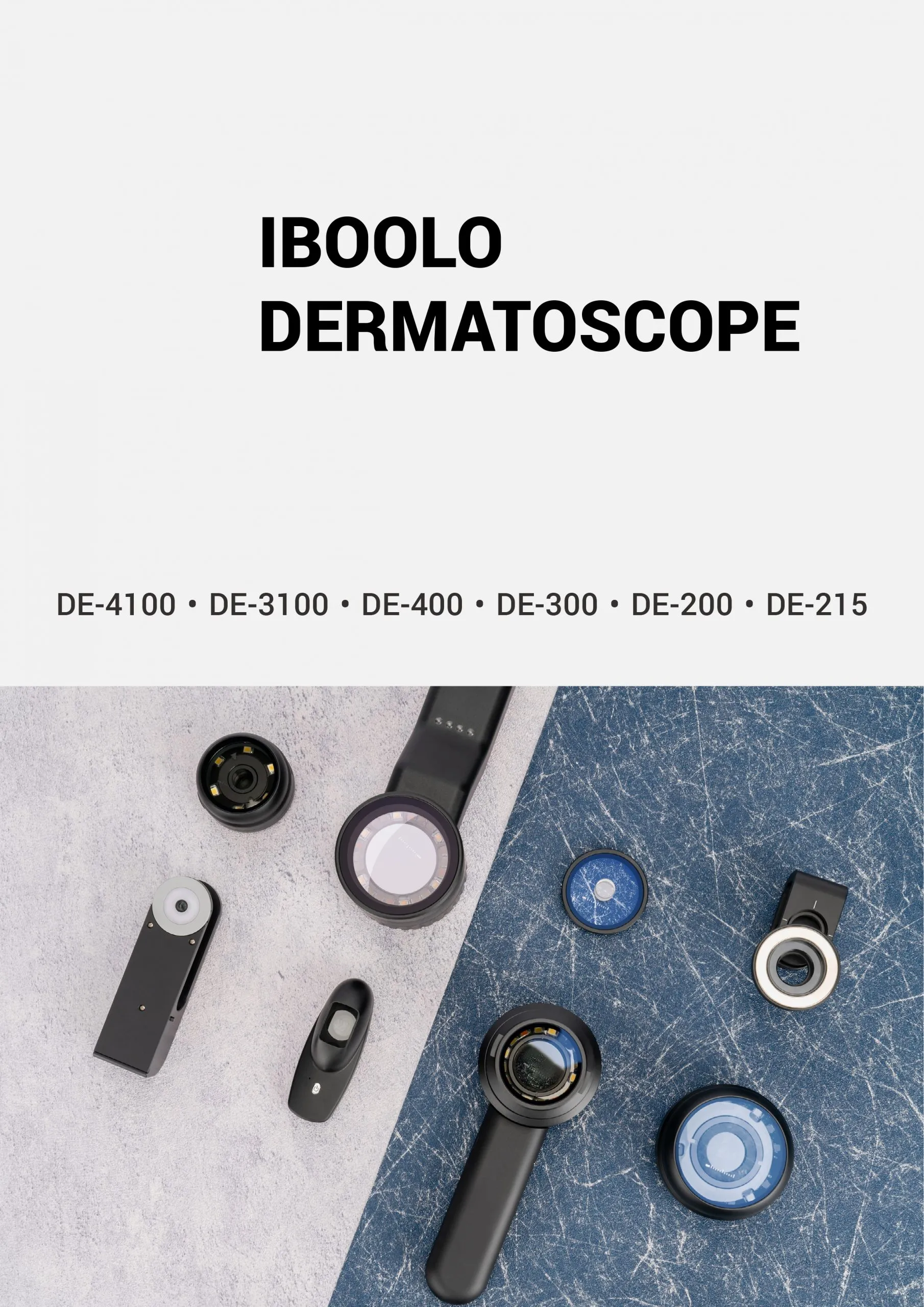
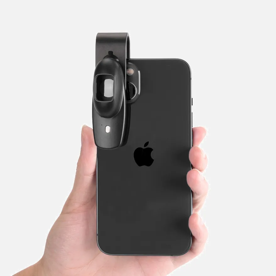
What Is Skin Cancer Dermoscopy?

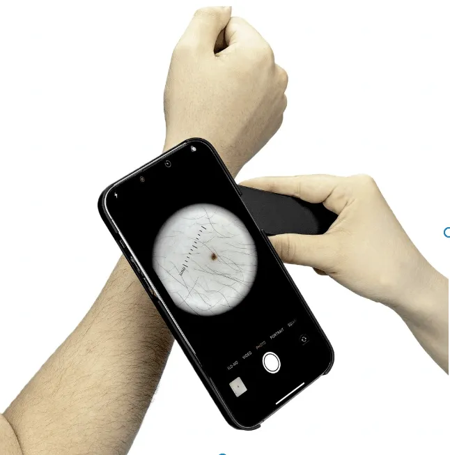
Dermoscopy is a non-invasive technique used to diagnose skin tumors. It involves using a handheld dermatoscope to visualize subsurface skin structures. These structures are usually not visible to the naked eye.

During a dermoscopy, a specialist puts oil or gel onto the skin and holds the dermatoscope onto it. This does not hurt or affect the skin.

Dermoscopy is used to diagnose both melanocytic and non-melanocytic tumors. It has been shown to increase the sensitivity and specificity for diagnosing melanoma. The dermoscopic diagnostic accuracy of basal cell carcinoma (BCC) is up to 95%–99%. Dermoscopy is also useful to distinguish pigmented basal cell carcinoma from other pigmented lesions.

How Does Skin Cancer Dermoscopy?

Dermoscopy is a noninvasive procedure that uses a handheld instrument called a dermatoscope to evaluate skin lesions. It allows the visualization of subsurface skin structures that are not usually visible to the naked eye.

Dermoscopy can help with:

  • Sensitivity: Dermoscopy can increase the sensitivity of skin cancer detection.
  • Biopsy ratio: Dermoscopy can decrease the ratio of benign to malignant biopsies.
  • Melanoma diagnosis: Dermoscopy can help diagnose thinner melanomas than a naked eye exam.
  • Accuracy: For basal cell carcinoma (BCC), dermoscopic diagnostic accuracy is up to 95%–99%.

Some characteristics of squamous cell carcinoma that can be seen using dermoscopy include:

  • Keratin clods surrounded by white circles
  • Linear irregular vessels heading to the center of the tumor
  • pinkish white background

To be classified as melanoma, a pigmented lesion must show an absence of pattern symmetry and color uniformity. It must also exhibit at least one of the following:

  • Blue-white veil
  • Multiple brown dots
  • pseudopods
  • Radial streaming
  • Scarlike depigmentation
  • peripheral block dots/globules
  • 5 to 6 colors

People May Ask

Can You Endure Years with Skin Cancer?

Survival in every stage of melanomaNinety percent or ninety out of one hundred patients will live with their melanoma for at least five years after being diagnosed. After being diagnosed with melanoma, almost 85 out of 100 individuals (more than 85%) will continue to live with the disease for ten years or more.

Which Nation Has The Greatest Rate of Cutaneous Cancer?

One of the highest rates of skin cancer worldwide is seen in Australia. We have clear skies and hot temperatures. Most of us are starting to realize the dangers of prolonged sun exposure in light of the summer's oppressive heat.

Could A BCC Develop into A Melanoma?

3,338 individuals in all received a BCC diagnosis during the study period; 82 (2.46%) of these patients went on to acquire melanoma (Fig. 1).

Can BCC Be Brought on by Stress?

Psychological stress may be a significant factor in the immunogenic tumor's environment and may have significant effects on BCC cancers in the future.

What Does BCC Look like in Its Early Stages?

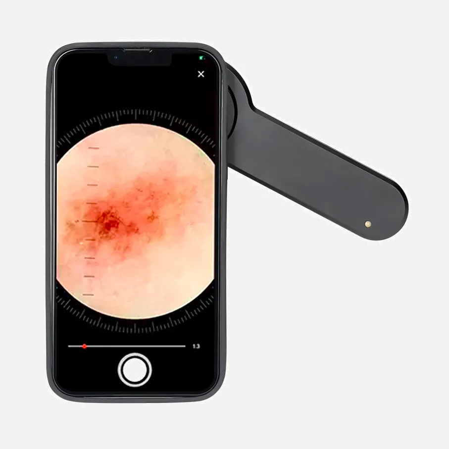
Open sores, red spots, pink growths, shiny bumps, scars, or growths with slightly elevated, rolling edges and/or a center indentation are some of the appearances of BCCs. BCCs have the potential to leak, crust, itch, or bleed. The lesions usually appear on body parts that are exposed to the sun.

Can Melanoma Be Present for 20 Years without Symptoms?

It is contingent upon the kind of melanoma. Radial melanoma, on the other hand, might spread slowly over a decade, whereas nodular melanoma spreads quickly over a few weeks. Similar to a cavity, a melanoma can proliferate for years without showing any noticeable symptoms.

At Thirty Years Old, How Frequent Is Skin Cancer?

When it is diagnosed, the average age of the patient is 66. However, even in individuals under 30, melanoma is not rare. Indeed, among young individuals, it's one of the most prevalent tumors (particularly young women). Check out Survival Rates for Melanoma Skin Cancer by Stage for survival data.

What Is The 20s BCC Prevalence?

The bulk of basal cell carcinoma cases affect elderly persons since the cancer frequently takes decades to grow. However, it is increasingly common in persons in their 20s and 30s and can also afflict younger adults. skin cancer in one's family or on oneself.

How Long May It Be before Skin Cancer Is Discovered?

For instance, a biopsy is required to confirm the diagnosis of some forms of skin cancer, which can first be identified only by visual inspection. However, another study discovered that some cancers can develop and spread for up to ten years without being seen, which further complicates diagnosis and therapy.

Can Skin Cancer Be Detected by A Dermoscopy?

Using a dermascope gives a skin cancer specialist an additional advantage even if they can still detect malignant indications with their eyes. a color and structural clarity that would be invisible to the unaided eye or even a low-powered microscope. Color, particularly the distribution of pigments, can serve as a diagnostic tool in and of itself.

Skin Cancer Dermoscopy Products

Cancer (Astrology) - Discovering Love and Harmony in Every Relationship: Boxed Set of Cancer Horoscopes (Relationship Books for Dating Couples) Kindle攵子书

Cancer: A Special Gift for Cancer Patients And 121 Pages of Notepad/Journal with Matte Finish, 6 x 9 平装 – 2020年 1月 6日

2019 Zodiac Sign: Cancer 平装 – 2018年 9月 12日

China customized skin cancer dermoscopy products supply

The Natural Cure for Basal and Squamous Cell Carcinomas and Keratoses Is the Answer to Skin Cancer January 1, 1998, paperback

China Professional skin cancer dermoscopy

Skin Cancer Diagnosis Made Simple: Over 220 full-color case discussions and a tutorial-based approach to skin cancer diagnosis for novices and experts alike. Kindle攵子书

China handheld skin cancer dermoscopy

Cancer: A Skin Care Schedule for Cancer Sign Signs 平装 – 大字版本, 2022年 1月 2日

China customized skin cancer dermoscopy

Cancer Horoscope 2022: Your go-to resource for matters of love, money, happiness, and moon magic Kindle攵子书

Top Reviews

sadeghani

The best Dermoscopy book. Very comprehensive and informative for dermatologist.

R.B. Culpepper

I avoided having to go through the typical "cut, burn, and poison procedure" to treat basal skin cancer by using the knowledge and research in this book.

Dan Zedek, MD

This Dermoscopy book has been enjoyable. Fantastic and very useful are the descriptions and photographs. This book will be an excellent primary source for dermoscopy education for our dermatology residents.

0

Have questions on gear or your order?

Our Gear Guides are here to help! Get personal advice from pro creatives

Name
Subject
Email address
How can we help?

Instant Answers

How Can Dermoscopy Images Be Captured?

Dermoscopy images can be captured and stored in different ways, such as: • Using a smartphone or tablet with dermoscopic adapter, which consisted in the package.• Using a digital camera

Dermoscopy images can be captured and stored in different ways, such as:

• Using a smartphone or tablet with dermoscopic adapter, which consisted in the package.
• Using a digital camera with dermoscopic adapter, there’s 49mm screw size camera adapter available to order now.

Compatible phone/tablet models:
All iPhone models, 95% Android phones, 90% tablet. For phone/tablet size in 5.25-14mm

Compatible camera models:
All camera with built 49mm filter screw, such as Canon EOS 70D, 80D, 90D; Canon EOS R7, R10, R50, R100; Canon M100, M200, M50, Mark II; Canon G7X Mark III, Sony ZV-1

How Can I Connect My Phone to My Dermatoscope?

There’s universal phone adapter for all our dermoscopes. Please check the installation procedure bellow or watch operation guide. Smartphone Connector (1) Place phone adapter screw in the center of smartphone’s

There’s universal phone adapter for all our dermoscopes. Please check the installation procedure bellow or watch operation guide.

Smartphone Connector

(1) Place phone adapter screw in the center of smartphone’s main camera.
(2) Screw magnet attachment on phone adapter.
(3) Put dermoscope’s back ring and magnet attachment together

Take The Best Images

You need to adjust the focus ring after the dermoscpe connected on smartphone to get the best images.

How Can I Clean My Dermoscopy after Usage?

Cleaning your dermoscopy after usage is important to prevent cross-contamination and infection. The cleaning method may vary depending on the type and model of your dermoscopy, so you should always

Cleaning your dermoscopy after usage is important to prevent cross-contamination and infection. The cleaning method may vary depending on the type and model of your dermoscopy, so you should always follow the manufacturer’s instructions. However, some general steps are:

• Turn off and disconnect your dermoscopy from any power source or device.

• Wipe off any visible dirt or debris from the dermoscopy with a soft cloth or tissue.

• Disinfect the dermoscopy with an alcohol-based wipe or spray, or a disinfectant solution recommended by the manufacturer. Make sure to cover all surfaces, especially the lens and contact plate.

• Let the dermoscopy air dry completely before storing it in a clean and dry place.

• Do not use abrasive or corrosive cleaners, solvents, or detergents that may damage the dermoscopy.

• Do not immerse the dermoscopy in water or any liquid, unless it is waterproof and designed for immersion.

You should clean your dermoscopy after each use, or at least once a day if you use it frequently. You should also check your dermoscopy regularly for any signs of damage or malfunction, and contact the manufacturer or service provider if needed.

Polarized VS Non-Polarized Dermoscopy

A dermoscopy is a device that allows the examination of skin lesions with magnificationand illumination. By revealing subsurface structures and patterns that are not visible tothe naked eye. It can

A dermoscopy is a device that allows the examination of skin lesions with magnificationand illumination. By revealing subsurface structures and patterns that are not visible tothe naked eye. It can improve the diagnose accuracy of skin lesions, such as melanoma,basal cell carcinoma, seborrheic keratosis, etc.

There are two main types of dermoscopy: Non polarized and polarized dermoscopy.We’ve fitted most of our dermoscopys with polarized and non-polarized light. They canbe used in multiple skin structures.

Non-polarized contact Mode

In non-polarized mode, the instrument can provide information about the superficialskin structures, such as milia-like cysts, comedo-like openings, and pigment in theepidemis.

The dermoscopy requires applying a liquid such as mineral oil or alcohol to the skin andplacing the lens in contact with the skin. This reduces surface reflection and enhancesthe view of subsurface structures.

Image with non-polarized light (DE-3100)

Polarized contact Mode

In polarized mode, the instrument allows for visualization for deeper skin structures,such as blood vessels, collagen, and pigment in the dermis.

The dermoscopy does not need to be in contact with the skin or use any liquid. Theirpolarized light can help to eliminate surface reflection and allow visualization ofvascular structures.

Image with polarized light (DE-3100)

Polarized non-contact Mode

The dermoscopy can also use polarized light to examine the skin without direct contact.

In polarized non-contact mode, the instrument allows for examination infected areasand lesions that are painful for the patient, or the difficult to contact pigmented lesions,such as nails and narrow areas.

The contact plate should be removed in this mode, and it does not require applying aliquid to the skin. As it doesn’t require pressure or fluid application on the skin, it canalso avoid cross-contamination and infection risk.

Image in polarized non-contact mode (DE-3100)

How Effectiveness Is Dermoscopy

Compared with visual inspection, the dermoscopy can be used to capture and store skin lesion photos, which play an important role in early skin cancer examination. The dermoscopy allows the

Compared with visual inspection, the dermoscopy can be used to capture and store skin lesion photos, which play an important role in early skin cancer examination.

The dermoscopy allows the examination of skin lesions with magnification and illumination. This can be greatly avoiding the factors that cause interference to visual detection. Such as lighting, skin color, hair and cosmetics.

Several studies have demonstrated that dermoscopy is useful in the identification of melanoma, when used by a trained professional.

It may improve the accuracy of clinical diagnosis by up to 35%
It may reduce the number of harmless lesions that are removed
In primary care, it may increase the referral of more worrisome lesions and reduce the referral of more trivial ones

A 2018 Cochrane meta-analysis published the accuracy of dermoscopy in the detection.

Table 1. Accuracy of dermoscopy in the detection of melanoma in adults
Detection Method Sensitivity, % Specificity, % Positive Likelihood Ratio NegativeLikelihood Ratio
Visual inspection alone (in person) 76 75 3.04 0.32
Dermoscopy with visual inspection (in person) 92 95 18 0.08
Image-based visual inspection alone (not in person) 47 42 0.81 1.3
Dermoscopy with image-based visual inspection (not in person) 81 82 4.5 0.23
ROC—receiver operating characteristic. *Estimated sensitivity calculated on the summary ROC curve at a fixed specificity of 80%.

As we can see, the dermoscope can improve the accuracy of diagnosis of skin lesions, especially melanoma.

Table 1. Accuracy of dermoscopy in the detection of melanoma in adults
Detection Method Sensitivity, % Specificity, % Positive Likelihood Ratio NegativeLikelihood Ratio
Visual inspection alone (in person) 79 77 3.4 0.27
Dermoscopy with visual inspection (in person) 93 99 93 0.07
Image-based visual inspection alone (not in person) 85 87 6.5 0.17
Dermoscopy with image-based visual inspection (not in person) 93 96 23 0.07
ROC—receiver operating characteristic. *Estimated sensitivity calculated on the summary ROC curve at a fixed specificity of 80%.

Characteristics of the dermatoscopic structure of the skin lesions include:

• Symmetry or asymmetry
• Homogeny/uniformity (sameness) or heterogeny (structural differences across the lesion)
• Distribution of pigment: brown lines, dots, clods and structureless areas
• Skin surface keratin: small white cysts, crypts, fissures
• Vascular morphology and pattern: regular or irregular
• Border of the lesion: fading, sharply cut off or radial streaks
• Presence of ulceration

There are specific dermoscopic patterns that aid in the diagnosis of the following pigmented skin lesions:

• Melanoma
• Moles (benign melanocytic naevus)
• Freckles (lentigos)
• Atypical naevi
• Blue naevi
• Seborrhoeic keratosis
• Pigmented basal cell carcinoma
• Haemangioma

No products in the cart.

No products in the cart.

Hot Search Terms