


Magnetic Ring for DE-3100 – IBOOLO
What Is Dermascope Vs Dermatoscope?
A dermatoscopy and a dermoscopy refer to the same handheld medical device used to perform dermoscopy examinations of skin lesions and screen for skin cancer. The terms are synonymous.
A dermatoscopy/dermoscopy utilizes magnification and light to visualize subtle morphological characteristics of pigmented lesions not visible to the naked eye. This non-invasive skin surface microscopy technique is known as dermatoscopy or chemiluminescence microscopy.
When applied properly, a dermatoscopy improves diagnostic accuracy and aids in differentiating benign moles from suspicious lesions requiring biopsy. It allows inspection of skin structures up to the reticular dermis layer to better assess disease risk. Some models also examine the scalp, hair, and nails.
In summary, a "dermatoscopy" and a "dermoscopy" describe the same specialized medical device designed for close-up skin examinations and enhanced visualization of lesions through magnification, illumination, and polarization filtering. Recognizing these two terms refer to the identical instrument prevents confusion when reading clinical literature on skin cancer screening methods.
Dermatoscope And Dermascope: The Same Device for Dermoscopy Skin Exams
A dermatoscope and a dermascope refer to the identical medical device used to conduct dermoscopy - the magnified inspection of skin lesions using light and magnification. This noninvasive technique goes by other names including dermatoscopy, epiluminescence microscopy, incident light microscopy and skin-surface microscopy.
The key principle involves lighting and magnifying lesions to uncover subtle visual characteristics impossible to see with the naked eye. Features like patterns of pigmentation, blood vessels, and skin texture become visible. When applied properly, dermoscopy improves diagnostic accuracy over visual inspection alone for assessing if a suspicious mole may be invasive melanoma or a more benign in situ melanoma.
In summary, "dermatoscope" and "dermascope" are two interchangeable terms for the same handheld device applied to the skin to allow magnified visualization of lesions. This dermoscopy technique lets dermatologists examine moles and other skin growths with greater precision to determine malignancy risks and guide biopsy decisions. Recognizing both terms refer to the same instrument prevents confusion when reading medical literature.
People May Ask
Your skin will first be treated by the physician with an ultrasonic gel or oil (like mineral oil). The dermatoscope can capture clearer images when it is coated with gel or oil. After applying the gel or oil, the medical professional will gently insert the dermatoscope into your skin.
A novel and intriguing morphological aspect of pigmented skin lesions has been revealed by the clinical application of dermoscopy. Through the visualization of morphological traits invisible to the unaided eye, dermoscopy is a non-invasive diagnostic method that connects clinical dermatology and dermatopathology.
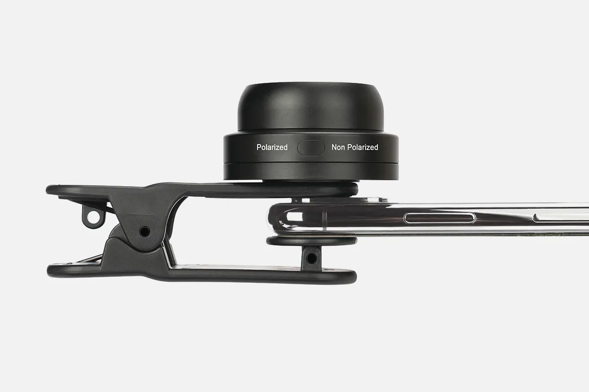
While Dermlite DL II only has cross-polarized mode, Heine Delta 20 only has non-polarized mode, which necessitates a contact fluid. The non-polarized dermatoscope aids in identifying the skin's surface structures, whereas the polarized dermatoscope permits more profound viewing.
Dermoscopy dates back to the middle of the modern era, with significant contributions by Ernst Karl Abbe, Unna, Muller, Saphier, and others, as well as Borel's discovery (1655–1656) that lay the groundwork.
The 10-fold magnification that handheld dermatoscopes typically offer is usually sufficient for daily use. Lower magnifications also have the advantage of offering a more comprehensive view of a big scalp area.
A handheld device known as a dermatoscope is used to perform dermoscopy. Subsurface skin structures in the epidermis, papillary dermis, and dermoepidermal junction-structures that are often invisible to the unaided eye-can be seen thanks to this method [2-4].
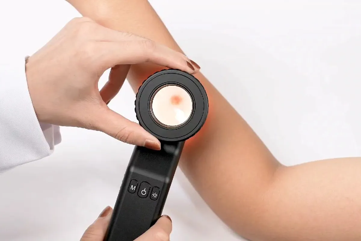
A doctor or individual can inspect and diagnose skin lesions and disorders, including melanoma, using a dermatoscope, a hand-held visual assistance equipment. Examining the nails, hair, and scalp can also be facilitated by it. A dermatologist's practice typically has a dermatoscope.
On the mole that bothers you, apply one drop of oil. Directly touch the skin with the dermatoscope, press down gently, and use your smartphone to take a picture. Touching the screen's center may be necessary to concentrate the mole's image. Take and preserve clear dermoscopic pictures.
Dermoscopy, sometimes referred to as dermatoscopy, skin surface microscopy, or epiluminescence microscopy, is a non-invasive in vivo method that has long proved helpful in the assessment of skin lesions that seem worrisome.
When diagnosing skin cancer, dermatoscopes, also known as dermascopes, are the most used type of skin surface microscopy. It is possible to identify benign lesions without a biopsy and better understand those that need further attention.
Dermascope Vs Dermatoscope Products
Portable Ear Light and Exam Kit for Home and Professional Use - Cynamed Mini Otoscope - 3X Magnifying Fiber Optic Scope with Extra Tips, Bulb, and Carrying Case - Pocket Diagnostic Tools (Black)

Disposable ear specula for adults and older children, Treela 200 PCS Otoscope Cover Plastic Otoscope Specula Tips (4.25 mm, 200 Pieces)
Otoscope Covers for Zyrev ZetaLife 50 pieces of disposable ear holder tips made of plastic. Otoscope Covers, Disposable, 2.5/3.5mm

KMDES Wireless Digital Microscope: Handheld USB HD Inspection Camera with 50X–1000X Magnification, Adjustable 8 LED Lights, and Stand Upgrade that Prevents Shake for PC Phone Compatibility. Available in White
Upgraded Shake-Free, Adjustable 50X–1000X Magnification Wireless Digital Microscope Handheld USB HD Inspection Camera on a Metal Stand (KMDES) 8-light LED portable microscope for computers, smartphones, and other devices - blue
A veterinary otoscope Pet Otoscope, Canine Otoscope, Ophthalmoscope Set, Ear Exam, Professional Diagnostic, Vet Otoscope Set, Otoscope for Dogs, Earache, Ear Canal Exam, Eardrum Exam, Premium

Handheld Microscope for Children and Students, HUTACT Pocket Microscope for Kids, 60X-135X Mini Portable Microscope with 5 Microscope Slides

With the BATCLIP (Black) - Premium Leather Handmade Clip-On Stethoscope Hip Holder, you can finally say goodbye to wearing around your neck, misplacing, or being misplaced. Display your luxury stethoscope with class, taste, and pride.

MEDCASE Radiance Otoscope with Light German Fiber Optic - LED Lighted Ear Scope with Speculum for Ear Assessment and Diagnosis - Perfect for Both Home and Professional Use - Available in Purple Color
Polarized Skin Dermatology Light 3Gen Lumio Dermlite
Hot Products


News & Blog


Top Reviews
C. Ellingson
I have three years old with this clip. It is incredible. Any type of stethoscope you use will fit into it with ease. I own a Welch Alyn with replaceable heads. I have no issue carrying either and I frequently swap between adult and pediatric diaphragms. I anticipate using this for many years to come because of the excellent craftsmanship. Pros: -The greatest benefit of this equipment is the shoulder relaxation it provides. The stethescope may start to weigh heavily on your shoulders after 12 to 30 hours. This is wonderful to have sitting on my hips. - It is simple to retrieve the stethescope. Just pull the strap back, allowing it to drop into your other hand. It only takes a few seconds, and you are set to go. "-The stethoscope that was hanging from the neck was no longer able to obstruct or strike patients in the face. -A lifetime warranty, I believe. Drawbacks: -The stethescope produces a loop by your side that may get entangled in objects. It takes roughly 5 seconds to replace the stethescope in the batclip, which is a little longer than it takes to wear it around your neck.
Ian Hopkins
How does going to a code feel when your steth is around your neck? Akin to attempting to revive a bowling ball while positioned like a bowling pin. You don't have an issue if it's in your jacket, but it may fall out at any minute (eek). Then, when you put your head to the patient's chest to listen to their heart, they say something like, "Hey, you're not a doctor," and things get messy. So, explain why you think you should carry a clip with your stethoscope attached. much like BATMAN. Think about this. With all the confidence you have from not getting slapped in the face with your own tools and always knowing where they are, you walk into that code with your neck unencumbered and, with one great thrust, you've cleared out that pesky triple coronary blockage and your patient now has ROSC. You may even pull it out, play it, and force it back to the video, but proceed with caution—the nurses might pass out from the sight of your incredible CPR skills. It has come to my attention that when I sit in an auditorium chair, the screen
AJ
After acquiring the USA-made Batclip Stethoscope Holder, I'm delighted I made the purchase. In my everyday work as a healthcare practitioner, this high-quality, useful tool has been a great addition. Design and Build Quality: The Batclip has a thoughtfully designed and superbly built product. This stethoscope holder is made in the USA and is composed of strong materials for long-term endurance. Usability: The Batclip Stethoscope Holder is very easy to use. It fits neatly into my pocket or waistline and holds my stethoscope firmly and without any trouble at all. Even during the busiest shifts, my stethoscope stays in place all day thanks to the sturdy and dependable clip mechanism. Compatibility: I like that several stethoscope models may be used with the Batclip. It has supported several brands that I have used with it, all of them comfortably and securely. I can utilize the Batclip because of this compatibility, which is a nice feature.


