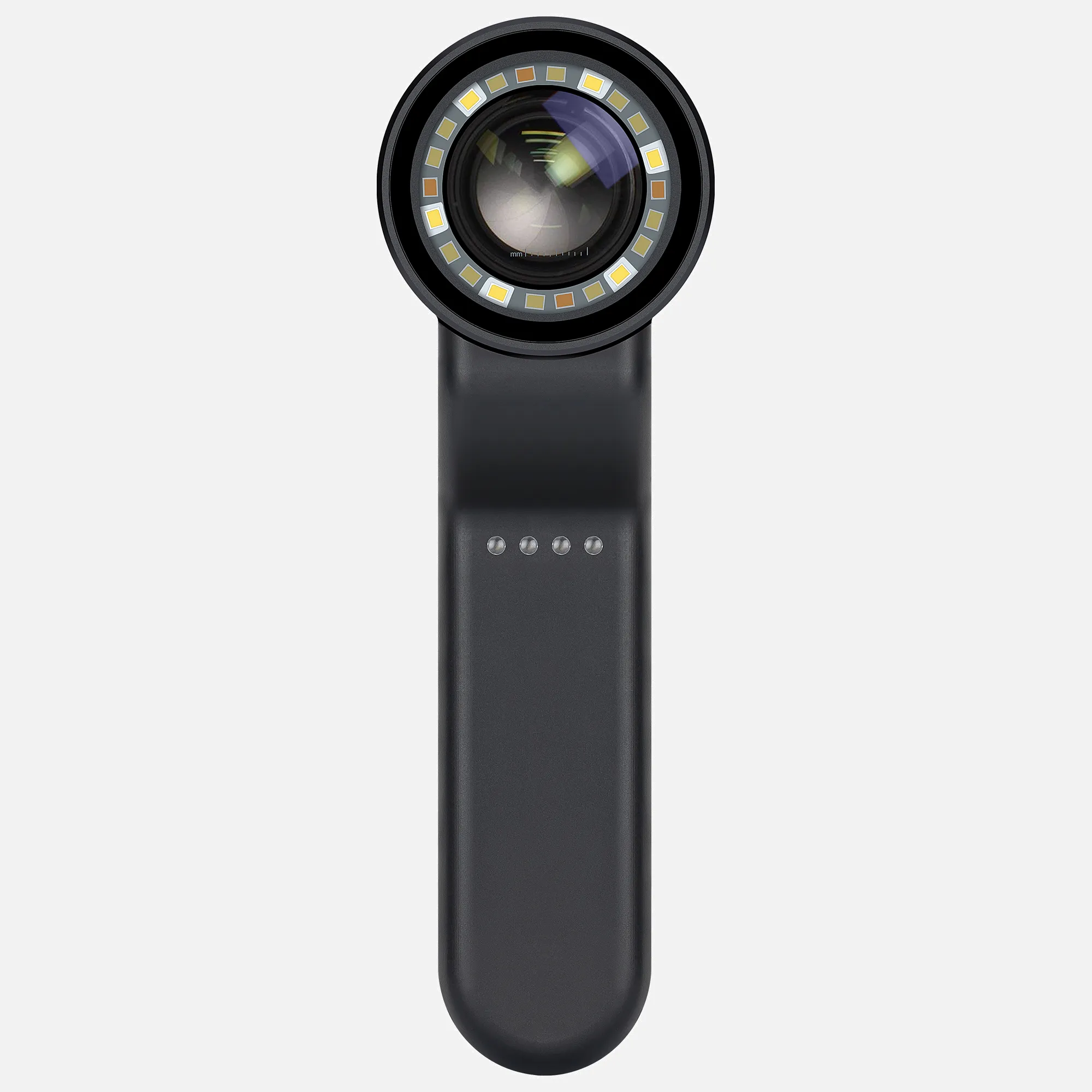
Utilizing Dermoscopy to Assess Characteristic Manifestations of Psoriasis

Dermoscopy is an effective tool to evaluate psoriasis across different affected areas of the skin. Typical dermoscopic features of psoriatic lesions include:

Scalp psoriasis: Red dots, globules, and twisted looping vessels are visible.

Nail psoriasis: Changes like onycholysis, salmon patches under the nail, and splinter hemorrhages can be observed.

Inverse psoriasis: Dotted vessels on a reddish backdrop are apparent.

Dotted vessels are the most common dermoscopic vascular marker in psoriatic plaques.

Psoriasis manifests as raised, scaly red inflammatory patches with distinct borders. It frequently affects the scalp, trunk, buttocks, and limbs, though it can appear anywhere on the skin. Using a dermatoscope facilitates detailed visualization of microscopic manifestations of this chronic condition. Dermoscopy is valuable for determining disease extent and monitoring treatments.

How Dermoscopy Can Be Utilized in Diagnosing And Evaluating Psoriasis

Dermoscopy is an important non-invasive technique that aids visual diagnosis of psoriasis across different body areas. Using a dermatoscope provides magnified visualization and documentation of distinct characteristics:

Scalp psoriasis: Red dots, globules and twisted vessels are visible

Nail psoriasis: Signs like salmon patches, onycholysis, splinter hemorrhages

pustular psoriasis: Dotted vessels surrounded by white/yellow pustules

palmoplantar: Yellow scales, orange dots, pronounced vessels

Other common dermoscopic markers seen in psoriatic plaques include: sharply demarcated borders, white scaling, dull red erythema.

Dermoscopic evaluation improves detection of subtle signs and extent of involvement. It helps determine if a skin biopsy is needed for further confirmation in ambiguous presentations. Overall, incorporating dermoscopy with clinical inspection grants superior assessment of this chronic papulosquamous disease. Sequential documentation aids in gauging treatment efficacy as well.

People May Ask

How Can A Dermoscopy Distinguish between Plaque Psoriasis And Pityriasis Rubra Pilaris?

On a light red backdrop, SD was identified by the patchy distribution of yellow scales and dotted vessels, while PR was distinguished by the peripherally placed white scales and the patchy distribution of dotted vessels on a yellowish background.

What Are The Finest Dermoscopy Features for Bowen's Disease?

Dermoscopy is used to identify diseases like Bowen's disease. Glomerular veins and a scaly surface are the most common dermoscopic characteristics of Bowen's disease.

Under Dermoscopy, How Does BCC Appear?

With their characteristic bluish-pink color, asymmetric arborizing vasculature, and focal ulceration, superficial basal cell carcinomas are often diagnosed by skilled dermoscopists. White regions of regression and little scaling could also exist.

Psoriasis And Seborrheic Dermatitis Are They The Same?

whereas scaly erythematous patches with itching can be the appearance of either disease1, psoriasis typically coexists with lesions on the extensor areas, whereas seborrheic dermatitis affects other skin regions rich in sebaceous glands, such as the ears, eyebrows, central chest, and intertriginous areas.

How Is The Presence of Psoriasis Diagnosed by A Dermatologist?

Biopsy of the SkinYour dermatologist can typically identify psoriasis simply by looking at your skin. In the event that additional information is required to validate the diagnosis and exclude alternative etiologies of symptoms, such as cutaneous lupus or eczema, a skin biopsy might be conducted.

How Can One Identify Psoriasis?

Your doctor will typically check your nails, scalp, and skin for symptoms of psoriasis in order to make the diagnosis. Inquiries concerning your health, medical history, and family history could also be made, including whether you: Feel sensations like skin that is burning or itching. had a recent illness or undergone a stressful event.

What Kind of Skin Pattern Does Psoriasis Cause?

The following symptoms may manifest as plaque psoriasis: Thick, elevated skin patches known as plaques. Certain plaques are covered in scale, a thin, dried layer of silvery white paint. Various-sized plaques.

In Pityriasis Rosea, What Are The Dermoscopic Findings?

characteristics of the dermis. Beautiful collarette of scales with brown globules (white arrow), middle yellow with a reddish background, peripheral dotted vessels with patchy distribution (black circle), and the plaque's periphery attached to red arrows.

What Are Scalp Psoriasis's Dermoscopic Findings?

Evaluation of scalp psoriasis by trichoscopic meansRed spots and uniformly distributed globules are seen at low magnifications. White or white-silver dry scales are a common discovery. Recently, indications supporting the diagnosis of psoriasis have been identified, including hidden hair and signet ring vessels.

How Can One Distinguish between Eczema And Psoriasis Using Dermoscopy?

When psoriasis lesions were examined under a dermoscopy, 76.3% (29/38) showed diffuse scaling, 60.5% (23/38) showed white scales, and 76.3% (29/38) showed dotted vessels along with a regular distribution of vessels; in hand eczema lesions, 78.5% (11/14) showed diffuse scaling, and 57.1% (8/...) showed white and yellow scales.

Dermoscopy of Psoriasis Products

First Edition, Kindle Edition of Surface Imaging for Biomedical Applications

Harper s Textbook of Pediatric Dermatology, Fourth Edition, Two Volume Set

Diagnosis and Treatment of Common Disorders in Clinical Dermatology, Second Edition 2第二 版本

China Professional dermoscopy of psoriasis

The Print Replica of the Alternative Dermoscopy Atlas (Minor Surgery Series) Kindle Version

China smartphone dermoscopy of psoriasis

Image analysis using dermoscopy (Digital Imaging and Computer Vision Book 10) 1第一 版本, Kindle电子书

China customized dermoscopy of psoriasis products supply

Review of Dermoscopy Criteria, First Edition

China customized dermoscopy of psoriasis

Dermoscopy of Hair and Scalp Disorders: Including Clinical and Pathological Correlations 1第一 퉈本, Kindle~子乪

Top Reviews

Miami man

I completed my dermatological training just as dermoscopy was starting to gain popularity. The evidence for improved diagnosing skills is now overwhelming. Throughout my multiple courses at the AAD and other locations, I consistently felt more perplexed and overwhelmed than before. I discovered there was no mechanism for me to "train" myself and receive feedback. This book is different in that it teaches you how to identify traits and then apply them in a self-directed course. It may be completed one chapter at a time and is well written. I feel like I've learned enough about this significant advancement in melanoma diagnosis after enough repetition.

Ritterburg

For those interested in dermatology, this book is helpful and contains many examples; nonetheless, it is overly dense with formulas and tables. An excellent dermatology atlas is also essential for learning dermatology.(z.B. Marghoob, Braun, Atlas of Dermoscopy..)

sach509

I self-trained using this book. It's quite explicit and colorful.

0

Have questions on gear or your order?

Our Gear Guides are here to help! Get personal advice from pro creatives

Name
Subject
Email address
How can we help?

Instant Answers

How Can Dermoscopy Images Be Captured?

Dermoscopy images can be captured and stored in different ways, such as: • Using a smartphone or tablet with dermoscopic adapter, which consisted in the package.• Using a digital camera

Dermoscopy images can be captured and stored in different ways, such as:

• Using a smartphone or tablet with dermoscopic adapter, which consisted in the package.
• Using a digital camera with dermoscopic adapter, there’s 49mm screw size camera adapter available to order now.

Compatible phone/tablet models:
All iPhone models, 95% Android phones, 90% tablet. For phone/tablet size in 5.25-14mm

Compatible camera models:
All camera with built 49mm filter screw, such as Canon EOS 70D, 80D, 90D; Canon EOS R7, R10, R50, R100; Canon M100, M200, M50, Mark II; Canon G7X Mark III, Sony ZV-1

How Can I Connect My Phone to My Dermatoscope?

There’s universal phone adapter for all our dermoscopes. Please check the installation procedure bellow or watch operation guide. Smartphone Connector (1) Place phone adapter screw in the center of smartphone’s

There’s universal phone adapter for all our dermoscopes. Please check the installation procedure bellow or watch operation guide.

Smartphone Connector

(1) Place phone adapter screw in the center of smartphone’s main camera.
(2) Screw magnet attachment on phone adapter.
(3) Put dermoscope’s back ring and magnet attachment together

Take The Best Images

You need to adjust the focus ring after the dermoscpe connected on smartphone to get the best images.

How Can I Clean My Dermoscopy after Usage?

Cleaning your dermoscopy after usage is important to prevent cross-contamination and infection. The cleaning method may vary depending on the type and model of your dermoscopy, so you should always

Cleaning your dermoscopy after usage is important to prevent cross-contamination and infection. The cleaning method may vary depending on the type and model of your dermoscopy, so you should always follow the manufacturer’s instructions. However, some general steps are:

• Turn off and disconnect your dermoscopy from any power source or device.

• Wipe off any visible dirt or debris from the dermoscopy with a soft cloth or tissue.

• Disinfect the dermoscopy with an alcohol-based wipe or spray, or a disinfectant solution recommended by the manufacturer. Make sure to cover all surfaces, especially the lens and contact plate.

• Let the dermoscopy air dry completely before storing it in a clean and dry place.

• Do not use abrasive or corrosive cleaners, solvents, or detergents that may damage the dermoscopy.

• Do not immerse the dermoscopy in water or any liquid, unless it is waterproof and designed for immersion.

You should clean your dermoscopy after each use, or at least once a day if you use it frequently. You should also check your dermoscopy regularly for any signs of damage or malfunction, and contact the manufacturer or service provider if needed.

Polarized VS Non-Polarized Dermoscopy

A dermoscopy is a device that allows the examination of skin lesions with magnificationand illumination. By revealing subsurface structures and patterns that are not visible tothe naked eye. It can

A dermoscopy is a device that allows the examination of skin lesions with magnificationand illumination. By revealing subsurface structures and patterns that are not visible tothe naked eye. It can improve the diagnose accuracy of skin lesions, such as melanoma,basal cell carcinoma, seborrheic keratosis, etc.

There are two main types of dermoscopy: Non polarized and polarized dermoscopy.We’ve fitted most of our dermoscopys with polarized and non-polarized light. They canbe used in multiple skin structures.

Non-polarized contact Mode

In non-polarized mode, the instrument can provide information about the superficialskin structures, such as milia-like cysts, comedo-like openings, and pigment in theepidemis.

The dermoscopy requires applying a liquid such as mineral oil or alcohol to the skin andplacing the lens in contact with the skin. This reduces surface reflection and enhancesthe view of subsurface structures.

Image with non-polarized light (DE-3100)

Polarized contact Mode

In polarized mode, the instrument allows for visualization for deeper skin structures,such as blood vessels, collagen, and pigment in the dermis.

The dermoscopy does not need to be in contact with the skin or use any liquid. Theirpolarized light can help to eliminate surface reflection and allow visualization ofvascular structures.

Image with polarized light (DE-3100)

Polarized non-contact Mode

The dermoscopy can also use polarized light to examine the skin without direct contact.

In polarized non-contact mode, the instrument allows for examination infected areasand lesions that are painful for the patient, or the difficult to contact pigmented lesions,such as nails and narrow areas.

The contact plate should be removed in this mode, and it does not require applying aliquid to the skin. As it doesn’t require pressure or fluid application on the skin, it canalso avoid cross-contamination and infection risk.

Image in polarized non-contact mode (DE-3100)

How Effectiveness Is Dermoscopy

Compared with visual inspection, the dermoscopy can be used to capture and store skin lesion photos, which play an important role in early skin cancer examination. The dermoscopy allows the

Compared with visual inspection, the dermoscopy can be used to capture and store skin lesion photos, which play an important role in early skin cancer examination.

The dermoscopy allows the examination of skin lesions with magnification and illumination. This can be greatly avoiding the factors that cause interference to visual detection. Such as lighting, skin color, hair and cosmetics.

Several studies have demonstrated that dermoscopy is useful in the identification of melanoma, when used by a trained professional.

It may improve the accuracy of clinical diagnosis by up to 35%
It may reduce the number of harmless lesions that are removed
In primary care, it may increase the referral of more worrisome lesions and reduce the referral of more trivial ones

A 2018 Cochrane meta-analysis published the accuracy of dermoscopy in the detection.

Table 1. Accuracy of dermoscopy in the detection of melanoma in adults
Detection Method Sensitivity, % Specificity, % Positive Likelihood Ratio NegativeLikelihood Ratio
Visual inspection alone (in person) 76 75 3.04 0.32
Dermoscopy with visual inspection (in person) 92 95 18 0.08
Image-based visual inspection alone (not in person) 47 42 0.81 1.3
Dermoscopy with image-based visual inspection (not in person) 81 82 4.5 0.23
ROC—receiver operating characteristic. *Estimated sensitivity calculated on the summary ROC curve at a fixed specificity of 80%.

As we can see, the dermoscope can improve the accuracy of diagnosis of skin lesions, especially melanoma.

Table 1. Accuracy of dermoscopy in the detection of melanoma in adults
Detection Method Sensitivity, % Specificity, % Positive Likelihood Ratio NegativeLikelihood Ratio
Visual inspection alone (in person) 79 77 3.4 0.27
Dermoscopy with visual inspection (in person) 93 99 93 0.07
Image-based visual inspection alone (not in person) 85 87 6.5 0.17
Dermoscopy with image-based visual inspection (not in person) 93 96 23 0.07
ROC—receiver operating characteristic. *Estimated sensitivity calculated on the summary ROC curve at a fixed specificity of 80%.

Characteristics of the dermatoscopic structure of the skin lesions include:

• Symmetry or asymmetry
• Homogeny/uniformity (sameness) or heterogeny (structural differences across the lesion)
• Distribution of pigment: brown lines, dots, clods and structureless areas
• Skin surface keratin: small white cysts, crypts, fissures
• Vascular morphology and pattern: regular or irregular
• Border of the lesion: fading, sharply cut off or radial streaks
• Presence of ulceration

There are specific dermoscopic patterns that aid in the diagnosis of the following pigmented skin lesions:

• Melanoma
• Moles (benign melanocytic naevus)
• Freckles (lentigos)
• Atypical naevi
• Blue naevi
• Seborrhoeic keratosis
• Pigmented basal cell carcinoma
• Haemangioma

No products in the cart.

No products in the cart.

Hot Search Terms