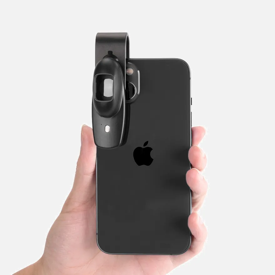
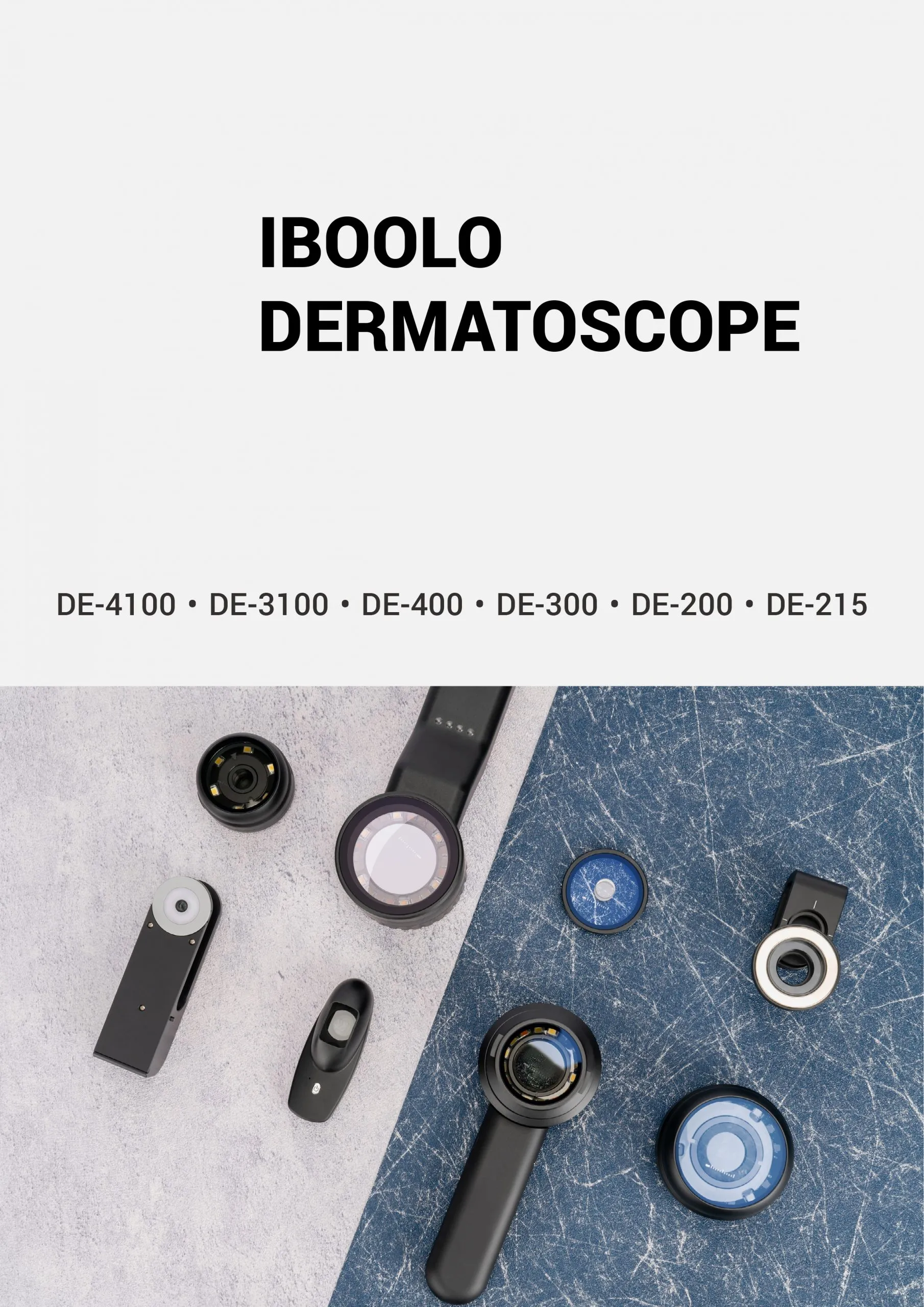
What Is Dermoscopy Meaning?

Dermoscopi are specialized medical devices for dermoscopy skin evaluations. These handheld instruments use optics and lighting to visualize subtle subsurface skin structures not visible to the naked eye.

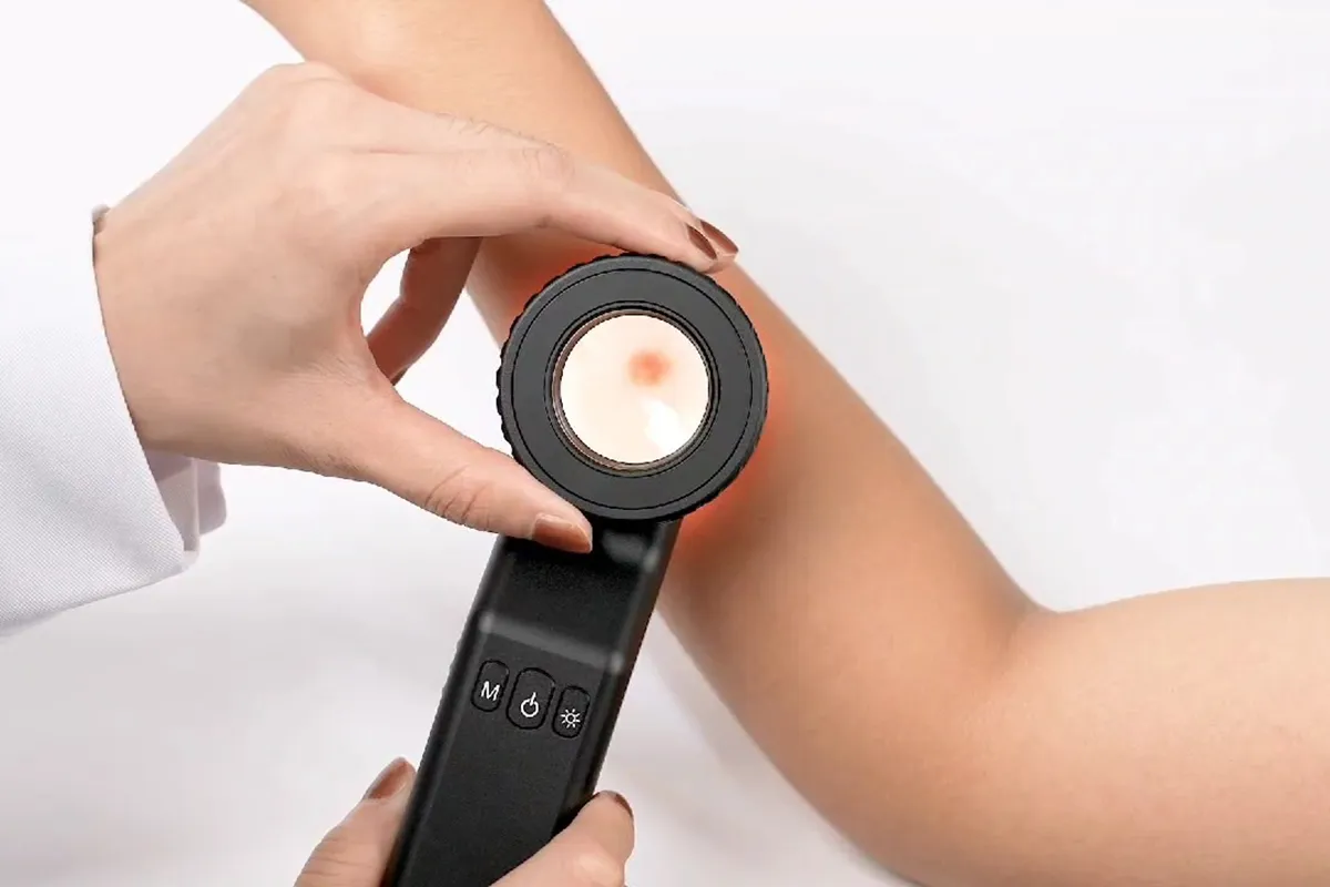
During an exam, a gel is first applied to allow improved optical transmission to the skin. The lens of the dermoscope is then held directly against the lesion while slowly scanning across the area of interest.

Inside the device, features like 10x magnification and polarized lighting transform diffuse transilluminated light from below the skin into a detailed microscopic view. Delicate morphological patterns and vascular details emerge.

This enhanced subsurface perspective facilitates expert evaluation of lesions for rapid characterization and accurate diagnosis, especially for melanoma and other skin cancers. Suspicious areas can be quickly identified.

Dermoscopi grants physicians an invaluable window beneath the skin's surface. Subtle visual clues amplify the clinician's vision dramatically through precision optics - empowering life-saving diagnoses.

What Does Dermoscopy Mean?

Dermoscopy, sometimes called dermatoscopy or chemiluminescence microscopy, is a non-invasive medical imaging technique that uses optical magnification and light to enable detailed visualization of subtle structures beneath the surface of skin lesions. It is performed using a handheld device called a dermatoscope (dermoscopy).

During a dermoscopic examination, the lens of the specialized device is placed on the skin after applying immersion fluid. Features like 10x magnification and polarized lighting transform transilluminated light from deeper tissue layers into a highly detailed subsurface perspective not visible to the naked human eye.

This enhanced in vivo microscopic perspective of the epidermis, dermo-epidermal junction, and upper dermis facilitates expert medical evaluation of skin lesions - especially important for detecting melanoma and ruling out malignant skin cancers. Suspicious subsurface morphological patterns and vessels become apparent.

Dermoscopy is an essential imaging technique that grants physicians a vital window beneath the skin's surface through precision optics. This magnified vision enables life-saving diagnoses by revealing nuanced visual indicators of disease.

People May Ask

Is A Biopsy Required for A Dermatologist to Detect Melanoma?

How a melanoma is identified. The sole reliable method for a clinician to determine whether a melanoma lesion-a suspicious skin area-is cancerous is to biopsy it. During a biopsy, a small sample of tissue is removed by the physician to be tested in a lab.

On A Dermoscopy, What Are The Red Dots?

Dotted vessels, which have a diameter of between 0.01 and 0.02 mm, are tiny red dots that show that the vessels are oriented perpendicular to the skin's surface. Dotted vessels might be observed in the skin that is inflammatory, damaged, or covers stasis. They are, nevertheless, also visible in cutaneous malignancies.

What Is Dermoscopy Known by Another Name?

A noninvasive in vivo method called dermoscopy is mainly employed to examine skin lesions [1]. Skin-surface microscopy, incident light microscopy, dermatoscopy, and epiluminescence microscopy are synonyms.

How Can Melanoma Be Verified?

A skin examination looks for birthmarks, moles, or other pigmented areas that don't seem normal in terms of size, shape, color, or texture. Your doctor may remove all or part of the abnormal skin during a biopsy, along with a tiny portion of surrounding normal tissue. Under a microscope, a pathologist examines the tissue to hunt for cancerous cells.

What Are The Signs of A Benign Or Malignant Lesion?

A benign tumor has edges that are smooth, regular, and distinct. In comparison to a benign tumor, a malignant tumor grows more quickly and has uneven borders. Moreover, a malignant tumor has the potential to spread to other bodily parts. Although a benign tumor can grow to be rather large, it won't travel to other parts of your body or infiltrate surrounding tissue.

How Can A Tumor' Malignant Status Be Determined?

A biopsy is the most accurate method of determining if a cyst or tumor is benign or malignant. In this process, a sample of the afflicted tissue-or, in certain situations, the whole suspect area-is taken, and it is examined under a microscope.

How Do Malignant Moles Appear?

Uneven color: There may be variations in tan, brown, and black tones. There may also be patches of red, pink, blue, white, or gray. Diameter: There is a size variation, typically an increase. The majority of melanomas are larger than peas, measuring more than 6 millimeters, or around 1/4 inch, although they can be much smaller.

For What Is Dermoscopy Used As A Screening Test?

Skin cancer diagnosis by dermoscopy, including melanoma and nonmelanoma, is a well-established procedure.

For What Purpose Is Dermatoscopy Used?

Using a portable instrument known as a dermatoscope, dermoscopy is a test used to examine skin lesions. Skin cancer diagnosis is most frequently aided by dermoscopy. It doesn't hurt and is non-invasive. Other names for this test include skin surface microscopy, epiluminescence microscopy, and dermatoscopy.

What Is Meant by Dermascope?

Dermoscopy is a non-invasive, in-vivo technique that has been traditionally helpful for the examination of suspected skin lesions. It is sometimes referred to as dermatoscopy, epiluminescence microscopy, or skin surface microscopy.

Dermoscopy Meaning Products

Personalities and horoscopes: The temperaments of the zodiac signs Kindle攵子书

Kindle~子书 The 2021 Horoscopes Bible: Daily horoscopes for every zodiac sign in 2021

China customized dermoscopy meaning products supply

Dermoscopy Color Atlas, First Edition, Kindle Edition

China handheld dermoscopy meaning

The Only Method to Gain Knowledge About Relationships: Synastry Methods (5) September 5, 2009, Paperback

China wholesale dermoscopy meaning products supply

Personal Growth With the Zodiac Oracle (Personal Growth Series) – Published in paperback on November 1, 2002

China affordable dermoscopy meaning products supply

Fuzzy Techniques for Dermoscopic Image Segmentation Cloth – December 13, 2012

China customized dermoscopy meaning

The socio-economic significance of PGMs, PGM preconcentration and spectroscopy, chemistry and spectroscopy, adsorption and preconcentration, and PGM separation and determination are all covered in this section.

China smartphone dermoscopy meaning products supply

Horoscopy: June 1, 2021, 200-page college lined (occultarum) paperback journal

Top Reviews

Ian Henry

The talk by Dr. Bowling is enhanced by this book (google PCDS). As an older physician, my only issue is that I have to observe a lot of dermoscoped lesions before I can get comfortable with their characteristics. So, the learning curve is high unless you are working in clinics with an expert on a regular basis and have access to histology feedback.

Mr. K. A. Boulton

A wonderful introduction to dermoscopy for novices that includes a ton of vibrant, high-quality images. It is presented with great illustrations in a very logical order. Compared to the widely available alternative, I like the organization of this book more.

RagsToRags

Review I saw was solely for the Kindle edition. took this paperback out of the medical reference section. I've been doing oscopy for almost ten years, so I would consider myself a quite competent dermoscopist. However, I try to read something new every year to stay current. I can't judge it as someone who is just learning dermoscopy, but it seems to provide a decent summary. This basic manual is unique in that it covers trichoscopy in addition to melanocytic lesions. I have better pictures of scabies. A few of the photos are not the best. 4.8 out of 5.0 overall.

0

Have questions on gear or your order?

Our Gear Guides are here to help! Get personal advice from pro creatives

Name
Subject
Email address
How can we help?

Instant Answers

How Can Dermoscopy Images Be Captured?

Dermoscopy images can be captured and stored in different ways, such as: • Using a smartphone or tablet with dermoscopic adapter, which consisted in the package.• Using a digital camera

Dermoscopy images can be captured and stored in different ways, such as:

• Using a smartphone or tablet with dermoscopic adapter, which consisted in the package.
• Using a digital camera with dermoscopic adapter, there’s 49mm screw size camera adapter available to order now.

Compatible phone/tablet models:
All iPhone models, 95% Android phones, 90% tablet. For phone/tablet size in 5.25-14mm

Compatible camera models:
All camera with built 49mm filter screw, such as Canon EOS 70D, 80D, 90D; Canon EOS R7, R10, R50, R100; Canon M100, M200, M50, Mark II; Canon G7X Mark III, Sony ZV-1

How Can I Connect My Phone to My Dermatoscope?

There’s universal phone adapter for all our dermoscopes. Please check the installation procedure bellow or watch operation guide. Smartphone Connector (1) Place phone adapter screw in the center of smartphone’s

There’s universal phone adapter for all our dermoscopes. Please check the installation procedure bellow or watch operation guide.

Smartphone Connector

(1) Place phone adapter screw in the center of smartphone’s main camera.
(2) Screw magnet attachment on phone adapter.
(3) Put dermoscope’s back ring and magnet attachment together

Take The Best Images

You need to adjust the focus ring after the dermoscpe connected on smartphone to get the best images.

How Can I Clean My Dermoscopy after Usage?

Cleaning your dermoscopy after usage is important to prevent cross-contamination and infection. The cleaning method may vary depending on the type and model of your dermoscopy, so you should always

Cleaning your dermoscopy after usage is important to prevent cross-contamination and infection. The cleaning method may vary depending on the type and model of your dermoscopy, so you should always follow the manufacturer’s instructions. However, some general steps are:

• Turn off and disconnect your dermoscopy from any power source or device.

• Wipe off any visible dirt or debris from the dermoscopy with a soft cloth or tissue.

• Disinfect the dermoscopy with an alcohol-based wipe or spray, or a disinfectant solution recommended by the manufacturer. Make sure to cover all surfaces, especially the lens and contact plate.

• Let the dermoscopy air dry completely before storing it in a clean and dry place.

• Do not use abrasive or corrosive cleaners, solvents, or detergents that may damage the dermoscopy.

• Do not immerse the dermoscopy in water or any liquid, unless it is waterproof and designed for immersion.

You should clean your dermoscopy after each use, or at least once a day if you use it frequently. You should also check your dermoscopy regularly for any signs of damage or malfunction, and contact the manufacturer or service provider if needed.

Polarized VS Non-Polarized Dermoscopy

A dermoscopy is a device that allows the examination of skin lesions with magnificationand illumination. By revealing subsurface structures and patterns that are not visible tothe naked eye. It can

A dermoscopy is a device that allows the examination of skin lesions with magnificationand illumination. By revealing subsurface structures and patterns that are not visible tothe naked eye. It can improve the diagnose accuracy of skin lesions, such as melanoma,basal cell carcinoma, seborrheic keratosis, etc.

There are two main types of dermoscopy: Non polarized and polarized dermoscopy.We’ve fitted most of our dermoscopys with polarized and non-polarized light. They canbe used in multiple skin structures.

Non-polarized contact Mode

In non-polarized mode, the instrument can provide information about the superficialskin structures, such as milia-like cysts, comedo-like openings, and pigment in theepidemis.

The dermoscopy requires applying a liquid such as mineral oil or alcohol to the skin andplacing the lens in contact with the skin. This reduces surface reflection and enhancesthe view of subsurface structures.

Image with non-polarized light (DE-3100)

Polarized contact Mode

In polarized mode, the instrument allows for visualization for deeper skin structures,such as blood vessels, collagen, and pigment in the dermis.

The dermoscopy does not need to be in contact with the skin or use any liquid. Theirpolarized light can help to eliminate surface reflection and allow visualization ofvascular structures.

Image with polarized light (DE-3100)

Polarized non-contact Mode

The dermoscopy can also use polarized light to examine the skin without direct contact.

In polarized non-contact mode, the instrument allows for examination infected areasand lesions that are painful for the patient, or the difficult to contact pigmented lesions,such as nails and narrow areas.

The contact plate should be removed in this mode, and it does not require applying aliquid to the skin. As it doesn’t require pressure or fluid application on the skin, it canalso avoid cross-contamination and infection risk.

Image in polarized non-contact mode (DE-3100)

How Effectiveness Is Dermoscopy

Compared with visual inspection, the dermoscopy can be used to capture and store skin lesion photos, which play an important role in early skin cancer examination. The dermoscopy allows the

Compared with visual inspection, the dermoscopy can be used to capture and store skin lesion photos, which play an important role in early skin cancer examination.

The dermoscopy allows the examination of skin lesions with magnification and illumination. This can be greatly avoiding the factors that cause interference to visual detection. Such as lighting, skin color, hair and cosmetics.

Several studies have demonstrated that dermoscopy is useful in the identification of melanoma, when used by a trained professional.

It may improve the accuracy of clinical diagnosis by up to 35%
It may reduce the number of harmless lesions that are removed
In primary care, it may increase the referral of more worrisome lesions and reduce the referral of more trivial ones

A 2018 Cochrane meta-analysis published the accuracy of dermoscopy in the detection.

Table 1. Accuracy of dermoscopy in the detection of melanoma in adults
Detection Method Sensitivity, % Specificity, % Positive Likelihood Ratio NegativeLikelihood Ratio
Visual inspection alone (in person) 76 75 3.04 0.32
Dermoscopy with visual inspection (in person) 92 95 18 0.08
Image-based visual inspection alone (not in person) 47 42 0.81 1.3
Dermoscopy with image-based visual inspection (not in person) 81 82 4.5 0.23
ROC—receiver operating characteristic. *Estimated sensitivity calculated on the summary ROC curve at a fixed specificity of 80%.

As we can see, the dermoscope can improve the accuracy of diagnosis of skin lesions, especially melanoma.

Table 1. Accuracy of dermoscopy in the detection of melanoma in adults
Detection Method Sensitivity, % Specificity, % Positive Likelihood Ratio NegativeLikelihood Ratio
Visual inspection alone (in person) 79 77 3.4 0.27
Dermoscopy with visual inspection (in person) 93 99 93 0.07
Image-based visual inspection alone (not in person) 85 87 6.5 0.17
Dermoscopy with image-based visual inspection (not in person) 93 96 23 0.07
ROC—receiver operating characteristic. *Estimated sensitivity calculated on the summary ROC curve at a fixed specificity of 80%.

Characteristics of the dermatoscopic structure of the skin lesions include:

• Symmetry or asymmetry
• Homogeny/uniformity (sameness) or heterogeny (structural differences across the lesion)
• Distribution of pigment: brown lines, dots, clods and structureless areas
• Skin surface keratin: small white cysts, crypts, fissures
• Vascular morphology and pattern: regular or irregular
• Border of the lesion: fading, sharply cut off or radial streaks
• Presence of ulceration

There are specific dermoscopic patterns that aid in the diagnosis of the following pigmented skin lesions:

• Melanoma
• Moles (benign melanocytic naevus)
• Freckles (lentigos)
• Atypical naevi
• Blue naevi
• Seborrhoeic keratosis
• Pigmented basal cell carcinoma
• Haemangioma

No products in the cart.

No products in the cart.

Hot Search Terms