Purchasing A Dermatoscope - Key Wholesalers And Selection Criteria

Reputable retailers to purchase dermatoscopes from include:

  • DermLite - An authorized distributor of Dermlite branded dermatoscopes
  • Delasco - Sells the DermLite DL100 model for $395 with medical license requirement
  • Mega Depot - Offers dermatoscopes at budget-friendly pricing
  • Hospital Store - Sells a variety of dermatoscopy models at wholesale prices

When selecting a specific dermatoscopy, key aspects to evaluate include:

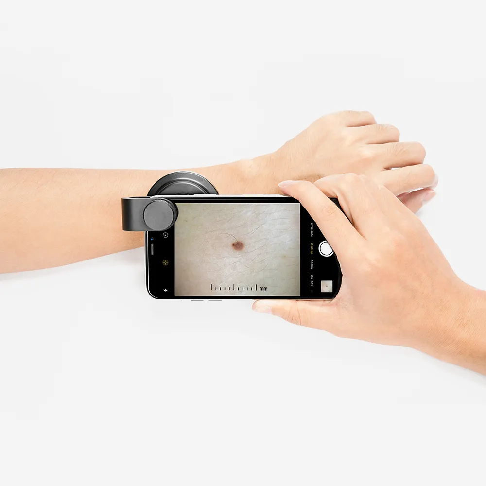
  • Smartphone/Camera Compatibility - For capturing images to track skin changes
  • Polarization Type - polarized lighting improves lesion visibility
  • Contact plate and Non-Contact Viewing Modes - Enable both close and wide examinations
  • Magnification power - 10x is standard; higher magnification identifies finer details
  • Budget - Contact manufacturers to try models; choose based on personal needs

Additional tips when buying a dermatoscopy:

  • Consider getting a camera to record findings visually
  • Explore features and pricing to find the optimal match for your practice
  • Try different models over time to determine the best fit

Carefully identifying the desired visualization, documentation, and pricing factors facilitates selecting a high-quality dermatoscopy suited for precise skin evaluations.

How Does A Dermatoscope Buy?

Here are some tips for buying a dermatoscopy:

Contact manufacturers, Try out different dermatoscopes over time and explore what suits you and your budget

Consider your budget; consider the cost of the dermatoscopy, as well as the quality of the images and the magnification

Consider other equipment; consider using fluid, alcohol hand gel, or streets, except around the eyes and nails

Consider compatibility, Consider compatibility with smartphones and cameras

Consider polarization; consider whether the dermatoscopy is polarized, linear-polarized, unpolarized, or variable

Consider viewing; consider whether the dermatoscopy offers contact or non-contact viewing, or both

Consider portability, Consider the dermatoscopic portability and ease of use

Consider mode, Consider whether the dermatoscopy has both non-polarizer and polarizer mode

Other things to consider when buying a dermatoscopy include:

  • Compatibility with smartphones and cameras
  • polarization
  • Contact or non-contact viewing
  • Magnification
  • Quality of images

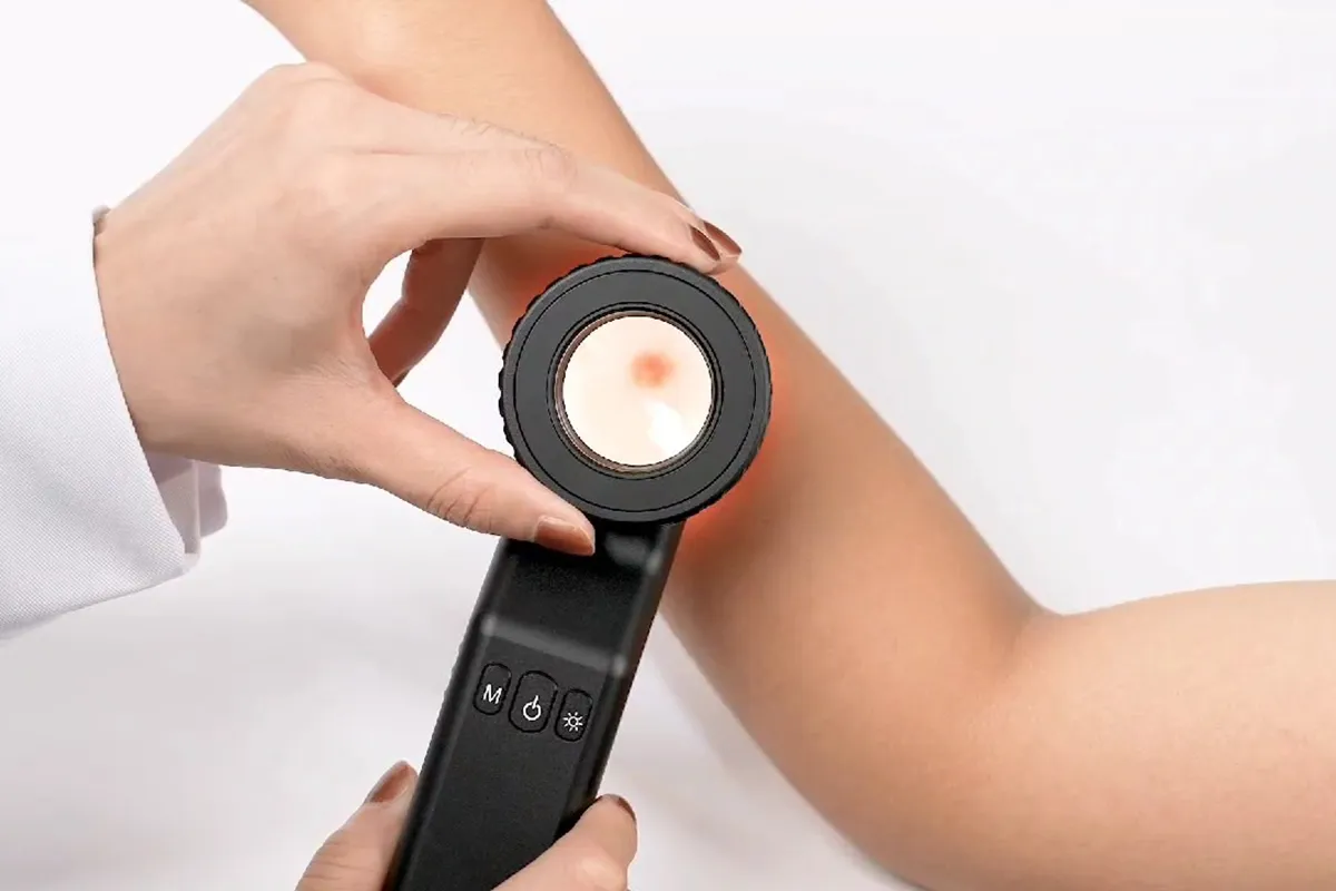
A dermatoscope is a handheld instrument that allows users to visualize subsurface skin structures in the epidermis, at the dermoepidermal junction, and in the papillary dermis.

People May Ask

In Dermoscopy, What Is The Starburst Pattern?

The characteristic dermoscopic signature of Spitz/Reed nevus is the [starburst" pattern. Confluent junctional melanocytic nests and histological radial growth are represented by pigmented, strongly concentrated streaks that are radially distributed at the periphery of a lesion.

Are Pigmented Lesions Harmful?

Darker-than-the-rest of your skin tone moles may be cause for concern. Moles, or nevi, become darker due to skin pigmentation, which also affects them. Certain extremely black moles may represent melanoma, a kind of skin cancer.

Raised Moles: Are They Harmful?

Most moles are benign. They could get wrinkled, raised, or covered in hair. If a mole changes in size or color, or if you experience discomfort, bleeding, inflammation, or itching, consult your doctor.

What Is The Appearance of A Malignant Mole?

Uneven color: There may be variations in tan, brown, and black tones. There may also be patches of red, pink, blue, white, or gray. Diameter: There is a size variation, typically an increase. The majority of melanomas are larger than peas, measuring more than 6 millimeters, or around 1/4 inch, although they can be much smaller.

On A Dermoscopy, What Are The Brown Dots?

The site is identified by the color: blue or grey dots are caused by melanophages in the dermis, brown globules are caused by junctional nests of melanocytes, and black dots are caused by free melanin in the stratum corneum.

What Do Melanoma's GREY Dots Represent?

Structures that are grey or blueMelanin in the dermis-found in melanophages or neoplastic cells-causes grey or blue lines, rings, clods, or spots. Dermal melanin indicates cancer in tumors exhibiting disarray. Melanoma, pBCC22, or pIEC may all have grey or blue structures.

How Frequently Are Melanoma Cases Overdiagnosed?

It's interesting to note that a recent study showed that 68% of 115 dermatopathologists concurred that overdiagnosis for atypical nevi, 47% for melanoma in situ, and 35% for invasive melanoma is a public health concern; dermatopathologists with longer practice histories, however, were significantly less likely to believe that...

How Frequently Do Dermatologists Overlook Melanoma?

Reversing these data, a physician would find that for every 50 individuals they assess without performing a complete physical examination, 1 skin cancer is missing and for every 400 patients, 1 melanoma is missed.

What Makes A Dermatoscope Different from A Dermoscope?

A noninvasive in vivo method called dermoscopy is mainly employed to examine skin lesions [1]. Skin-surface microscopy, incident light microscopy, dermatoscopy, and epiluminescence microscopy are synonyms. A handheld device known as a dermatoscope is used to perform dermoscopy.

Can Melanoma Be Found Using A Dermoscope?

When melanoma is diagnosed by professionals, dermoscopy is a more accurate method than examining a worrisome skin lesion with the unaided eye. Interpreting dermoscopy with the patient in person yields better results than using dermoscopy images alone.

Dermatoscope Buy Products

China $ dermatoscope buy

Carson MicroBrite Plus LED Illuminated 60x-120x Compact Microscope (MM-300)

dermatoscope buy $

The APEXEL is a handheld digital microscope featuring a 2 LCD screen, providing 800x magnification and coin-style illumination. It can be connected to a PC via USB and supports SD cards.

China $ dermatoscope buy $

3Gen DermLite IV DL4 PigmentBoost Plus Polarized Dermoscope

3-Gen Handyscope Mobile Device DermLite Dermatoscope

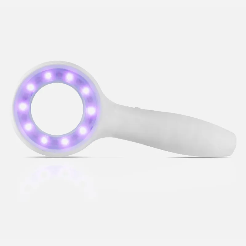
Polarized Skin Dermatology with Pocket Epiluminescence Dermascope with Rechargeable Light

Dermatology, trichology, skin care, and telehealth are all served by the Firefly DE300 digital dermotscope. Accredited by the FDA and the C.E.

Skin Diagnostic Dermatoscope Set Dermal Instruments Dermascope Dermatology

Ten completely completed folding mirror paper microscopes are included in the classroom assembly kit for folding mirrors.

Carson eFlex 75x/300x Effective Magnification (Based on 21 Monitor) Flexible Stand and Base LED Illuminated USB Digital Microscope (MM-840), White

The MW10084 pocket microscope is equipped with an LED light that can be adjusted, a focus wheel, a flashlight, and a microscope mode. It has a 30x powerful magnification.

Top Reviews

Billy

Performs as planned, has good quality, and a clear image. I wanted to let you know that: a) the built-in light is really bright, but it's an excellent method to use up the remaining energy in nearly dead batteries; b) the 60X magnification is too great to view whole tiny insects, but it isn't stated as much in the advertisement. I read the reviews on the camera adapter before purchasing it.

Jessica

We have all been captivated for hours by this small handheld microscope, which was purchased for my six-year-old. Get this! Honestly, ideal for all ages as a Christmas stuffer

PG

lovely small pocket microscope. The led light only requires one AA battery, so I'm assuming it will last for a long time—unless you simply keep it on. Both the zooming in and focusing are smooth and effortless. It doesn't zoom in very far, so don't expect to see moving cells or anything, but it will bring even the tiniest dust mite up close and personal. It seems well-built and the price was reasonable. I'd definitely buy this for both of my granddaughters if I were to buy it again.

0

Have questions on gear or your order?

Our Gear Guides are here to help! Get personal advice from pro creatives

Name
Subject
Email address
How can we help?

Instant Answers

How Can Dermoscopy Images Be Captured?

Dermoscopy images can be captured and stored in different ways, such as: • Using a smartphone or tablet with dermoscopic adapter, which consisted in the package.• Using a digital camera

Dermoscopy images can be captured and stored in different ways, such as:

• Using a smartphone or tablet with dermoscopic adapter, which consisted in the package.
• Using a digital camera with dermoscopic adapter, there’s 49mm screw size camera adapter available to order now.

Compatible phone/tablet models:
All iPhone models, 95% Android phones, 90% tablet. For phone/tablet size in 5.25-14mm

Compatible camera models:
All camera with built 49mm filter screw, such as Canon EOS 70D, 80D, 90D; Canon EOS R7, R10, R50, R100; Canon M100, M200, M50, Mark II; Canon G7X Mark III, Sony ZV-1

How Can I Connect My Phone to My Dermatoscope?

There’s universal phone adapter for all our dermoscopes. Please check the installation procedure bellow or watch operation guide. Smartphone Connector (1) Place phone adapter screw in the center of smartphone’s

There’s universal phone adapter for all our dermoscopes. Please check the installation procedure bellow or watch operation guide.

Smartphone Connector

(1) Place phone adapter screw in the center of smartphone’s main camera.
(2) Screw magnet attachment on phone adapter.
(3) Put dermoscope’s back ring and magnet attachment together

Take The Best Images

You need to adjust the focus ring after the dermoscpe connected on smartphone to get the best images.

How Can I Clean My Dermoscopy after Usage?

Cleaning your dermoscopy after usage is important to prevent cross-contamination and infection. The cleaning method may vary depending on the type and model of your dermoscopy, so you should always

Cleaning your dermoscopy after usage is important to prevent cross-contamination and infection. The cleaning method may vary depending on the type and model of your dermoscopy, so you should always follow the manufacturer’s instructions. However, some general steps are:

• Turn off and disconnect your dermoscopy from any power source or device.

• Wipe off any visible dirt or debris from the dermoscopy with a soft cloth or tissue.

• Disinfect the dermoscopy with an alcohol-based wipe or spray, or a disinfectant solution recommended by the manufacturer. Make sure to cover all surfaces, especially the lens and contact plate.

• Let the dermoscopy air dry completely before storing it in a clean and dry place.

• Do not use abrasive or corrosive cleaners, solvents, or detergents that may damage the dermoscopy.

• Do not immerse the dermoscopy in water or any liquid, unless it is waterproof and designed for immersion.

You should clean your dermoscopy after each use, or at least once a day if you use it frequently. You should also check your dermoscopy regularly for any signs of damage or malfunction, and contact the manufacturer or service provider if needed.

Polarized VS Non-Polarized Dermoscopy

A dermoscopy is a device that allows the examination of skin lesions with magnificationand illumination. By revealing subsurface structures and patterns that are not visible tothe naked eye. It can

A dermoscopy is a device that allows the examination of skin lesions with magnificationand illumination. By revealing subsurface structures and patterns that are not visible tothe naked eye. It can improve the diagnose accuracy of skin lesions, such as melanoma,basal cell carcinoma, seborrheic keratosis, etc.

There are two main types of dermoscopy: Non polarized and polarized dermoscopy.We’ve fitted most of our dermoscopys with polarized and non-polarized light. They canbe used in multiple skin structures.

Non-polarized contact Mode

In non-polarized mode, the instrument can provide information about the superficialskin structures, such as milia-like cysts, comedo-like openings, and pigment in theepidemis.

The dermoscopy requires applying a liquid such as mineral oil or alcohol to the skin andplacing the lens in contact with the skin. This reduces surface reflection and enhancesthe view of subsurface structures.

Image with non-polarized light (DE-3100)

Polarized contact Mode

In polarized mode, the instrument allows for visualization for deeper skin structures,such as blood vessels, collagen, and pigment in the dermis.

The dermoscopy does not need to be in contact with the skin or use any liquid. Theirpolarized light can help to eliminate surface reflection and allow visualization ofvascular structures.

Image with polarized light (DE-3100)

Polarized non-contact Mode

The dermoscopy can also use polarized light to examine the skin without direct contact.

In polarized non-contact mode, the instrument allows for examination infected areasand lesions that are painful for the patient, or the difficult to contact pigmented lesions,such as nails and narrow areas.

The contact plate should be removed in this mode, and it does not require applying aliquid to the skin. As it doesn’t require pressure or fluid application on the skin, it canalso avoid cross-contamination and infection risk.

Image in polarized non-contact mode (DE-3100)

How Effectiveness Is Dermoscopy

Compared with visual inspection, the dermoscopy can be used to capture and store skin lesion photos, which play an important role in early skin cancer examination. The dermoscopy allows the

Compared with visual inspection, the dermoscopy can be used to capture and store skin lesion photos, which play an important role in early skin cancer examination.

The dermoscopy allows the examination of skin lesions with magnification and illumination. This can be greatly avoiding the factors that cause interference to visual detection. Such as lighting, skin color, hair and cosmetics.

Several studies have demonstrated that dermoscopy is useful in the identification of melanoma, when used by a trained professional.

It may improve the accuracy of clinical diagnosis by up to 35%
It may reduce the number of harmless lesions that are removed
In primary care, it may increase the referral of more worrisome lesions and reduce the referral of more trivial ones

A 2018 Cochrane meta-analysis published the accuracy of dermoscopy in the detection.

Table 1. Accuracy of dermoscopy in the detection of melanoma in adults
Detection Method Sensitivity, % Specificity, % Positive Likelihood Ratio NegativeLikelihood Ratio
Visual inspection alone (in person) 76 75 3.04 0.32
Dermoscopy with visual inspection (in person) 92 95 18 0.08
Image-based visual inspection alone (not in person) 47 42 0.81 1.3
Dermoscopy with image-based visual inspection (not in person) 81 82 4.5 0.23
ROC—receiver operating characteristic. *Estimated sensitivity calculated on the summary ROC curve at a fixed specificity of 80%.

As we can see, the dermoscope can improve the accuracy of diagnosis of skin lesions, especially melanoma.

Table 1. Accuracy of dermoscopy in the detection of melanoma in adults
Detection Method Sensitivity, % Specificity, % Positive Likelihood Ratio NegativeLikelihood Ratio
Visual inspection alone (in person) 79 77 3.4 0.27
Dermoscopy with visual inspection (in person) 93 99 93 0.07
Image-based visual inspection alone (not in person) 85 87 6.5 0.17
Dermoscopy with image-based visual inspection (not in person) 93 96 23 0.07
ROC—receiver operating characteristic. *Estimated sensitivity calculated on the summary ROC curve at a fixed specificity of 80%.

Characteristics of the dermatoscopic structure of the skin lesions include:

• Symmetry or asymmetry
• Homogeny/uniformity (sameness) or heterogeny (structural differences across the lesion)
• Distribution of pigment: brown lines, dots, clods and structureless areas
• Skin surface keratin: small white cysts, crypts, fissures
• Vascular morphology and pattern: regular or irregular
• Border of the lesion: fading, sharply cut off or radial streaks
• Presence of ulceration

There are specific dermoscopic patterns that aid in the diagnosis of the following pigmented skin lesions:

• Melanoma
• Moles (benign melanocytic naevus)
• Freckles (lentigos)
• Atypical naevi
• Blue naevi
• Seborrhoeic keratosis
• Pigmented basal cell carcinoma
• Haemangioma

No products in the cart.

No products in the cart.

Hot Search Terms