


DE-400 Dermatoscope – IBOOLO
Dermoscopi Technology: The Future Guardian of Skin Health
In today's era of rapid technological advancement, dermoscopi technology has emerged as a revolutionary tool in skin health. From improving diagnostic accuracy to enhancing patient care, the applications of dermatoscopy are constantly expanding. This article will delve into the innovative applications of dermoscopi in various areas, including medicine, cosmetics, environmental monitoring, and mental health, and how it is poised to become the future guardian of skin health.
The Intersection of Dermatoscopy And Dermatopathology
The application of dermoscopi in dermatopathology gives pathologists a fresh perspective on observing and analyzing skin lesions. Compared to traditional visual examination, dermoscopi offers magnification up to several dozen times, revealing details beneath the skin's surface and enabling pathologists to identify lesion types more accurately. Furthermore, the use of dermoscopi in pathological research has led to breakthrough discoveries, such as improved early detection of melanoma, significantly increasing patient survival rates.
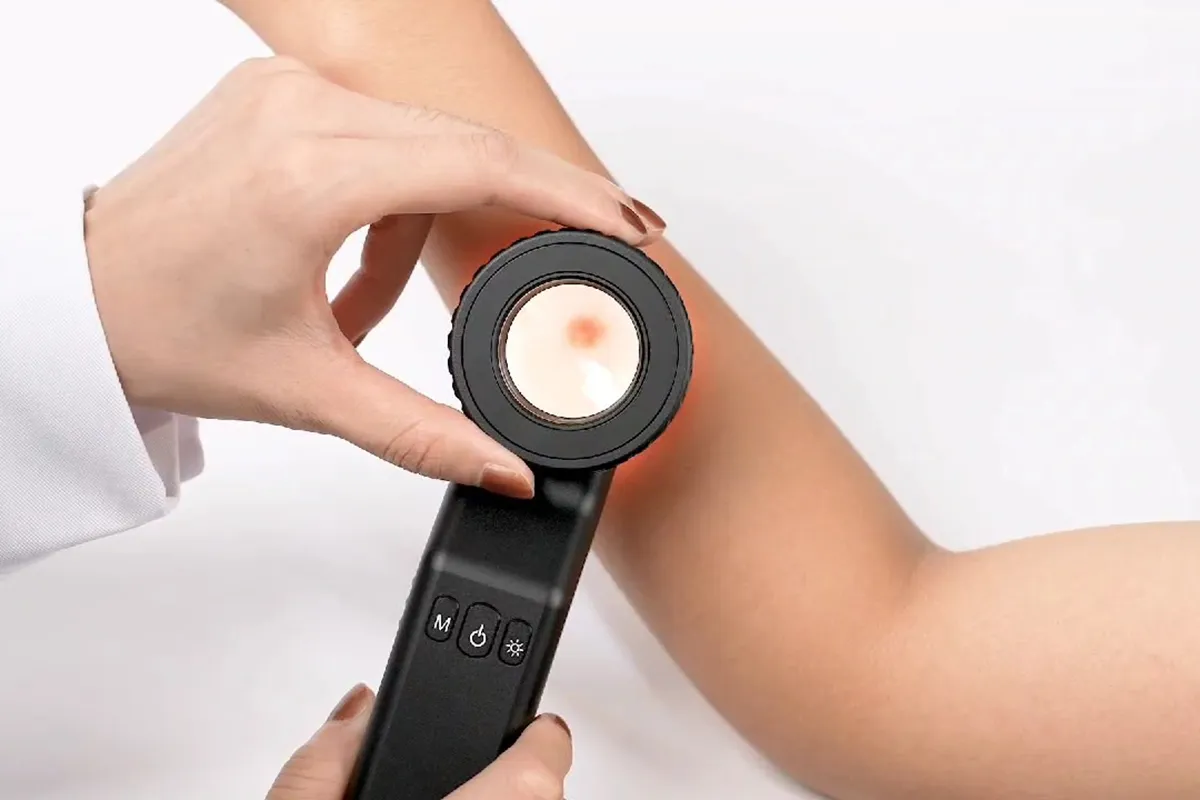
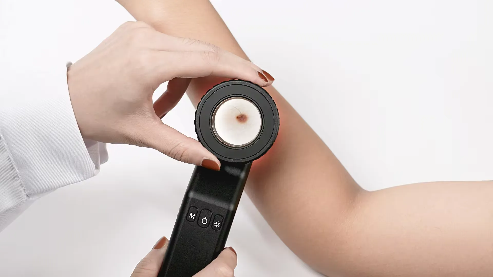
Dermoscopi for Skin Examination
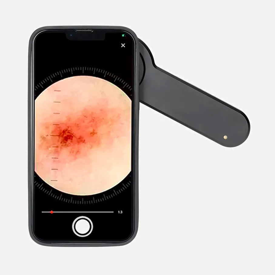
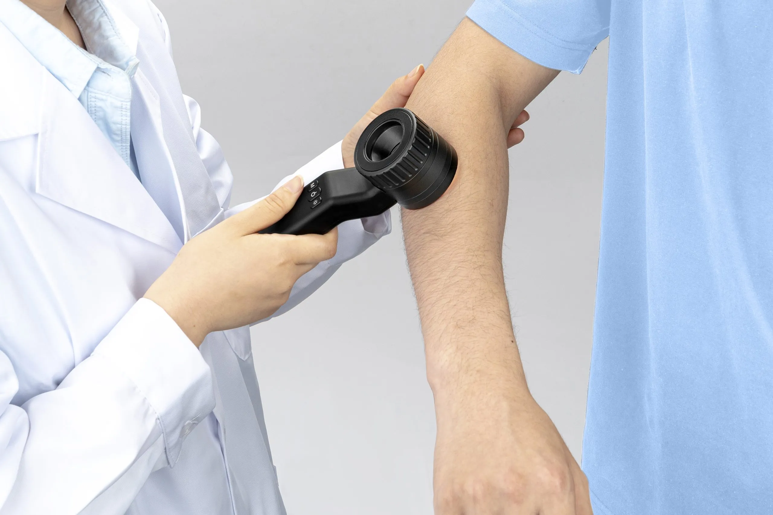
A dermatoscopy examination, also known as a chemiluminescence or skin surface microscopy examination, is a non-invasive examination using a dermatoscope to examine skin lesions. The dermoscopi is a camera-like tool that allows for the unobstructed examination of skin lesions by eliminating surface reflections. Dermatoscopy examinations can magnify skin structures and help differentiate between various types of lesions, such as melanoma, dysplastic nevi, and non-melanoma skin cancers. It can also be used to diagnose skin infections and infestations, such as cutaneous larva migrans, a parasitic worm infection that affects the skin. Dermatoscopy examinations are safe and suitable for all skin types and ages. During the examination, the doctor will apply ultrasound gel or oil to the skin to improve image clarity and gently press the dermatoscope onto the skin. The examination is painless, but you may experience slight pressure.
Dermoscopi in Telemedicine
With the rise of telemedicine, dermoscopi technology has played a crucial role. Through high-quality image transmission, doctors can provide remote preliminary skin diagnoses for patients. This not only offers convenience for patients in remote areas but also alleviates the pressure on urban medical centers. However, remote dermoscopi diagnosis also faces technical challenges, such as ensuring image quality, protecting patient privacy, and data security. To address these issues, the medical industry is exploring advanced image compression techniques, encryption methods, and patient identity verification methods.
Innovative Dermoscopi Applications in The Cosmetics Industry
The cosmetics industry has a high demand for precise skin analysis, and dermoscopi technology meets this need. Through dermoscopi cosmetic professionals can observe skin conditions in depth, enabling them to provide more personalized skincare solutions for their clients. Whether assessing the extent of skin aging, identifying acne types, or monitoring changes in pigmentation, dermatoscopy offers a scientific and objective approach. As technology advances, the applications of dermoscopi in the cosmetics industry will become even more widespread.
Dermoscopi Technology And Environmental Factors
Environmental factors, such as ultraviolet radiation and air pollution, have a significant impact on skin health. Dermoscopi technology can help us observe the effects of these environmental factors on the skin more directly. For instance, through dermoscopi, doctors can monitor UV-induced skin damage and provide appropriate protection advice accordingly. Additionally, dermoscopi data can be used in environmental monitoring projects, helping scientists assess the long-term impact of environmental pollution on skin health.
Accessibility And Popularization of Dermoscopi Technology
Despite the immense potential of dermoscopi technology, its widespread adoption remains limited by various factors. To improve the accessibility of dermoscopi technology, several measures need to be taken, including reducing equipment costs, increasing awareness among doctors and patients, and implementing supportive policies. Community health programs can integrate dermoscopi technology to provide regular skin health screenings for residents. policymakers can encourage the adoption of dermoscopi technology in medical institutions through funding support, tax incentives, and other measures.
The development and application of dermoscopi technology offer us a fresh perspective on understanding and protecting skin health. From medical diagnosis to cosmetic care, from environmental monitoring to mental health, the potential applications of dermatoscopy are boundless. As technology continues to advance and innovate, we have reason to believe that dermoscopi will become the future guardian of skin health.
People May Ask
The pigmented lesion must lack pattern symmetry and color uniformity in addition to at least one of the following characteristics in order to be diagnosed as melanoma: numerous brown dots, pseudopods, radial streaming, scar-like depigmentation, peripheral block spots/globules, five to six colors, a blue-white veil,...
It is not able to confirm for you that you have it. A biopsy test is the sole method available for diagnosing the illness.
When melanoma is diagnosed by professionals, dermoscopy is a more accurate method than examining a worrisome skin lesion with the unaided eye. Interpreting dermoscopy with the patient in person yields better results than using dermoscopy images alone.
In order to diagnose skin cancer, a skin biopsy is always necessary.A skin biopsy is what your dermatologist will do to get rid of the spot. It is imperative to have a skin biopsy. It's the sole method for determining if you have skin cancer. There is no other way to be certain.
According to reports, dermoscopy's sensitivity can vary from 60% to 100%, depending on a number of variables including the examiners' level of experience and the lesions' diagnostic complexity. Dermoscopy can help diagnose melanoma more accurately, but it cannot take the place of a histopathologic examination.
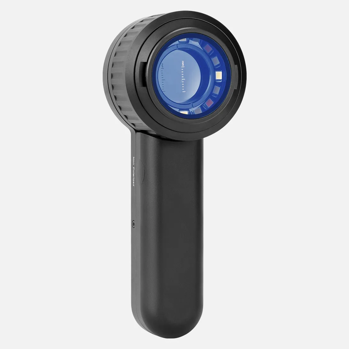
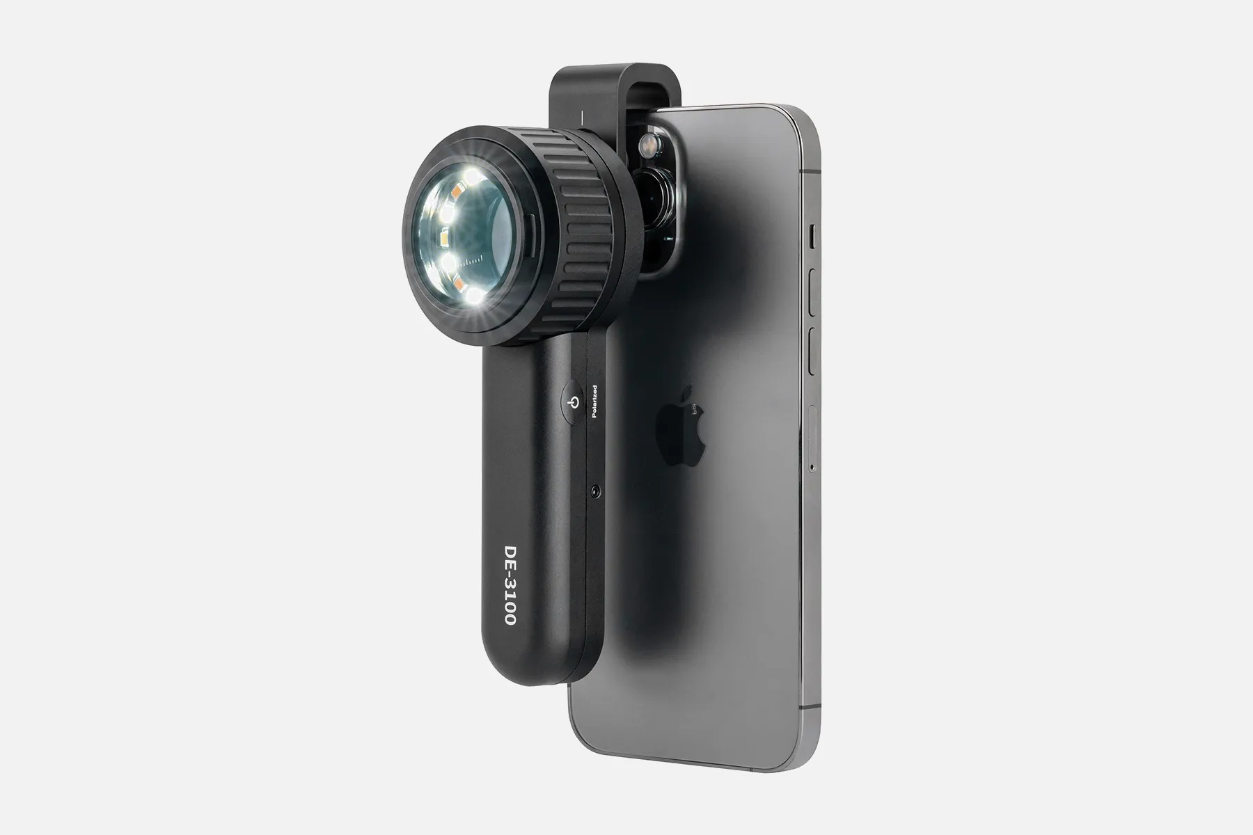
The quality, construction, and features of dermatoscopes vary, as does their price. Dermatoscopes are produced and supplied by numerous companies.
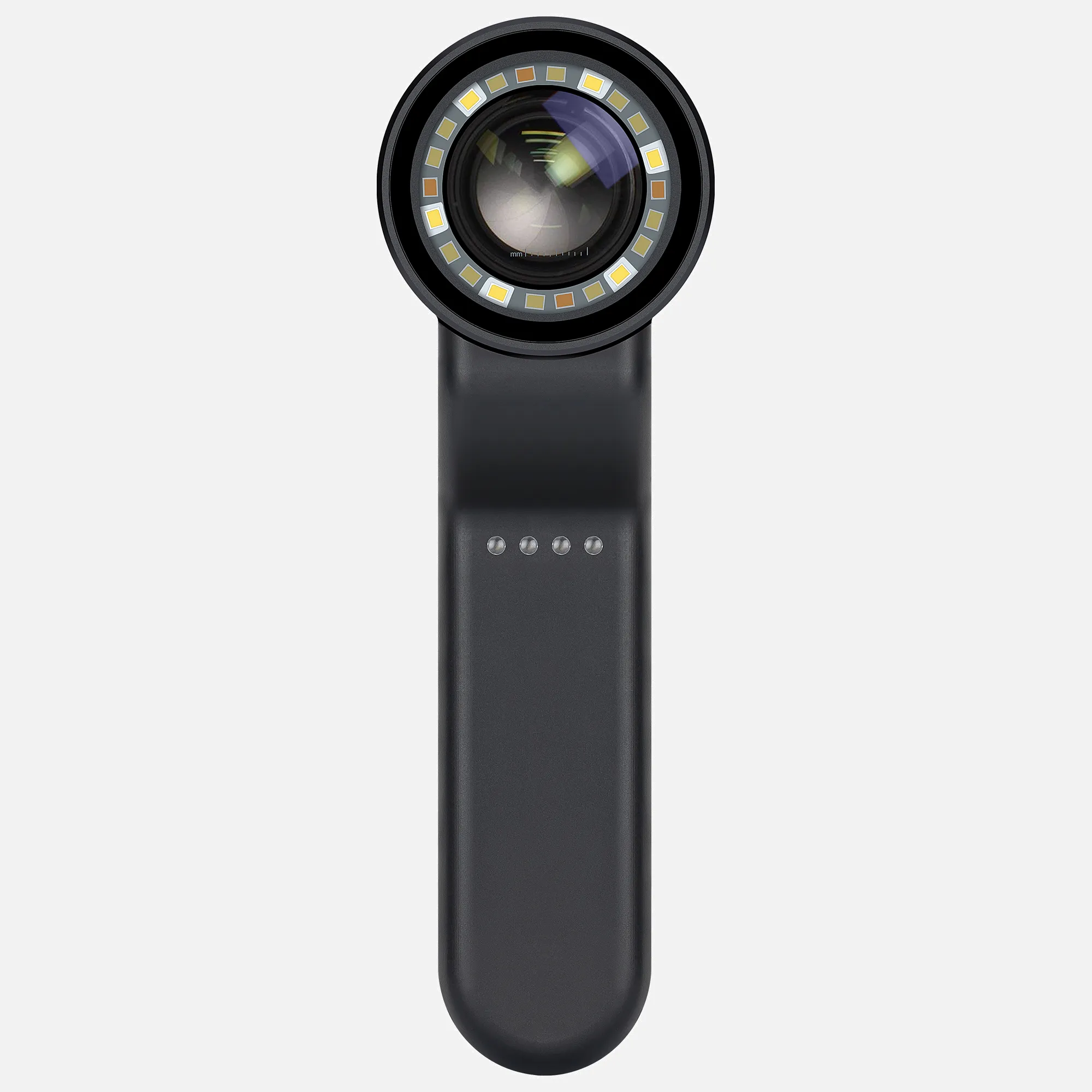
Using a portable instrument known as a dermatoscope, dermoscopy is a test performed to examine skin lesions. Skin cancer diagnosis is most frequently aided by dermoscopy. It doesn't hurt and is non-invasive. Other names for this test include skin surface microscopy, epiluminescence microscopy, and dermatoscopy.
Skin cancer diagnosis by dermoscopy, including melanoma and nonmelanoma, is a well-established procedure.
Dermoscopy, in the hands of a skilled practitioner, can be useful in detecting melanomas and verifying malignant skin tumors. This may lessen the quantity of benign lesions that are needlessly removed. Using a dermoscope helps identify melanomas more accurately than one could simply looking at them with the unaided eye.
Dermoscopy is a non-invasive, in-vivo technique that has been traditionally helpful for the examination of suspected skin lesions. It is sometimes referred to as dermatoscopy, epiluminescence microscopy, or skin surface microscopy.
Dermoscopi Products
Dermascope, 3Gen V DL5 Polarized
3Gen 偏光 Dermascope DermLite DL200 HR
DL1 3GEN 脱毛皮肤敜
Three-generation DermLite GL Polarized Light Beautifier
DERMATALOGY DERMATOSCOPE Set Gray OdontoMed 2011

The 3Gen Lumio S Dermlite 恏光皮肤皮肤皮肤科 Dermascope 灪
Third Generation DermLite Carbon Microscope Dermascope
3Gen Polarized Dermascope DermLite IV DL4W
Handheld digital dermatoscope/microscope, wifi polarization FIREFLY de350
With its original zipped pouch, the HEINE DELTA 30 Dermatoscope LED
Hot Products


News & Blog


Top Reviews
Kristen Overcash
superiority, ease of use, and mobility. It's possible to spend a lot more money on a dermatoscope, but I don't understand why. In terms of teledermatology, this device also functions nicely with my cell phone. I can now email my pathologist high-quality dermoscopic images and use them for training.
Tech Geek Reviews
small and simple to operate! Excellent for examining skin lesions in the clinic. Long-lasting battery that fits neatly in your pocket. Five points!
Aimée
Five stars: simple to use, efficient. Excellent communication with the vendor. Extremely suggested.


