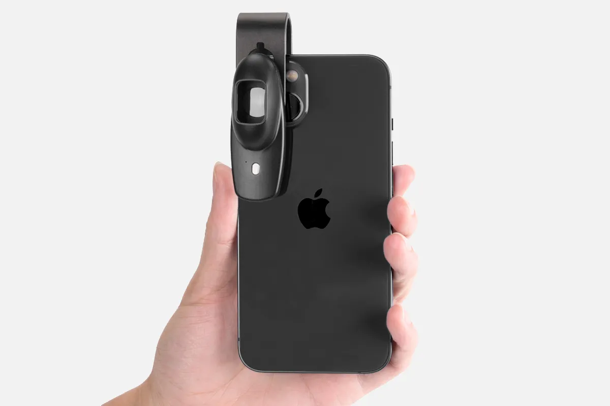
What Is A Polarized Dermatoscope?

A polarized dermatoscope is a handheld instrument that views deeper layers of skin. It uses polarized light to visualize deep skin structures without requiring a liquid interface or direct skin contact.

Dermoscopy is a non-invasive, in vivo technique to examine cutaneous lesions. It's performed with a dermatoscope, a handheld instrument containing a transilluminating light source, and a magnifying optic.

There are two main types of dermatoscopes: Handheld portable and stationary mounted.

Non-polarized dermoscopy is used to view superficial layers of skin. The use of both polarized and non-polarized methods can provide complementary information.

How Does A Polarized Dermatoscope?

Polarized dermatoscopes utilize specialized polarized lighting and filters to optimize the visualization of subsurface skin structures. Light-reflecting off the skin's surface can obscure structures beneath it. Polarized configurations block these surface reflections.

During an exam, cross-polarized lighting modes emit polarized light onto skin lesions, penetrating surface layers and scattering off deeper tissues before reflecting back. A second polarizing filter orthogonally on the returning light pathway effectively blocks distracting surface glare.

This enhanced polarization allows improved visualization of delicate morphological details and pigment networks in the epidermal, dermal-epidermal junction, and papillary dermis levels. Features like blood vessel arrangements emerge more clearly.

By reducing shine and glare, polarized dermatoscopes reveal vital subsurface clues for diagnosing melanoma and differentiating conditions like seborrheic keratosis. The technology offers superior anatomical assessment compared to standard non-polarized devices. This facilitates better clinical interpretation of lesions through dermoscopic evaluation.

Polarized configurations filter surface reflections to grant sharper magnification of the micro-universe within skin architecture - a valuable perspective for enhanced screening, inspection, and biopsy decisions.

People May Ask

What Makes Polarization Distinct from Polarized States?

Light that is not polarized can be made polarized by transformation. Waves of light that vibrate only in one plane are called polarized waves. Polarization is the process of converting unpolarized light into polarized light....
Physics Lesson: Polarizationhttp://physicsclassroom.comphysicsclassroom.com › light › Lesson-1 › P...

What Uses Does A Dermatoscope Serve?

A doctor or individual can inspect and diagnose skin lesions and disorders, including melanoma, using a dermatoscope, a hand-held visual assistance equipment. Examining the nails, hair, and scalp can also be facilitated by it. A dermatologist's practice typically has a dermatoscope.18 March 2021...
What is visible through a dermatoscope? - Medical News Today
available at medicalnewstoday.com
https://www.medicalnewstoday.com › articles › dermatos...

How Precise Is The Dermatoscope?

Compared to not using dermoscopy, the diagnosis accuracy for melanoma was significantly greater (log odds ratio 4.0 [95% CI 3.0 to 5.1] versus 2.7 [1.9 to 3.4]; an improvement of 49%, p = 0.001). The level of experience of the examiners had a substantial impact on the accuracy of the dermoscopy diagnosis.Dermoscopy's diagnostic accuracy - PubMed
nih.govhttps://pubmed.ncbi.nlm.nih.gov ..

What Is Dermoscopy's Underlying Principle?

A dermatoscope can take pictures for later comparison and evaluate features down to the reticular dermis. Transillumination of a lesion to analyze it at high magnification and see subtle details is the fundamental idea behind dermoscopy.August 8, 2023The passage continues.Overview of Dermoscopy and Its Extradiagnostic Uses - NCBINBK537131 can be found under books at nih.gov.

What Distinguishes Images That Are Polarized from Those That Are Not?

In contrast to non-polarized sunglasses, polarized lenses include a chemical coating that lowers glare. It may therefore be more challenging to see in bright light when using non-polarized sunglasses. In order to reduce glare, polarized glasses filter horizontal light waves while allowing vertical light waves to pass through the lens.April 8, 2022...
Sunglasses with or without polarization: What's the difference?Instead,www.myvision.orgPolarized vs non-polarized eyeglasses: https://myvision.org/...

Why Does Being Polarized Matter?

Sunglasses with polarized, or anti-glare, lenses help prevent eyestrain and glare from light. They thereby enhance safety and vision in the sun. Glare and reflected light can cause temporary blindness and frustration when working or playing outside.Jun. 15, 2022What Uses Do Polarized Lenses Serve? The American Academy of Ophthalmology
aao.org
https://www.aao.org › eye-health › glasses-contacts › pol...

What Separates Polarized from Non-Polarized Materials?

You will not experience glare or any of its more uncomfortable or potentially harmful consequences if you use polarized sunglasses. Comparing your eyesight with non-polarized lenses, you should be able to see more contrast and clarity. The coating that polarized sunglasses have to block glare is absent from non-polarized sunglasses.26 May 2022The Difference Between Polarized and Non-Polarized Sunglasses | Warby Parkerwww.warbyparker.comwww.warbyparker.com › education › polarized-vs-non...

How Does Polarized Imaging Work?

In addition to helping to identify undesired flaws, polarization imaging can aid to improve image contrast and provide a clearer view of the subject. Abnormalities such as surface damage and foreign object presence can be highlighted by this kind of high-speed photography....
Technoimaging.com/techimaging.com/Polarization ImagingView this page: https://techimaging.com/applications/polarization-ima...

It Is Called Polarized, But Why?

Because light moves in waves, it vibrates. Light typically vibrates in a variety of directions. However, light only vibrates in one direction-horizontally-when it hits a horizontal surface, such as snow, water, or a car roof. Polarization is the term for this procedure.Instead,Instead,Is It Better for Your Eyes to Wear Polarized Sunglasses?Keyecare at Stoney CreekWelcome to https://stoneycreekeyecare.com › What Are Polarized Sunglasses?

How Does Polarized Dermoscopy Work?

Polarized light dermoscopy, or PD, is different from nonpolarized light dermoscopy (NPD) in that it can visualize deep skin structures without requiring a liquid interface or direct skin contact with the device....
The Distinctions Between Polarized Light Dermoscopy and...The complete article can be found at JAMA Network at https://jamanetwork.com/jamadermatology.

Polarized Dermatoscope Products

China smartphone polarized dermatoscope manufacturer & factory

AMR, EDOF, WF-20 Compatible Dino-Lite USB Digital Microscope AF4915ZT - 1.3MP, 20x - 220x Optical Magnification, Measurement, Polarized Light

China portable polarized dermatoscope

Dino-Lite USB Digital Microscope AM4113ZTL - 1.3MP, Measurement, Polarized Light, 10x - 90x Optical Magnification, Long Working Distance

China affordable polarized dermatoscope manufacturer & factory

Dino-Lite AM5216ZT-720p VGA Digital Microscope with 20x - 220x Optical Magnification and Polarized Light

China smartphone polarized dermatoscope

Dino-Lite USB digital microscope AF7115MZT: Measurement, polarized light, 5MP, 10x - 220x optical magnification

China customized polarized dermatoscope manufacturer & factory

Dino-Lite AF4915ZTL USB Digital Microscope - 1.3 MP, 10x - 140x Optical Magnification, Measurement, Polarized Light, AMR, EDOF, Extended Working Range, Compatibility with WF-20

China customized polarized dermatoscope

Polarized light, long working distance, 5x to 140x optical magnification, and Dino-Lite VGA digital microscope AM5216ZTL-720p

China high quality polarized dermatoscope manufacturer & factory

Dino-Lite 1.3MP, 10x - 50x, 220x Optical Magnification, Measurement, Polarized Light USB Digital Microscope AM4113ZT

China affordable polarized dermatoscope

Dino-lite PREMIER 4116zt Pocket Digital Microscope 10 x ~ 50 x 240 x Polarizing VGA

polarized dermatoscope manufacturer & factory

Dino-Lite AM4116ZTL VGA Digital Microscope: 800 x 600 Resolution, 10x - 90x Optical Magnification, Polarized Light, Extended Working Range?

China handheld polarized dermatoscope

The 200X Microscope Mini Pocket Microscope with LED Light is a portable digital microscope accessory for smartphones. It comes in white.

Top Reviews

NAVET

These are older models and work great! We have been using them for about 4 years and bought another just because we expanded one of our operations that uses them. These are used on our production areas and I have not heard of anything negative about them. We have not had any of them fail to function or even have issues! I hope this is still the case for upcoming updates.

WDJ

excellent camera for looking at small items. used it for work to inspect medical components.

Bryan Burch

The resolution on this digital microscope is fantastic. I really appreciate that I can still zoom in without having to have the lens touching the sample.

0

Have questions on gear or your order?

Our Gear Guides are here to help! Get personal advice from pro creatives

Name
Subject
Email address
How can we help?

Instant Answers

How Can Dermoscopy Images Be Captured?

Dermoscopy images can be captured and stored in different ways, such as: • Using a smartphone or tablet with dermoscopic adapter, which consisted in the package.• Using a digital camera

Dermoscopy images can be captured and stored in different ways, such as:

• Using a smartphone or tablet with dermoscopic adapter, which consisted in the package.
• Using a digital camera with dermoscopic adapter, there’s 49mm screw size camera adapter available to order now.

Compatible phone/tablet models:
All iPhone models, 95% Android phones, 90% tablet. For phone/tablet size in 5.25-14mm

Compatible camera models:
All camera with built 49mm filter screw, such as Canon EOS 70D, 80D, 90D; Canon EOS R7, R10, R50, R100; Canon M100, M200, M50, Mark II; Canon G7X Mark III, Sony ZV-1

How Can I Connect My Phone to My Dermatoscope?

There’s universal phone adapter for all our dermoscopes. Please check the installation procedure bellow or watch operation guide. Smartphone Connector (1) Place phone adapter screw in the center of smartphone’s

There’s universal phone adapter for all our dermoscopes. Please check the installation procedure bellow or watch operation guide.

Smartphone Connector

(1) Place phone adapter screw in the center of smartphone’s main camera.
(2) Screw magnet attachment on phone adapter.
(3) Put dermoscope’s back ring and magnet attachment together

Take The Best Images

You need to adjust the focus ring after the dermoscpe connected on smartphone to get the best images.

How Can I Clean My Dermoscopy after Usage?

Cleaning your dermoscopy after usage is important to prevent cross-contamination and infection. The cleaning method may vary depending on the type and model of your dermoscopy, so you should always

Cleaning your dermoscopy after usage is important to prevent cross-contamination and infection. The cleaning method may vary depending on the type and model of your dermoscopy, so you should always follow the manufacturer’s instructions. However, some general steps are:

• Turn off and disconnect your dermoscopy from any power source or device.

• Wipe off any visible dirt or debris from the dermoscopy with a soft cloth or tissue.

• Disinfect the dermoscopy with an alcohol-based wipe or spray, or a disinfectant solution recommended by the manufacturer. Make sure to cover all surfaces, especially the lens and contact plate.

• Let the dermoscopy air dry completely before storing it in a clean and dry place.

• Do not use abrasive or corrosive cleaners, solvents, or detergents that may damage the dermoscopy.

• Do not immerse the dermoscopy in water or any liquid, unless it is waterproof and designed for immersion.

You should clean your dermoscopy after each use, or at least once a day if you use it frequently. You should also check your dermoscopy regularly for any signs of damage or malfunction, and contact the manufacturer or service provider if needed.

Polarized VS Non-Polarized Dermoscopy

A dermoscopy is a device that allows the examination of skin lesions with magnificationand illumination. By revealing subsurface structures and patterns that are not visible tothe naked eye. It can

A dermoscopy is a device that allows the examination of skin lesions with magnificationand illumination. By revealing subsurface structures and patterns that are not visible tothe naked eye. It can improve the diagnose accuracy of skin lesions, such as melanoma,basal cell carcinoma, seborrheic keratosis, etc.

There are two main types of dermoscopy: Non polarized and polarized dermoscopy.We’ve fitted most of our dermoscopys with polarized and non-polarized light. They canbe used in multiple skin structures.

Non-polarized contact Mode

In non-polarized mode, the instrument can provide information about the superficialskin structures, such as milia-like cysts, comedo-like openings, and pigment in theepidemis.

The dermoscopy requires applying a liquid such as mineral oil or alcohol to the skin andplacing the lens in contact with the skin. This reduces surface reflection and enhancesthe view of subsurface structures.

Image with non-polarized light (DE-3100)

Polarized contact Mode

In polarized mode, the instrument allows for visualization for deeper skin structures,such as blood vessels, collagen, and pigment in the dermis.

The dermoscopy does not need to be in contact with the skin or use any liquid. Theirpolarized light can help to eliminate surface reflection and allow visualization ofvascular structures.

Image with polarized light (DE-3100)

Polarized non-contact Mode

The dermoscopy can also use polarized light to examine the skin without direct contact.

In polarized non-contact mode, the instrument allows for examination infected areasand lesions that are painful for the patient, or the difficult to contact pigmented lesions,such as nails and narrow areas.

The contact plate should be removed in this mode, and it does not require applying aliquid to the skin. As it doesn’t require pressure or fluid application on the skin, it canalso avoid cross-contamination and infection risk.

Image in polarized non-contact mode (DE-3100)

How Effectiveness Is Dermoscopy

Compared with visual inspection, the dermoscopy can be used to capture and store skin lesion photos, which play an important role in early skin cancer examination. The dermoscopy allows the

Compared with visual inspection, the dermoscopy can be used to capture and store skin lesion photos, which play an important role in early skin cancer examination.

The dermoscopy allows the examination of skin lesions with magnification and illumination. This can be greatly avoiding the factors that cause interference to visual detection. Such as lighting, skin color, hair and cosmetics.

Several studies have demonstrated that dermoscopy is useful in the identification of melanoma, when used by a trained professional.

It may improve the accuracy of clinical diagnosis by up to 35%
It may reduce the number of harmless lesions that are removed
In primary care, it may increase the referral of more worrisome lesions and reduce the referral of more trivial ones

A 2018 Cochrane meta-analysis published the accuracy of dermoscopy in the detection.

Table 1. Accuracy of dermoscopy in the detection of melanoma in adults
Detection Method Sensitivity, % Specificity, % Positive Likelihood Ratio NegativeLikelihood Ratio
Visual inspection alone (in person) 76 75 3.04 0.32
Dermoscopy with visual inspection (in person) 92 95 18 0.08
Image-based visual inspection alone (not in person) 47 42 0.81 1.3
Dermoscopy with image-based visual inspection (not in person) 81 82 4.5 0.23
ROC—receiver operating characteristic. *Estimated sensitivity calculated on the summary ROC curve at a fixed specificity of 80%.

As we can see, the dermoscope can improve the accuracy of diagnosis of skin lesions, especially melanoma.

Table 1. Accuracy of dermoscopy in the detection of melanoma in adults
Detection Method Sensitivity, % Specificity, % Positive Likelihood Ratio NegativeLikelihood Ratio
Visual inspection alone (in person) 79 77 3.4 0.27
Dermoscopy with visual inspection (in person) 93 99 93 0.07
Image-based visual inspection alone (not in person) 85 87 6.5 0.17
Dermoscopy with image-based visual inspection (not in person) 93 96 23 0.07
ROC—receiver operating characteristic. *Estimated sensitivity calculated on the summary ROC curve at a fixed specificity of 80%.

Characteristics of the dermatoscopic structure of the skin lesions include:

• Symmetry or asymmetry
• Homogeny/uniformity (sameness) or heterogeny (structural differences across the lesion)
• Distribution of pigment: brown lines, dots, clods and structureless areas
• Skin surface keratin: small white cysts, crypts, fissures
• Vascular morphology and pattern: regular or irregular
• Border of the lesion: fading, sharply cut off or radial streaks
• Presence of ulceration

There are specific dermoscopic patterns that aid in the diagnosis of the following pigmented skin lesions:

• Melanoma
• Moles (benign melanocytic naevus)
• Freckles (lentigos)
• Atypical naevi
• Blue naevi
• Seborrhoeic keratosis
• Pigmented basal cell carcinoma
• Haemangioma

No products in the cart.

No products in the cart.

Hot Search Terms