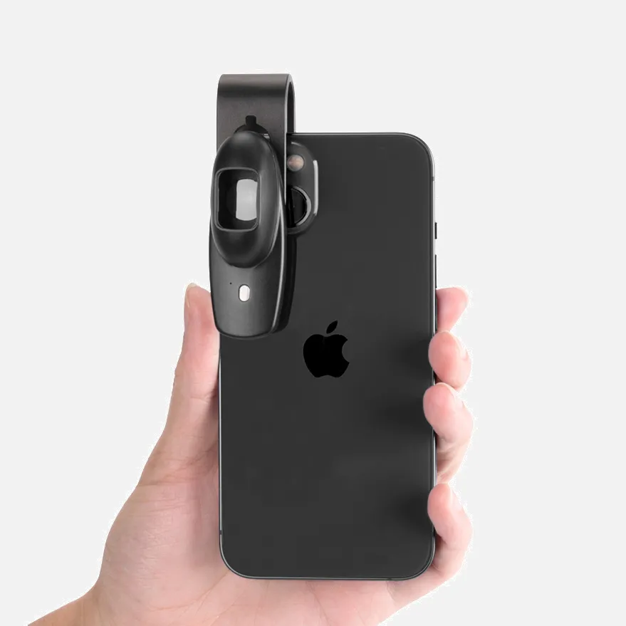
What Is A Handheld Dermatoscope?

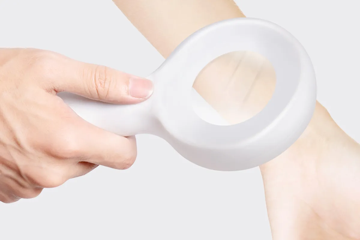
A handheld dermatoscope is a visual aid device that helps doctors and others examine and diagnose skin diseases and lesions. It's also used to inspect hair, skin, and nails for possible symptoms of underlying conditions.

A dermatoscope consists of a magnifying optic and a transilluminating light source, usually with 10-fold magnification. It works by transilluminating a lesion to study it with high magnification and visualize subtle features.

Dermoscopy is using a dermatoscope to examine the skin. It allows the visualization of subsurface skin structures in the epidermis, dermo-epidermal junction, and papillary dermis. These structures are usually not visible to the naked eye.

Explanation of How A Handheld Dermatoscope Works

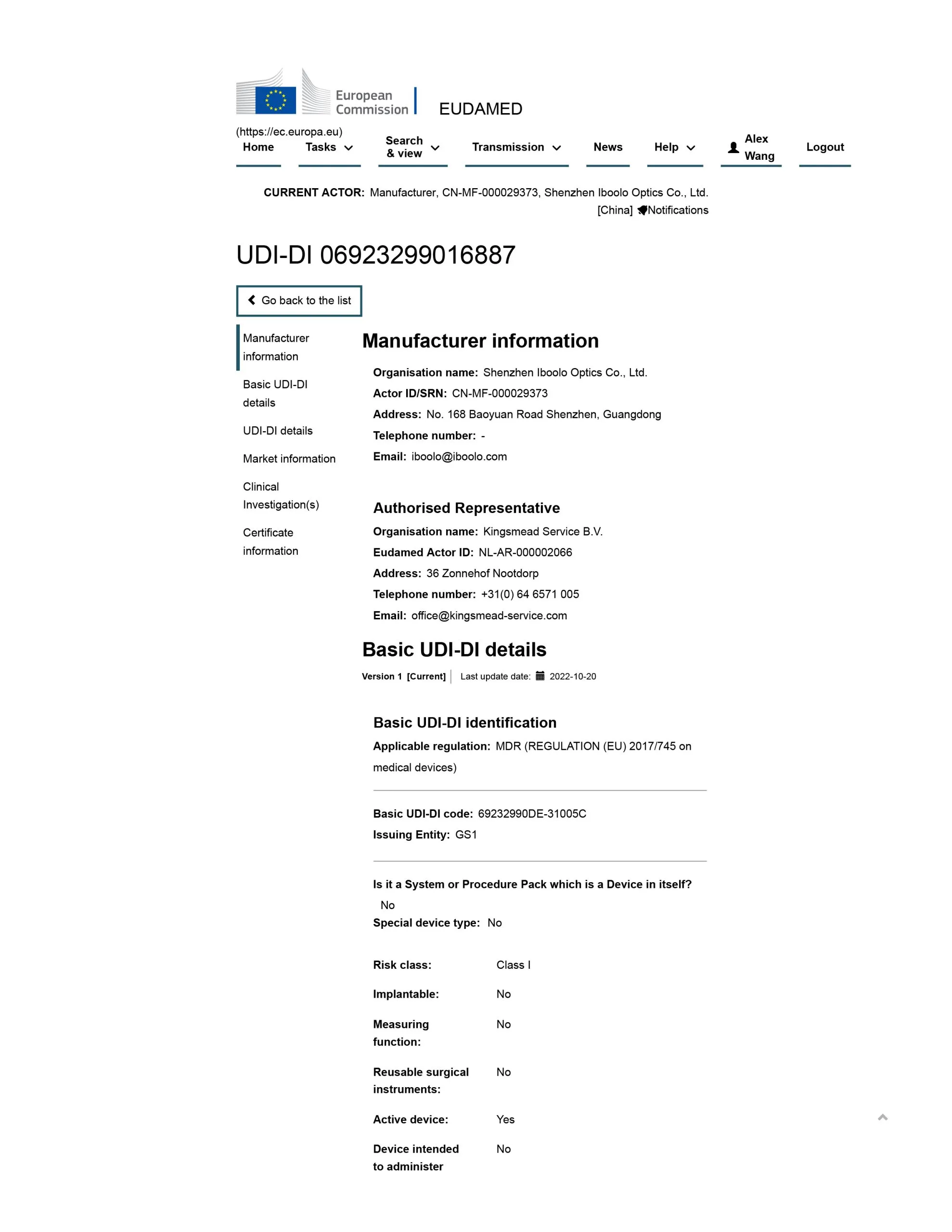
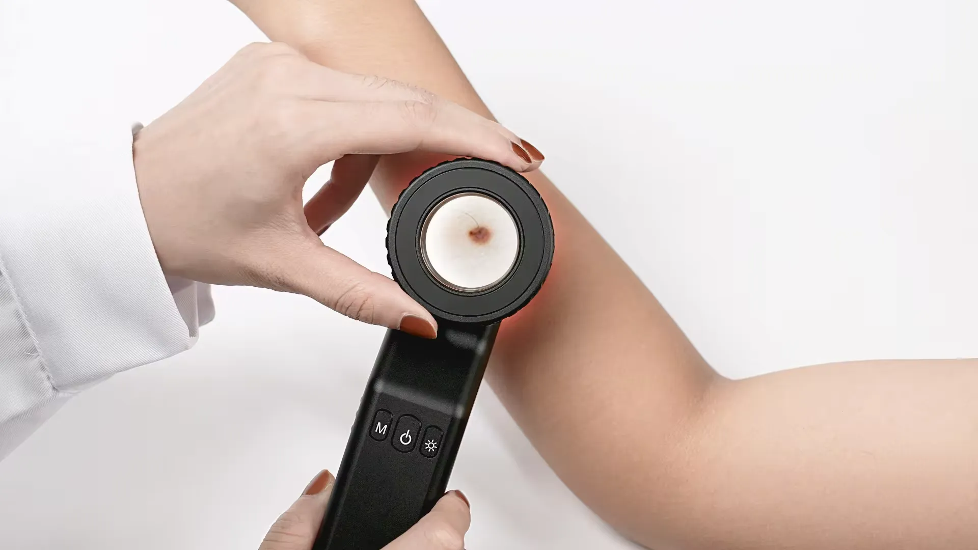
A handheld dermatoscope is a portable medical device that utilizes optical magnification and illumination to perform skin examinations through a technique called dermoscopy.

To use a handheld dermatoscope, a gel is first applied to the skin to allow improved light transmission to subsurface layers. The lens head is then held steadily over the lesion while carefully scanning. Magnification is typically set to 10x power, enabling inspection of delicate morphological patterns not visible to the naked human eye.

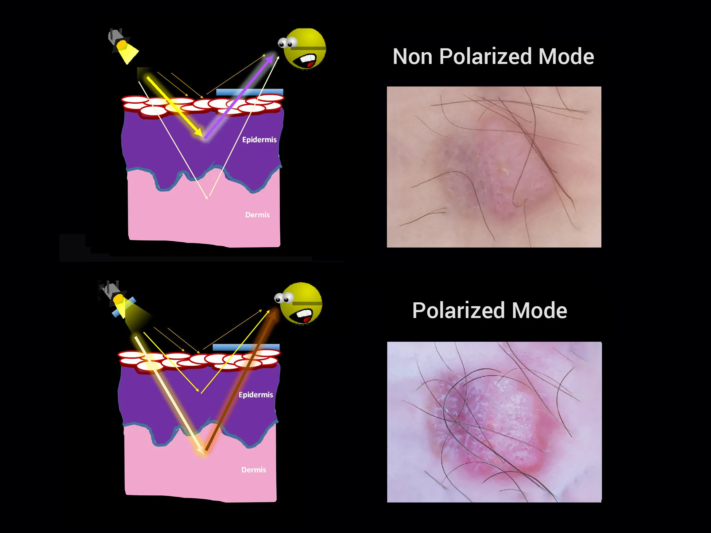
Inside the device, specialized lighting like polarized or LED modes transform diffusely backscattered light from under the skin into a highly detailed perspective of structures in the epidermal, dermal-epidermal junction, and upper dermal levels.

This enhanced visual data grants dermatologists a vital subsurface view to evaluate skin growths and determine malignancy risk. Handheld models are designed to provide customizable ergonomic utility during patient exams. Combined with expertise, they facilitate accurate screening and documentation of suspicious moles. Some versions allow connectivity with smartphones for image capture and tracking over time.

People May Ask

What Is The Lifespan of A Malignant Mole?

Survival in every stage of melanomaFollowing a diagnosis, nearly all patients (nearly 100%) will continue to live with their melanoma for at least a year. Ninety percent or ninety out of one hundred patients will live with their melanoma for at least five years after being diagnosed.

What Hue Is The Skin of A BCC?

Because of its rarity and pigmentation, BCC can be challenging to diagnose in skin of color. Over half of BCCs in people with color-dyed skin have pigmentation that appears "pearly" brown or black. In skin of color, nodular BCCs are the most common subtype; morphoeic BCCs are less common.

Can A Skin Scrape Be Used to Diagnose Scabies?

A conclusive diagnosis of scabies infection depends on the identification of mites, eggs, or feces in skin scrapings or biopsies, even though the history, study of the morphology, and location of skin lesions can provide hints.

To Observe Scabies Mites, What Kind of Magnification Is Required?

To identify the mite, its eggs, or its excrement, apply scrapings to a glass slide, cover with a coverslip, and magnify the sample 10–40 times.

What Uses Does A Dermatoscope Serve?

A dermatoscope is a tool used for mapping moles and diagnosing skin cancer. Its superior magnifying lens allows for a detailed inspection of the skin's structure. A handheld device that captures high-resolution images is called a dermatoscope.

What Feature of A Malignant Tumor Is Most Feared?

The most terrifying aspect of malignant tumors must be their ability to spread. They develop and spread swiftly to other healthy tissues in the body, and they are malignant in origin. Metastasis is what this is.

Can I Use An Otoscope at Home Safely?

Never diagnose or treat ear issues with a home otoscope. Get an ear examination from your doctor if you're worried about an ear issue.

What Makes Otoscopes So Costly?

When it comes to the design of the otoscope or its battery light source, the more sophisticated technology that is built into the device, the more expensive the otoscope will be.

Which Three Otoscope Types Are There?

Categories of OtoscopesThree varieties of otoscopes can be distinguished: 1) pocket; 2) full-size; and 3) video. Intentionally made to be smaller and lighter than regular otoscopes, pocket otoscopes are meant to fit in a pocket.

How Does A Portable Dermatoscope Work?

A doctor or individual can inspect and diagnose skin lesions and disorders, including melanoma, using a dermatoscope, a hand-held visual assistance equipment.

Handheld Dermatoscope Products

China customized handheld dermatoscope suppliers & manufacturers

Pen-style magnifier with a 100x magnification and adjustable clear look, portable, multifunctional, and two LED lights for use in jewelry shops and offices.

China high quality handheld dermatoscope suppliers & manufacturers

Mikikit 3-piece Pocket Dermatoscope and Phone Microscope Set LED Magnifying Glass Magnifier with White Handheld Camera Lens

China portable handheld dermatoscope

Handheld 400x Fiber Optic Magnifier for Fiber Optical Inspection Microscope with LED Illumination and Non-Slip Rubber 1.25mm LC Adapter / 2.5mm SC/FC/ST /

China affordable handheld dermatoscope

Practical USB Digital Microscope with 50x–1600x Magnification, a movable stand, eight LED lights, and an HD camera for adults and children that works with Android, iPad, Windows, and Mac computers

China high quality handheld dermatoscope

Handheld digital microscope q-scope QS.20500 2 MP 500 X

China Professional handheld dermatoscope suppliers & manufacturers

Supereyes B005 Portable USB Digital Microscope Endoscope Magnifier with 11mm Tripod LED Diameter

China Professional handheld dermatoscope

Portable Optical Fiber Microscope, 200X 400X Magnification Inspection Tool with 2.5mm Adapter, White LED Optical Fiber Microscope

China portable handheld dermatoscope suppliers & manufacturers

The smartlux DIGITAL is a 1650 portable video magnifier with one eschenbach.

handheld dermatoscope suppliers & manufacturers

Eschenbach Foldable Inspection Loupe with Metal Magnifier, ESCHENBACH 1176-10, 10x Magnification

China smartphone handheld dermatoscope suppliers & manufacturers

Small Microscope, Telescopic Design, 60x Portable Handheld USB Microscope Camera, Broad Irradiation Range (9882(RD) silver)

0

Have questions on gear or your order?

Our Gear Guides are here to help! Get personal advice from pro creatives

Name
Subject
Email address
How can we help?

Instant Answers

How Can Dermoscopy Images Be Captured?

Dermoscopy images can be captured and stored in different ways, such as: • Using a smartphone or tablet with dermoscopic adapter, which consisted in the package.• Using a digital camera

Dermoscopy images can be captured and stored in different ways, such as:

• Using a smartphone or tablet with dermoscopic adapter, which consisted in the package.
• Using a digital camera with dermoscopic adapter, there’s 49mm screw size camera adapter available to order now.

Compatible phone/tablet models:
All iPhone models, 95% Android phones, 90% tablet. For phone/tablet size in 5.25-14mm

Compatible camera models:
All camera with built 49mm filter screw, such as Canon EOS 70D, 80D, 90D; Canon EOS R7, R10, R50, R100; Canon M100, M200, M50, Mark II; Canon G7X Mark III, Sony ZV-1

How Can I Connect My Phone to My Dermatoscope?

There’s universal phone adapter for all our dermoscopes. Please check the installation procedure bellow or watch operation guide. Smartphone Connector (1) Place phone adapter screw in the center of smartphone’s

There’s universal phone adapter for all our dermoscopes. Please check the installation procedure bellow or watch operation guide.

Smartphone Connector

(1) Place phone adapter screw in the center of smartphone’s main camera.
(2) Screw magnet attachment on phone adapter.
(3) Put dermoscope’s back ring and magnet attachment together

Take The Best Images

You need to adjust the focus ring after the dermoscpe connected on smartphone to get the best images.

How Can I Clean My Dermoscopy after Usage?

Cleaning your dermoscopy after usage is important to prevent cross-contamination and infection. The cleaning method may vary depending on the type and model of your dermoscopy, so you should always

Cleaning your dermoscopy after usage is important to prevent cross-contamination and infection. The cleaning method may vary depending on the type and model of your dermoscopy, so you should always follow the manufacturer’s instructions. However, some general steps are:

• Turn off and disconnect your dermoscopy from any power source or device.

• Wipe off any visible dirt or debris from the dermoscopy with a soft cloth or tissue.

• Disinfect the dermoscopy with an alcohol-based wipe or spray, or a disinfectant solution recommended by the manufacturer. Make sure to cover all surfaces, especially the lens and contact plate.

• Let the dermoscopy air dry completely before storing it in a clean and dry place.

• Do not use abrasive or corrosive cleaners, solvents, or detergents that may damage the dermoscopy.

• Do not immerse the dermoscopy in water or any liquid, unless it is waterproof and designed for immersion.

You should clean your dermoscopy after each use, or at least once a day if you use it frequently. You should also check your dermoscopy regularly for any signs of damage or malfunction, and contact the manufacturer or service provider if needed.

Polarized VS Non-Polarized Dermoscopy

A dermoscopy is a device that allows the examination of skin lesions with magnificationand illumination. By revealing subsurface structures and patterns that are not visible tothe naked eye. It can

A dermoscopy is a device that allows the examination of skin lesions with magnificationand illumination. By revealing subsurface structures and patterns that are not visible tothe naked eye. It can improve the diagnose accuracy of skin lesions, such as melanoma,basal cell carcinoma, seborrheic keratosis, etc.

There are two main types of dermoscopy: Non polarized and polarized dermoscopy.We’ve fitted most of our dermoscopys with polarized and non-polarized light. They canbe used in multiple skin structures.

Non-polarized contact Mode

In non-polarized mode, the instrument can provide information about the superficialskin structures, such as milia-like cysts, comedo-like openings, and pigment in theepidemis.

The dermoscopy requires applying a liquid such as mineral oil or alcohol to the skin andplacing the lens in contact with the skin. This reduces surface reflection and enhancesthe view of subsurface structures.

Image with non-polarized light (DE-3100)

Polarized contact Mode

In polarized mode, the instrument allows for visualization for deeper skin structures,such as blood vessels, collagen, and pigment in the dermis.

The dermoscopy does not need to be in contact with the skin or use any liquid. Theirpolarized light can help to eliminate surface reflection and allow visualization ofvascular structures.

Image with polarized light (DE-3100)

Polarized non-contact Mode

The dermoscopy can also use polarized light to examine the skin without direct contact.

In polarized non-contact mode, the instrument allows for examination infected areasand lesions that are painful for the patient, or the difficult to contact pigmented lesions,such as nails and narrow areas.

The contact plate should be removed in this mode, and it does not require applying aliquid to the skin. As it doesn’t require pressure or fluid application on the skin, it canalso avoid cross-contamination and infection risk.

Image in polarized non-contact mode (DE-3100)

How Effectiveness Is Dermoscopy

Compared with visual inspection, the dermoscopy can be used to capture and store skin lesion photos, which play an important role in early skin cancer examination. The dermoscopy allows the

Compared with visual inspection, the dermoscopy can be used to capture and store skin lesion photos, which play an important role in early skin cancer examination.

The dermoscopy allows the examination of skin lesions with magnification and illumination. This can be greatly avoiding the factors that cause interference to visual detection. Such as lighting, skin color, hair and cosmetics.

Several studies have demonstrated that dermoscopy is useful in the identification of melanoma, when used by a trained professional.

It may improve the accuracy of clinical diagnosis by up to 35%
It may reduce the number of harmless lesions that are removed
In primary care, it may increase the referral of more worrisome lesions and reduce the referral of more trivial ones

A 2018 Cochrane meta-analysis published the accuracy of dermoscopy in the detection.

Table 1. Accuracy of dermoscopy in the detection of melanoma in adults
Detection Method Sensitivity, % Specificity, % Positive Likelihood Ratio NegativeLikelihood Ratio
Visual inspection alone (in person) 76 75 3.04 0.32
Dermoscopy with visual inspection (in person) 92 95 18 0.08
Image-based visual inspection alone (not in person) 47 42 0.81 1.3
Dermoscopy with image-based visual inspection (not in person) 81 82 4.5 0.23
ROC—receiver operating characteristic. *Estimated sensitivity calculated on the summary ROC curve at a fixed specificity of 80%.

As we can see, the dermoscope can improve the accuracy of diagnosis of skin lesions, especially melanoma.

Table 1. Accuracy of dermoscopy in the detection of melanoma in adults
Detection Method Sensitivity, % Specificity, % Positive Likelihood Ratio NegativeLikelihood Ratio
Visual inspection alone (in person) 79 77 3.4 0.27
Dermoscopy with visual inspection (in person) 93 99 93 0.07
Image-based visual inspection alone (not in person) 85 87 6.5 0.17
Dermoscopy with image-based visual inspection (not in person) 93 96 23 0.07
ROC—receiver operating characteristic. *Estimated sensitivity calculated on the summary ROC curve at a fixed specificity of 80%.

Characteristics of the dermatoscopic structure of the skin lesions include:

• Symmetry or asymmetry
• Homogeny/uniformity (sameness) or heterogeny (structural differences across the lesion)
• Distribution of pigment: brown lines, dots, clods and structureless areas
• Skin surface keratin: small white cysts, crypts, fissures
• Vascular morphology and pattern: regular or irregular
• Border of the lesion: fading, sharply cut off or radial streaks
• Presence of ulceration

There are specific dermoscopic patterns that aid in the diagnosis of the following pigmented skin lesions:

• Melanoma
• Moles (benign melanocytic naevus)
• Freckles (lentigos)
• Atypical naevi
• Blue naevi
• Seborrhoeic keratosis
• Pigmented basal cell carcinoma
• Haemangioma

No products in the cart.

No products in the cart.

Hot Search Terms