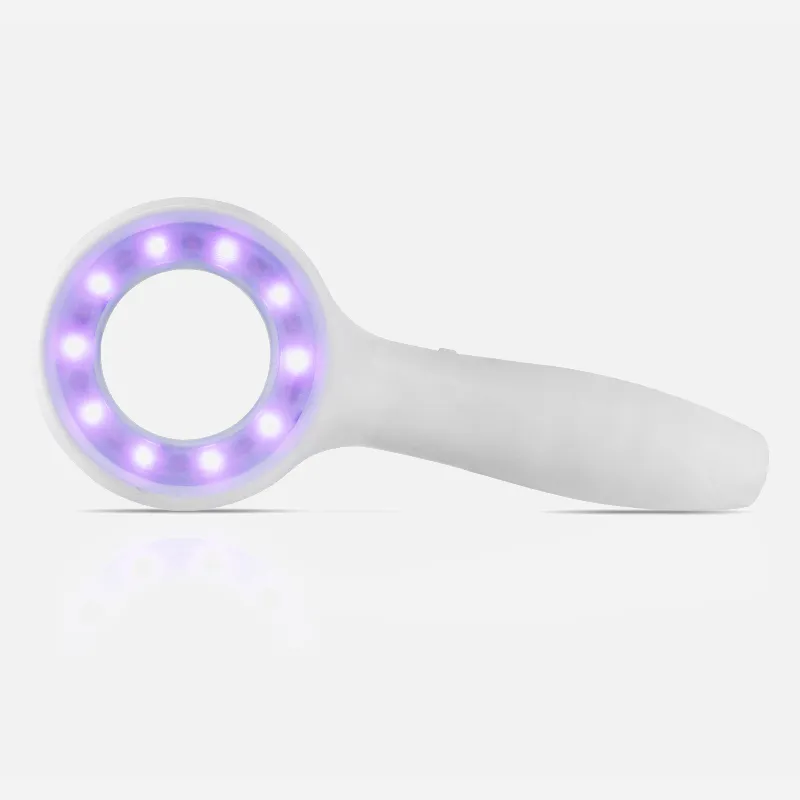
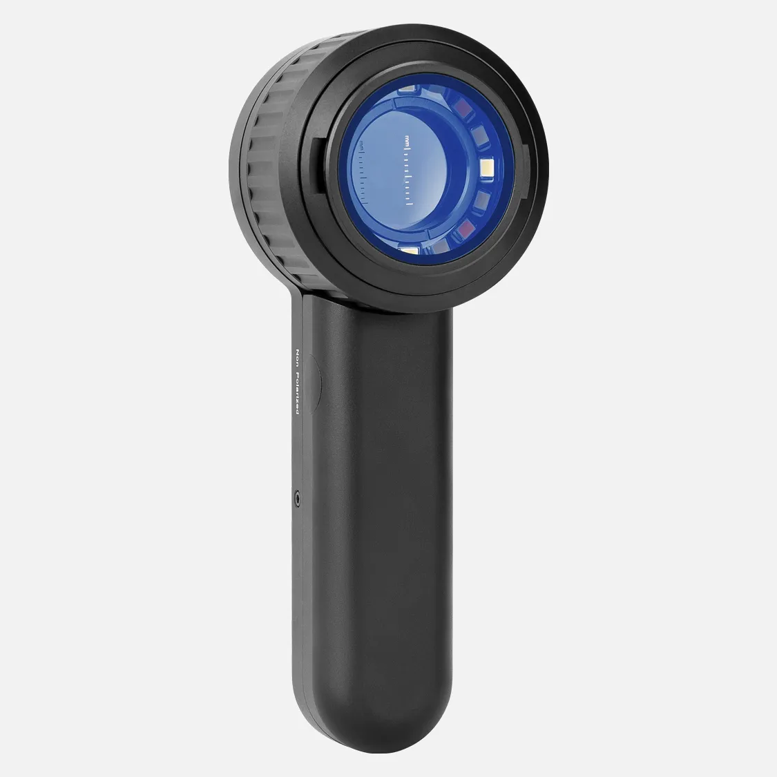
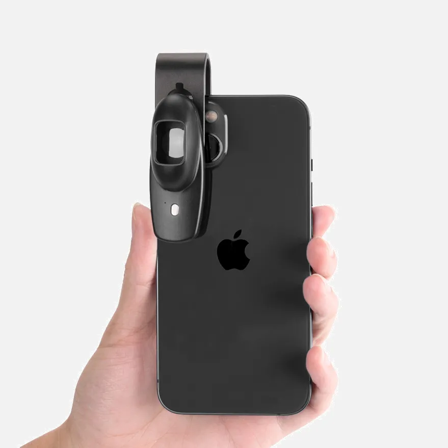
Dermoscopy Basal Cell Carcinoma

Dermoscopy is a non-invasive technique that can help diagnose basal cell carcinoma (BCC) early. Dermoscopy can accurately diagnose BCC up to 95–99% of the time.

Dermoscopic features of superficial BCC include:

  • Ulceration and adherent fiber
  • Leaf-like areas, which are brown to grey-blue discrete bulbous blobs that often form a pattern shaped like a leaf

*Comma vessels

  • Hemorrhage
  • Small ulcerations
  • Hypopigmented areas
  • Telangiectasias
  • Blue-gray ovoid nests
  • Asymmetrical arborising vessels

Dermoscopy can also help identify BCC by the presence of typical vascular structures, pigmented structures, and ulceration, and the absence of specific melanocytic structures.

BCC is one of the most common cancers in white people, and its worldwide incidence is continuously increasing. Lesions from BCC usually have one of the following characteristics:

  • A shiny, skin-colored bump that's translucent
  • A bump that can look pearly white or pink on white skin
  • A bump that often looks brown or glossy black on brown and Black skin

How Does Dermoscopy Basal Cell Carcinoma?

Dermoscopy is a non-invasive technique that can help diagnose basal cell carcinoma (BCC) early. It works by transilluminating a lesion, which allows it to be studied at high magnification to see subtle features.

Dermoscopic criteria associated with BCCs include:

  • Absence of a pigment network
  • Arborizing vessels
  • Large blue-gray ovoid nests
  • Multiple blue-gray globules
  • Leaf-like areas
  • Spoke wheel areas
  • Ulceration

Other dermoscopic features of BCC include:

  • Unfocused peripheral arborizing vessels
  • Keratin masses
  • Superficial scales
  • Ulceration or blood crusts
  • White structures
  • Blue-gray blotches

BCC's Dermoscopic diagnostic accuracy is 95%–99%. However, a diagnosis of BCC should not be based on a single feature but on a set of dermoscopic features in combination with the clinical context.

Experienced dermoscopists can often diagnose superficial BCCs by their typical bluish-pink color.

People May Ask

Which Factors Are Crucial for A Dermoscopic Diagnosis of Lentigo Maligna?

Asymmetric pigmented follicular openings, dark rhomboidal structures, slate gray dots, and slate gray globules are the four criteria that together have produced an 89% and 96% diagnostic sensitivity and specificity.

How Does Dermoscopy Fit into The Evaluation Process for Basal Cell Carcinoma?

The Use of Dermoscopy in BCC ManagementFor the assessment of residual tumor in BCC patients, dermoscopic examination following nonablative treatment has also shown promise. Remaining dermoscopic characteristics such as arborizing telangiectasias, ulceration, and pigmented formations are good markers of tumor persistence.

What Happens If You Don'T Treat A Basal Cell?

In the absence of therapy, a basal cell carcinoma has the potential to slowly spread throughout a significant portion of your body's skin. Furthermore, ulceration and long-term harm to the skin and adjacent tissues are potential side effects of basal cell carcinoma.

What Characteristics of BCC Are Macroscopic?

relative mass confinement.There is varying degrees of epidermal or follicular attachment.Large nuclear palisade-surrounded basaloid lobules.Because of the overproduction of mucin, loculules may be solid or exhibit central cyst development.Scleromyxoid stroma.A cleft forms in the stroma between the tumor lobules.Instead,Additional things...

How Does A BCC Begin to Take Shape?

a translucent, skin-colored pimple that is glossy and partially transparent. When compared to pink skin, the bump can appear pearly white. The bump frequently appears brown or glossy black on skin that is brown or black.

How Does A Basal Cell Carcinoma Typically Look?

How does a BCC look? Open sores, red spots, pink growths, shiny bumps, scars, or growths with slightly elevated, rolling edges and/or a center indentation are some of the appearances of BCCs. BCCs have the potential to leak, crust, itch, or bleed. The lesions usually appear on body parts that are exposed to the sun.

How Can Basal Cell Carcinoma Be Verified?

It is known as a skin biopsy and can be completed in the course of an office visit. A dermatologist can easily, swiftly, and securely execute this straightforward operation. The only way to be positive for any kind of skin cancer is to undergo a skin biopsy.

What Are The Squamous Cell Carcinoma's Dermoscopic Findings?

Common dermoscopic characteristics of carcinomas with cutaneous squamous cell carcinoma are:Instead,White circles.
White patches devoid of structure.Vessels with loops.Keratin in the center.When a tumor is poorly differentiated or growing quickly, the background may be pink or red.

What Are The Superficial BCC's Dermoscopic Characteristics?

Furthermore, comma vessels, hemorrhage, minor ulcerations, hypopigmented regions, telangiectasias, and blue-gray ovoid nests are characteristics of superficial BCCs, whereas arborizing vessels are also characteristic of nodular BCCs.

Can Basal Cell Carcinoma Be Found Via A Dermoscopy?

With their characteristic bluish-pink color, asymmetric arborizing vasculature, and focal ulceration, superficial basal cell carcinomas are often diagnosed by skilled dermoscopists. White regions of regression and little scaling could also exist.

Dermoscopy Basal Cell Carcinoma Products

dermoscopy basal cell carcinoma products supply

Actinic Keratosis: Learn how to reduce your risk of skin cancer and avoid recurrence to replace your anxiety and uncertainty. Kindle攵子书

China Professional dermoscopy basal cell carcinoma

Aloe vera and tea tree oil are among the skin-rejuvenating ingredients in Lane Labs SunSpot ES, a natural exfoliating gel (0.5 ounce).

China affordable dermoscopy basal cell carcinoma products supply

Kindle Edition of Practical Dermoscopy

China high quality dermoscopy basal cell carcinoma products supply

Pink-Think-Blink, First Edition, Kindle Edition: Dermatoscopy of Non-Pigmented Skin Tumors

China Professional dermoscopy basal cell carcinoma products supply

Ebook Enhanced Segmentation-Based Method Utilizing PSO and K-MEANS Algorithm for Basal Cell Carcinoma Identification from Skin Lesions

China customized dermoscopy basal cell carcinoma

First Edition of Dermoscopy in Darker Skin, Kindle Edition

China portable dermoscopy basal cell carcinoma

Sunspot ES, Natural Exfoliating Gel with Aloe Vera and Tea Tree Oil for Skin Renewal Ingredients by Lane Innovations (0.5 oz, 2 pack)

China customized dermoscopy basal cell carcinoma products supply

A Clear Guide to Medical Conditions: Diagnosis and Treatment of Basal Cell Carcinoma Kindle攵子书

China wholesale dermoscopy basal cell carcinoma products supply

The 30-Day Raw Vegan Plant-Based Detoxification & Regeneration Journal & Tracker: Healing Nevoid Basal Cell Carcinoma Syndrome A Reversing Conditions Journal and Tracker. Journal 2 - February 28, 2020 - Paperback

China handheld dermoscopy basal cell carcinoma products supply

January 1, 2017: The Natural and Quick Cure for Staple Bound Basal Cell Carcinoma

Top Reviews

ampm247

My second tube of Sunspot Es is this one. Since my first tube's results were so good, I bought a second one after a friend convinced me not to use it for her issue. Regrettably, the FDA is refusing to let the firm disclose that its true benefit is treating minor skin malignancies. I mean business. My personal experience was with a basal cell carcinoma on my ankle that the doctor wanted to remove. Despite my hesitation, I tried this Sunspot. Es product. Over the course of two weeks, it performed flawlessly except from occasional stinging and itching. On his cheek, my spouse had a large area of keratoses. It looked terrible, and it bled every now and again. Reluctantly, he gave the Sunspot Es a try, and was astonished to discover how powerful it was. The spot took around a month to vanish since he neglected to apply a bandage. When applied correctly, it is an excellent product for treating kerotases, squamous cell, or basal cell carcinomas. I was aware that removing a malignancy using traditional techniques results in significant harm or destruction of the surrounding tissue, which

Dawn McKenna

I was pleasantly surprised to find this book contained much more than the dull, elementary content I had assumed based on its cover. This book has an easy-to-read, pleasant, and sympathetic tone, but its substance is jam-packed with reliable scientific data and practical advice. Rather than merely wearing sunscreen and waiting for my next skin check, I'm so happy to finally have information that I can truly utilize to improve myself. I now have a ton of good actions that I can do to strengthen my immunity and maybe stop future eruptions, in addition to the therapeutic alternatives that this book explained. For the first time, I've been informed of alternatives to avoiding the sun, and I find that information to be really valuable.

Cliente

Written for the BEST dermatoscopist, it's an excellent book with appropriate content, good photos, and helpful outlines. It's essential, in my opinion, for anybody with an interest in dermoscopy.

0

Have questions on gear or your order?

Our Gear Guides are here to help! Get personal advice from pro creatives

Name
Subject
Email address
How can we help?

Instant Answers

How Can Dermoscopy Images Be Captured?

Dermoscopy images can be captured and stored in different ways, such as: • Using a smartphone or tablet with dermoscopic adapter, which consisted in the package.• Using a digital camera

Dermoscopy images can be captured and stored in different ways, such as:

• Using a smartphone or tablet with dermoscopic adapter, which consisted in the package.
• Using a digital camera with dermoscopic adapter, there’s 49mm screw size camera adapter available to order now.

Compatible phone/tablet models:
All iPhone models, 95% Android phones, 90% tablet. For phone/tablet size in 5.25-14mm

Compatible camera models:
All camera with built 49mm filter screw, such as Canon EOS 70D, 80D, 90D; Canon EOS R7, R10, R50, R100; Canon M100, M200, M50, Mark II; Canon G7X Mark III, Sony ZV-1

How Can I Connect My Phone to My Dermatoscope?

There’s universal phone adapter for all our dermoscopes. Please check the installation procedure bellow or watch operation guide. Smartphone Connector (1) Place phone adapter screw in the center of smartphone’s

There’s universal phone adapter for all our dermoscopes. Please check the installation procedure bellow or watch operation guide.

Smartphone Connector

(1) Place phone adapter screw in the center of smartphone’s main camera.
(2) Screw magnet attachment on phone adapter.
(3) Put dermoscope’s back ring and magnet attachment together

Take The Best Images

You need to adjust the focus ring after the dermoscpe connected on smartphone to get the best images.

How Can I Clean My Dermoscopy after Usage?

Cleaning your dermoscopy after usage is important to prevent cross-contamination and infection. The cleaning method may vary depending on the type and model of your dermoscopy, so you should always

Cleaning your dermoscopy after usage is important to prevent cross-contamination and infection. The cleaning method may vary depending on the type and model of your dermoscopy, so you should always follow the manufacturer’s instructions. However, some general steps are:

• Turn off and disconnect your dermoscopy from any power source or device.

• Wipe off any visible dirt or debris from the dermoscopy with a soft cloth or tissue.

• Disinfect the dermoscopy with an alcohol-based wipe or spray, or a disinfectant solution recommended by the manufacturer. Make sure to cover all surfaces, especially the lens and contact plate.

• Let the dermoscopy air dry completely before storing it in a clean and dry place.

• Do not use abrasive or corrosive cleaners, solvents, or detergents that may damage the dermoscopy.

• Do not immerse the dermoscopy in water or any liquid, unless it is waterproof and designed for immersion.

You should clean your dermoscopy after each use, or at least once a day if you use it frequently. You should also check your dermoscopy regularly for any signs of damage or malfunction, and contact the manufacturer or service provider if needed.

Polarized VS Non-Polarized Dermoscopy

A dermoscopy is a device that allows the examination of skin lesions with magnificationand illumination. By revealing subsurface structures and patterns that are not visible tothe naked eye. It can

A dermoscopy is a device that allows the examination of skin lesions with magnificationand illumination. By revealing subsurface structures and patterns that are not visible tothe naked eye. It can improve the diagnose accuracy of skin lesions, such as melanoma,basal cell carcinoma, seborrheic keratosis, etc.

There are two main types of dermoscopy: Non polarized and polarized dermoscopy.We’ve fitted most of our dermoscopys with polarized and non-polarized light. They canbe used in multiple skin structures.

Non-polarized contact Mode

In non-polarized mode, the instrument can provide information about the superficialskin structures, such as milia-like cysts, comedo-like openings, and pigment in theepidemis.

The dermoscopy requires applying a liquid such as mineral oil or alcohol to the skin andplacing the lens in contact with the skin. This reduces surface reflection and enhancesthe view of subsurface structures.

Image with non-polarized light (DE-3100)

Polarized contact Mode

In polarized mode, the instrument allows for visualization for deeper skin structures,such as blood vessels, collagen, and pigment in the dermis.

The dermoscopy does not need to be in contact with the skin or use any liquid. Theirpolarized light can help to eliminate surface reflection and allow visualization ofvascular structures.

Image with polarized light (DE-3100)

Polarized non-contact Mode

The dermoscopy can also use polarized light to examine the skin without direct contact.

In polarized non-contact mode, the instrument allows for examination infected areasand lesions that are painful for the patient, or the difficult to contact pigmented lesions,such as nails and narrow areas.

The contact plate should be removed in this mode, and it does not require applying aliquid to the skin. As it doesn’t require pressure or fluid application on the skin, it canalso avoid cross-contamination and infection risk.

Image in polarized non-contact mode (DE-3100)

How Effectiveness Is Dermoscopy

Compared with visual inspection, the dermoscopy can be used to capture and store skin lesion photos, which play an important role in early skin cancer examination. The dermoscopy allows the

Compared with visual inspection, the dermoscopy can be used to capture and store skin lesion photos, which play an important role in early skin cancer examination.

The dermoscopy allows the examination of skin lesions with magnification and illumination. This can be greatly avoiding the factors that cause interference to visual detection. Such as lighting, skin color, hair and cosmetics.

Several studies have demonstrated that dermoscopy is useful in the identification of melanoma, when used by a trained professional.

It may improve the accuracy of clinical diagnosis by up to 35%
It may reduce the number of harmless lesions that are removed
In primary care, it may increase the referral of more worrisome lesions and reduce the referral of more trivial ones

A 2018 Cochrane meta-analysis published the accuracy of dermoscopy in the detection.

Table 1. Accuracy of dermoscopy in the detection of melanoma in adults
Detection Method Sensitivity, % Specificity, % Positive Likelihood Ratio NegativeLikelihood Ratio
Visual inspection alone (in person) 76 75 3.04 0.32
Dermoscopy with visual inspection (in person) 92 95 18 0.08
Image-based visual inspection alone (not in person) 47 42 0.81 1.3
Dermoscopy with image-based visual inspection (not in person) 81 82 4.5 0.23
ROC—receiver operating characteristic. *Estimated sensitivity calculated on the summary ROC curve at a fixed specificity of 80%.

As we can see, the dermoscope can improve the accuracy of diagnosis of skin lesions, especially melanoma.

Table 1. Accuracy of dermoscopy in the detection of melanoma in adults
Detection Method Sensitivity, % Specificity, % Positive Likelihood Ratio NegativeLikelihood Ratio
Visual inspection alone (in person) 79 77 3.4 0.27
Dermoscopy with visual inspection (in person) 93 99 93 0.07
Image-based visual inspection alone (not in person) 85 87 6.5 0.17
Dermoscopy with image-based visual inspection (not in person) 93 96 23 0.07
ROC—receiver operating characteristic. *Estimated sensitivity calculated on the summary ROC curve at a fixed specificity of 80%.

Characteristics of the dermatoscopic structure of the skin lesions include:

• Symmetry or asymmetry
• Homogeny/uniformity (sameness) or heterogeny (structural differences across the lesion)
• Distribution of pigment: brown lines, dots, clods and structureless areas
• Skin surface keratin: small white cysts, crypts, fissures
• Vascular morphology and pattern: regular or irregular
• Border of the lesion: fading, sharply cut off or radial streaks
• Presence of ulceration

There are specific dermoscopic patterns that aid in the diagnosis of the following pigmented skin lesions:

• Melanoma
• Moles (benign melanocytic naevus)
• Freckles (lentigos)
• Atypical naevi
• Blue naevi
• Seborrhoeic keratosis
• Pigmented basal cell carcinoma
• Haemangioma

No products in the cart.

No products in the cart.

Hot Search Terms