Selecting An Optimal Dermatoscope

Dermatoscopes are specialized medical devices used to examine skin lesions and screen for signs of skin cancer. When selecting the best dermatoscopy, key factors to consider include:

1、Connectivity - Seek out models compatible with smartphones and cameras to enable capturing and telemedicine.
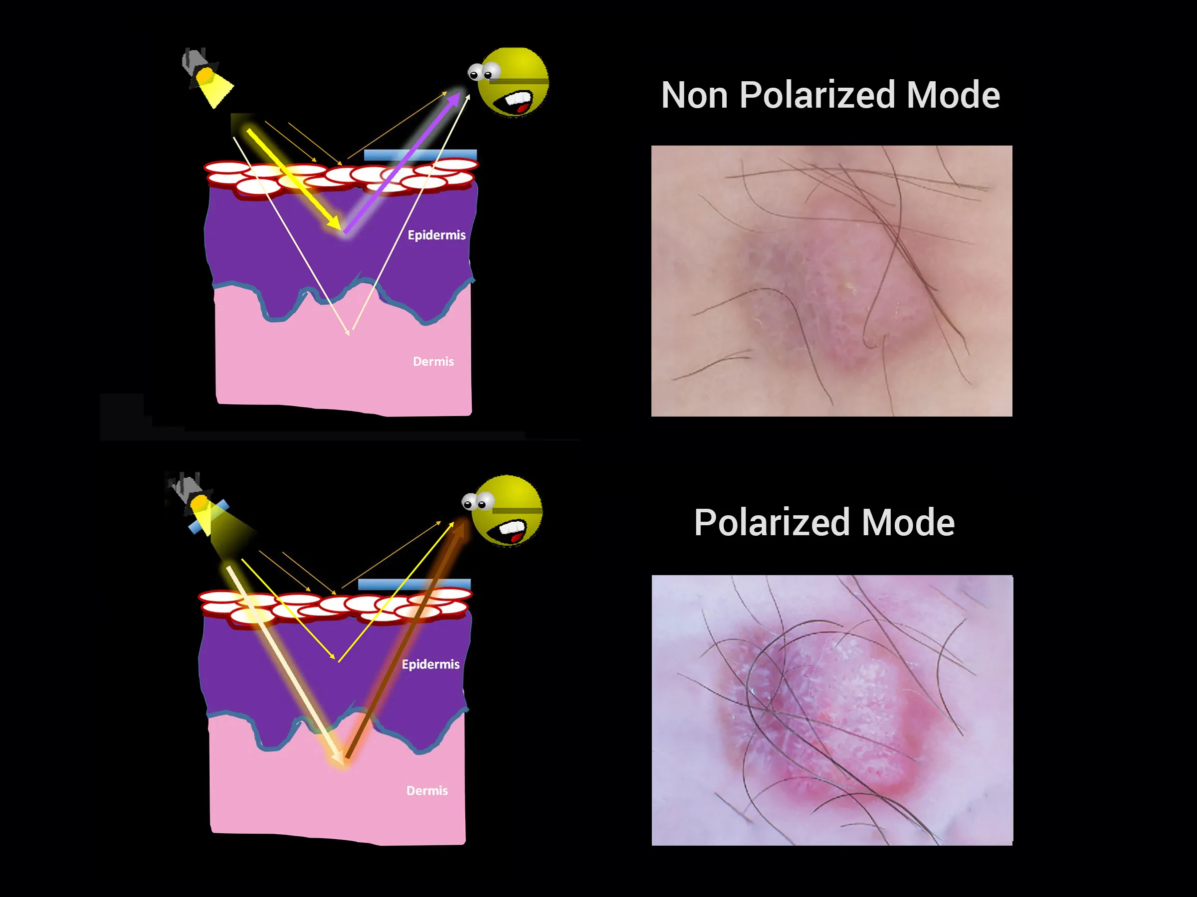
2、polarization - polarized light reduces skin surface reflections. Linear polarization alone or variable polarization modes may offer superior visualization.

3、Viewing Method - Contact (chemiluminescence) or non-contact dermatoscopes suit different examination needs. Dual modes allow switching techniques.

4、Magnification power - Higher magnification from 10x up to 40x allows identifying finer structural details based on diagnostic complexity.

Additional parameters are field of view size, presence of measurement tools, battery-powered or USB-recharged illumination, sturdy device materials, and bundled software. Determining the intended dermoscopy application and patient demographics helps identify the most fitting solution capabilities. Selecting an optimal dermatoscope that matches functional requirements facilitates enhanced skin screening, documentation, and analysis.

Key Features of A Premium Dermatoscope

A quality dermatoscopy allows a detailed inspection of skin structures up to the reticular dermis layer. It captures images to track changes over time. Ideal models offer smartphone connectivity, polarized lighting, scalable magnification from 10x to 40x, and contact and non-contact viewing modes.

Specifically, smartphone and camera compatibility enable telemedicine and documentation. Polarized lighting reduces surface reflections, improving the visibility of deeper skin tissues. Variable magnification suits everything from scanning a lesion to finely inspecting abnormalities. Finally, contact plates provide chemiluminescence to eliminate artifacts, while non-contact modes allow quick surveys.

A leading dermatoscope connects to mobile devices, reduces glare, adjusts magnification, and facilitates contact or non-contact analysis. Combined, these features empower medical professionals with enhanced visualization and insights for assessing lesions and skin conditions. Careful selection of these key criteria allows for matching a dermatoscopy model to diagnostic settings and workflow.

People May Ask

What Is The Appearance of Melanoma in Stage 0?

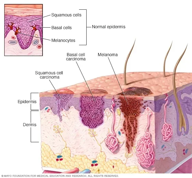
Stage 0 melanoma refers to a malignant tumor that is still limited to the epidermis, the top layer of skin. This indicates that the cancer cells have only spread to the skin's outer layer and have not penetrated the dermis, the skin's second layer.

What Is A Melanoma Warning Sign?

The first warning indications of melanoma are frequently changes in a mole or skin lesion's size, shape, color, or feel. A distinct region of skin from the surrounding skin is called a skin lesion. These alterations may take place in an already-existing lesion or mole, or melanoma may manifest as a strange-looking lesion or mole.

Melanoma: Is It Elevated Or Flat?

As they expand, melanomas may begin as flat patches. 4. If you can feel it, you should get it checked out, even if some moles can also be raised. Occasionally, when assessing melanoma, the "E" in the ABCDE guidance refers to "evolving." This is a result of the gradual changes in melanomas' size, shape, and color.

When You Have Melanoma, How Does Your Body Feel?

general signs and symptomsfirm or enlarged lymph nodes. a solid bump on your body. mysterious discomfort. feeling really drained or sick.

What Tools Do Dermatologists Utilize to Examine Skin?

Dermatoscopes are tools for examining skin lesions that use light and magnification. The majority of them have an otoscope-like shape; one kind of dermatoscope is seen in the article by Dr.

Would Dermatologists Overlook Melanoma?

Andy Jacobs is aware of how simple it is to overlook a malignant area on your skin. He was unaware that he had a malignant growth on his skin until a few days prior to receiving a diagnosis of melanoma, the most severe form of skin cancer.

Which Melanoma Scan Is The Best?

Computerized tomography (CT) scanCT scans, as opposed to standard x-rays, are able to display the fine detail in soft tissues (such internal organs). This examination might reveal whether any lymph nodes are swollen or whether there are any worrisome spots on organs like the liver or lungs that could indicate melanoma spreading.

To What Extent Can Melanoma Spread?

Tumor thickness: The Breslow measurement is the term used to describe the thickness of the melanoma. The likelihood of melanomas spreading is generally very low when they are less than 1 millimeter (mm) thick, or roughly 1/25 of an inch. The likelihood of the melanoma spreading increases with thickness.

Can You Develop Cancer from A Mole You'Ve Had for Years?

Is melanoma possible from a common mole? Melanoma, the most dangerous kind of skin cancer, is extremely uncommon to develop from a common mole. Those with numerous little or multiple large moles are more likely to acquire melanoma, even though common moles are not malignant (1).

What Distinguishes Heine from DermLite Dermatoscopes?

While Dermlite DL II only has cross-polarized mode, Heine Delta 20 only has non-polarized mode, which necessitates a contact fluid. The non-polarized dermatoscope aids in identifying the skin's surface structures, whereas the polarized dermatoscope permits more profound viewing.

Best Dermatoscope Products

China smartphone best dermatoscope suppliers & wholesalers

Digital otoscope including a 4.3-inch screen, SKYEAR ear wax removal tool camera with lights, a built-in 32GB card, a long 5-hour working life, and the ability to capture both photos and videos - color white

China top quality best dermatoscope

Low Vision Aid: Bierley Maggie 5 Portable Ultra Slim Electronic Magnifier

China portable best dermatoscope suppliers & wholesalers

Supereyes 11mm Diameter, B007, USB Digital Microscope Camera with 300X Zoom, Handheld Endoscope, Portable Magnifier Otoscope, and IP67 Waterproof for Windows, Mac, and Linux

China top quality best dermatoscope suppliers & wholesalers

UKCOCO Tiny Camera Digital Microscope Pocket Microscope Dermatoscope Microscope for Pocket Magnifying Glass Phone for Hand Led Phone Microscope Portable White Small Camera

China portable best dermatoscope

可互捚敜头手持数字显微敜放大镜 Supereyes B011 5.0MP 500X

China Professional best dermatoscope suppliers & wholesalers

12 inch color HD portable video magnifier, Eschenbach Visolux Digital XL FHD

China affordable best dermatoscope

the battery-operated Richter 3777 Ri-derma Dermatoscope

China smartphone best dermatoscope

With its 8 spectrum analysis, 28 million pixels, and a skin sensor scanner machine for skin problems, the FXNFXLA Skin Analysis System is a 3D skin tester with an LCD display screen.

China Professional best dermatoscope

The MicroMax LED-lighted 60x-75x Carson Pocket Microscope (MM-200) and the Pocket Micro 20x-60x LED-lighted Zoom Field Microscope with Aspheric Lens System (MM-450) are both available in blue.

China affordable best dermatoscope suppliers & wholesalers

OdontoMed2011 Gray DERMATOSCOPE Free CASE ODM Dermatology Examination

Top Reviews

Rosa guerrero

For usage at home, this is a really great otoscope. I adore that it has ear cleaning equipment in addition to a monitor. For the price, the camera quality is excellent. It's never been easy to clean my husband's and my ears. In addition, I have used it to verify sure my toddler's tubes are still in their proper places and to check on his ears. He has a history of ear infections, so I'm glad I have this device to check for those. When it comes to actual medical instruments used in healthcare, this digital otoscope is the best available overall.

J Biz

Excellent product in a lovely case that is just as advertised. Delivery was prompt. Extremely suggested.

Dustin Aunkst

Greetings, fellow fans of clear ears! Have you ever wondered why your ears seem so full? The amazing Digital Otoscope for Ear Wax Removal holds the key to the solution. Exceptionally Clear Views: Dive into the depths of your ears and see everything in rich detail thanks to cutting-edge camera technology. It's similar to setting out on a little journey within your ear canal! Mild & Secure: Is it a concern to poke and prod? Not anymore! Because of its lullaby-soft design, this otoscope is sure to provide everyone with a secure and comfortable experience. Bid Farewell to Guesswork: With the accuracy this equipment provides, you can quickly detect and get rid of wax buildup, putting an end to those unexplained ear pains. Friendly to Users: Ear examinations are made easy with the otoscope, which is user-friendly for both novice and expert gadget users. An Educational Event It's about getting a deeper understanding of your body, not just about cleaning your ears. Give your family a look at the images and use them t

0

Have questions on gear or your order?

Our Gear Guides are here to help! Get personal advice from pro creatives

Name
Subject
Email address
How can we help?

Instant Answers

How Can Dermoscopy Images Be Captured?

Dermoscopy images can be captured and stored in different ways, such as: • Using a smartphone or tablet with dermoscopic adapter, which consisted in the package.• Using a digital camera

Dermoscopy images can be captured and stored in different ways, such as:

• Using a smartphone or tablet with dermoscopic adapter, which consisted in the package.
• Using a digital camera with dermoscopic adapter, there’s 49mm screw size camera adapter available to order now.

Compatible phone/tablet models:
All iPhone models, 95% Android phones, 90% tablet. For phone/tablet size in 5.25-14mm

Compatible camera models:
All camera with built 49mm filter screw, such as Canon EOS 70D, 80D, 90D; Canon EOS R7, R10, R50, R100; Canon M100, M200, M50, Mark II; Canon G7X Mark III, Sony ZV-1

How Can I Connect My Phone to My Dermatoscope?

There’s universal phone adapter for all our dermoscopes. Please check the installation procedure bellow or watch operation guide. Smartphone Connector (1) Place phone adapter screw in the center of smartphone’s

There’s universal phone adapter for all our dermoscopes. Please check the installation procedure bellow or watch operation guide.

Smartphone Connector

(1) Place phone adapter screw in the center of smartphone’s main camera.
(2) Screw magnet attachment on phone adapter.
(3) Put dermoscope’s back ring and magnet attachment together

Take The Best Images

You need to adjust the focus ring after the dermoscpe connected on smartphone to get the best images.

How Can I Clean My Dermoscopy after Usage?

Cleaning your dermoscopy after usage is important to prevent cross-contamination and infection. The cleaning method may vary depending on the type and model of your dermoscopy, so you should always

Cleaning your dermoscopy after usage is important to prevent cross-contamination and infection. The cleaning method may vary depending on the type and model of your dermoscopy, so you should always follow the manufacturer’s instructions. However, some general steps are:

• Turn off and disconnect your dermoscopy from any power source or device.

• Wipe off any visible dirt or debris from the dermoscopy with a soft cloth or tissue.

• Disinfect the dermoscopy with an alcohol-based wipe or spray, or a disinfectant solution recommended by the manufacturer. Make sure to cover all surfaces, especially the lens and contact plate.

• Let the dermoscopy air dry completely before storing it in a clean and dry place.

• Do not use abrasive or corrosive cleaners, solvents, or detergents that may damage the dermoscopy.

• Do not immerse the dermoscopy in water or any liquid, unless it is waterproof and designed for immersion.

You should clean your dermoscopy after each use, or at least once a day if you use it frequently. You should also check your dermoscopy regularly for any signs of damage or malfunction, and contact the manufacturer or service provider if needed.

Polarized VS Non-Polarized Dermoscopy

A dermoscopy is a device that allows the examination of skin lesions with magnificationand illumination. By revealing subsurface structures and patterns that are not visible tothe naked eye. It can

A dermoscopy is a device that allows the examination of skin lesions with magnificationand illumination. By revealing subsurface structures and patterns that are not visible tothe naked eye. It can improve the diagnose accuracy of skin lesions, such as melanoma,basal cell carcinoma, seborrheic keratosis, etc.

There are two main types of dermoscopy: Non polarized and polarized dermoscopy.We’ve fitted most of our dermoscopys with polarized and non-polarized light. They canbe used in multiple skin structures.

Non-polarized contact Mode

In non-polarized mode, the instrument can provide information about the superficialskin structures, such as milia-like cysts, comedo-like openings, and pigment in theepidemis.

The dermoscopy requires applying a liquid such as mineral oil or alcohol to the skin andplacing the lens in contact with the skin. This reduces surface reflection and enhancesthe view of subsurface structures.

Image with non-polarized light (DE-3100)

Polarized contact Mode

In polarized mode, the instrument allows for visualization for deeper skin structures,such as blood vessels, collagen, and pigment in the dermis.

The dermoscopy does not need to be in contact with the skin or use any liquid. Theirpolarized light can help to eliminate surface reflection and allow visualization ofvascular structures.

Image with polarized light (DE-3100)

Polarized non-contact Mode

The dermoscopy can also use polarized light to examine the skin without direct contact.

In polarized non-contact mode, the instrument allows for examination infected areasand lesions that are painful for the patient, or the difficult to contact pigmented lesions,such as nails and narrow areas.

The contact plate should be removed in this mode, and it does not require applying aliquid to the skin. As it doesn’t require pressure or fluid application on the skin, it canalso avoid cross-contamination and infection risk.

Image in polarized non-contact mode (DE-3100)

How Effectiveness Is Dermoscopy

Compared with visual inspection, the dermoscopy can be used to capture and store skin lesion photos, which play an important role in early skin cancer examination. The dermoscopy allows the

Compared with visual inspection, the dermoscopy can be used to capture and store skin lesion photos, which play an important role in early skin cancer examination.

The dermoscopy allows the examination of skin lesions with magnification and illumination. This can be greatly avoiding the factors that cause interference to visual detection. Such as lighting, skin color, hair and cosmetics.

Several studies have demonstrated that dermoscopy is useful in the identification of melanoma, when used by a trained professional.

It may improve the accuracy of clinical diagnosis by up to 35%
It may reduce the number of harmless lesions that are removed
In primary care, it may increase the referral of more worrisome lesions and reduce the referral of more trivial ones

A 2018 Cochrane meta-analysis published the accuracy of dermoscopy in the detection.

Table 1. Accuracy of dermoscopy in the detection of melanoma in adults
Detection Method Sensitivity, % Specificity, % Positive Likelihood Ratio NegativeLikelihood Ratio
Visual inspection alone (in person) 76 75 3.04 0.32
Dermoscopy with visual inspection (in person) 92 95 18 0.08
Image-based visual inspection alone (not in person) 47 42 0.81 1.3
Dermoscopy with image-based visual inspection (not in person) 81 82 4.5 0.23
ROC—receiver operating characteristic. *Estimated sensitivity calculated on the summary ROC curve at a fixed specificity of 80%.

As we can see, the dermoscope can improve the accuracy of diagnosis of skin lesions, especially melanoma.

Table 1. Accuracy of dermoscopy in the detection of melanoma in adults
Detection Method Sensitivity, % Specificity, % Positive Likelihood Ratio NegativeLikelihood Ratio
Visual inspection alone (in person) 79 77 3.4 0.27
Dermoscopy with visual inspection (in person) 93 99 93 0.07
Image-based visual inspection alone (not in person) 85 87 6.5 0.17
Dermoscopy with image-based visual inspection (not in person) 93 96 23 0.07
ROC—receiver operating characteristic. *Estimated sensitivity calculated on the summary ROC curve at a fixed specificity of 80%.

Characteristics of the dermatoscopic structure of the skin lesions include:

• Symmetry or asymmetry
• Homogeny/uniformity (sameness) or heterogeny (structural differences across the lesion)
• Distribution of pigment: brown lines, dots, clods and structureless areas
• Skin surface keratin: small white cysts, crypts, fissures
• Vascular morphology and pattern: regular or irregular
• Border of the lesion: fading, sharply cut off or radial streaks
• Presence of ulceration

There are specific dermoscopic patterns that aid in the diagnosis of the following pigmented skin lesions:

• Melanoma
• Moles (benign melanocytic naevus)
• Freckles (lentigos)
• Atypical naevi
• Blue naevi
• Seborrhoeic keratosis
• Pigmented basal cell carcinoma
• Haemangioma

No products in the cart.

No products in the cart.

Hot Search Terms