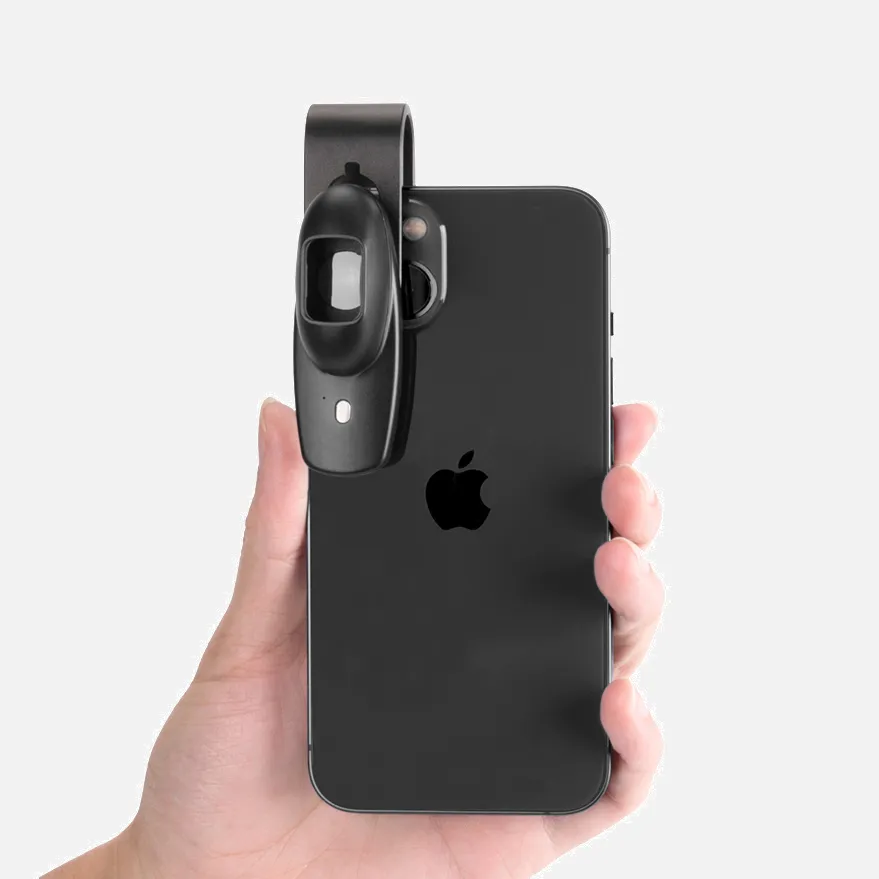
What Is Dermatoscopio Comprar?

A dermatoscope is a handheld visual aid device that can be used to examine and diagnose skin lesions and diseases. It's a common feature at a dermatologist's office.

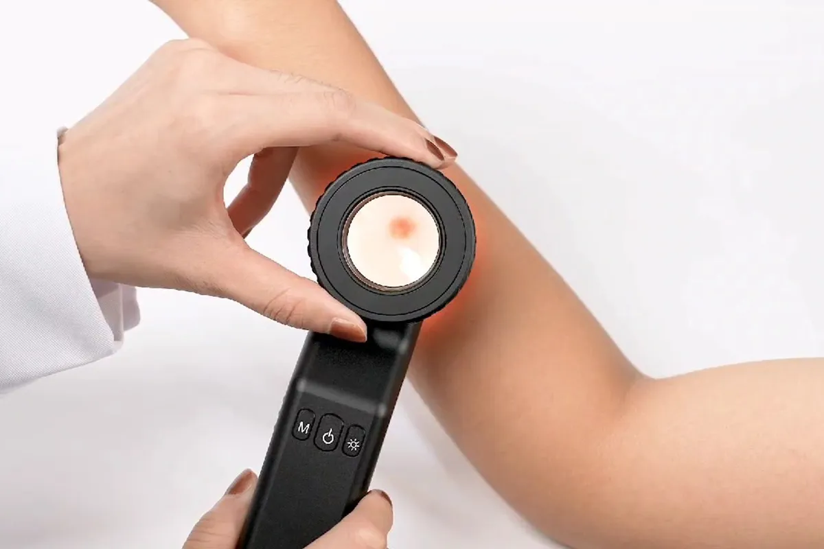
A dermatoscope is similar to a magnifying glass and can magnify up to 10 times. A specialist will apply oil or gel to the skin and then hold the dermatoscope on to it to examine the area.

Dermoscopy is also known as dermatoscopy, chemiluminescence, or skin surface microscopy. It's a non-invasive, in-vivo technique that can effectively identify melanomas and confirm malignant skin cancers.

Dermoscopy can also help identify subtle structures such as hemorrhagic areas and vascular structures. This can expand its application scope to evaluate non-pigmented skin disorders, including inflammatory diseases.

how does dermatoscopio comprar?

When purchasing a dermatoscope, buyers should understand key features and options to select the right model for their needs and setting.

Dermatoscopes come in various styles, magnification levels, attachment options, and integrated capabilities. Handheld models work well for flexible applications, while digital devices offer added imaging and tracking functionality.

Important considerations for purchasing include:

  • Magnification level (typically 10x or higher)
  • Illumination method (polarized, non-polarized)
  • Contact or non-contact evaluation
  • Digital attachments for photo/video capture
  • Smartphone connectivity and software apps
  • Image management and tracking software

Reputable dermatoscope suppliers can help buyers compare products and determine the appropriate solution based on factors like intended use, price range, and advanced features required.

Key Considerations When Purchasing A Dermatoscope

When buying a dermatoscopy, it is important to understand the options and select a model suited to your needs. Key purchasing considerations include:

Magnification Level: Dermatoscopes can magnify up to 10x or higher skin structures. Higher magnification improves the visualization of fine details.

Light Source: Models feature polarized or non-polarized lighting to illuminate skin lesions.

Contact or Non-Contact: Contact dermatoscopes have a plate that touches the skin. Non-contact versions allow a short working distance above the skin.

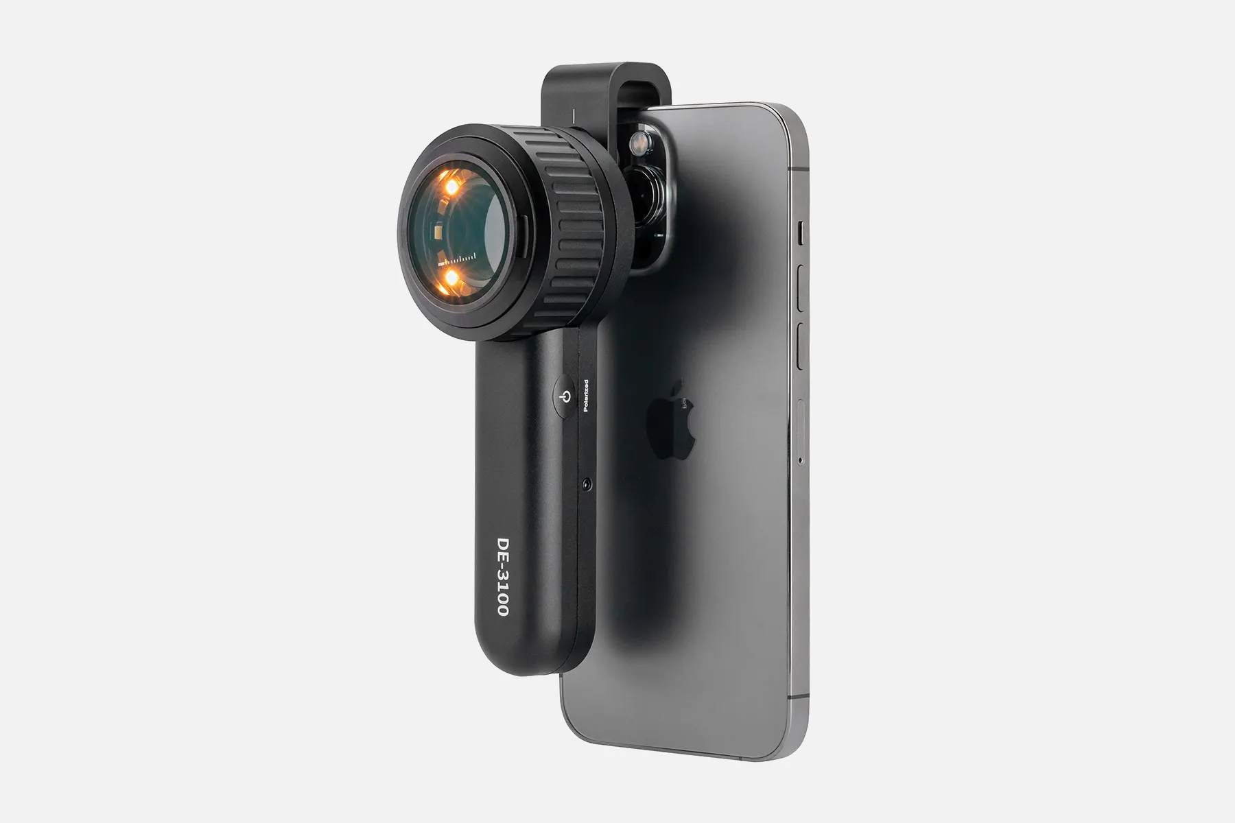
Attachment Options: Some dermatoscopes offer smartphone or camera adapters to capture dermoscopic images and video.

Software Features: Digital systems may integrate tracking programs, image analysis tools, and computer-assisted diagnosis.

Budget: prices range from $100 for simple manual models to over $5,000 for advanced digital systems.

Reputable medical equipment suppliers can guide buyers in selecting the appropriate dermatoscopy based on clinical setting, types of evaluations performed, patient volume, and budget. Proper training is key to utilizing these devices effectively for evaluating skin conditions.

People May Ask

Que Investigaciones Se Realizan Para Identificar CáNcer De Piel?

It's possible that your doctor will recommend that you see a dermatólogo, a physician who specializes in skin diseases, if they suspect you have skin cancer. They will examine the affected area more thoroughly.

¿CóMo Encontrar Un Melanoma in Tiempo Real?

Dermatoscopy, also known as epiluminescence microscopy, is an unconventional medical technique used to detect melanoma early. En la piel pigmentada, un médico puede examinar los patrones de tamaño, forma y pigmentación de las lesiones mediante el uso de un dispositivo manual.

What Is The Name of The Lunar Map?

What is a digital dermatoscopy or lunar mapping?It is possible to see a number of lunar structures that are invisible to the unaided eye thanks to this non-invasive technique.

What Does Topography Mean in Dermatology?

The goal of topografia is to count the areas of the skin affected by dermatosis in order to determine whether it is localized in a specific body part, such as the hands, feet, or legs, or if it is dispersed among many body parts, or generalized, meaning it affects more than 80% of the cutaneous surface.

How Much Does A Dermatologist Charge?

How much does a dermatologist in Mexico earn? El promedio salario de un dermatólogo en México is de $246 por hour o $480,000 al año. Professionals with more experience earn up to $525,000 annually, while entry-level positions start at $138,191 annually.

¿Qué Llaman Los Profesionales Que Examinan La Piel?

medical professional with a focus on diagnosing and treating skin issues. Se conoce como experto en dermatología además.

Who Can Diagnose A Dermatologist?

Professionals in health care who diagnose and treat skin diseases are known as dermatologists. De tal manera, sus funciones principales consisten en: Diagnozar enfermedades o alteraciones que pueden afectar a la piel, pelo, uñas o mucosas.

¿CóMo Se Realiza Una Fotomicroscopy Digital?

Se aplica un gel líquido específico en el área de interés durante una dermatoscopia y coloca el dermatoscopio sobre la lesión para realizar el examen. This gel enables the physician to see structures beneath the skin's surface. The need to use the gel has been eliminated by the most recent dermatoscopy models.

How Much Growth Does A Dermatoscopio Have?

To provide illumination, the majority of non-polarized dermatoscopes (DNP) have diode light emistors, and all of them are equipped with a 10-x magnification lens.

What Is Seen during A Dermatoscopy?

A non-invasive in vivo diagnostic technique called dermatoscopia was developed to study skin lesions. enhances early diagnosis of potentially malignant lesions, including melanoma, and improves the accuracy of diagnosis of hyperpigmented lesions.

Dermatoscopio Comprar Products

Student-proof biological microscope for use in labs and homes: Compound Microscope EXM-150 with Monocular Head, 40–400X Magnification, Disc Diaphragm, and Cordless LED Illumination

YOCTOSUN Magnifying Glasses with 4 Detachable Lenses, 2 LED Lights, Storage Case, Head Strap, Hands-Free Headband Magnifier for Crafts, Jewelry, and Close Work

20 Pcs DC 1.5-3V Micro Electric Motor 23000 RPM Car Toys Electric Motor Rectangular Small Electric Motor for Do It Yourself Projects Scientific Initiatives Metal Engine Motor Assembly

Portable Ear Light and Exam Kit for Home and Professional Use - Cynamed Mini Otoscope - 3X Magnifying Fiber Optic Scope with Extra Tips, Bulb, and Carrying Case - Pocket Diagnostic Tools (Maroon)

For the 3GEN, Cameron Sino New 1150mAh Replacement Battery DL2 Dermatoscope, DL3 Dermatoscope, DermLite Pro, DermLite II, DermLite III, and so on

Windows/Mac OS/TV Compatible, 32GB TF Card Included, Dcorn 10 HDMI LCD Digital Microscope 1500X, Coin Microscope for Adults with 16MP Camera Sensor, Soldering Microscope with LED Lights Touch Control

ZTEEERS Compound Binocular Microscope,40X-2500X Magnification with 10X/25X Eyepiece, LED Illumination, Abbe Condenser, Double-Layer Mechanical Stage,Microscope for Adults.

For iPhone, Android, and PC, Ninyoon 4K WiFi Microscope with Expert Stand, 50-1000X Digital USB Microscope Wireless Endoscope HD Camera Compatible with All Cellphones Windows Macintosh Tablet Android iPad Glimmer Windows

3Gen Dermatology Dermascope, DermLite DL100

[1080P HD & Full Lighted View] TOMLOV DM1 Wireless Digital Microscope 50X-1000X Wireless Charging Mini USB Trichome Coin Microscope Camera Magnifier for iPhone, iPad, Android Phone, and Computer via WiFi

Top Reviews

nth Solutions

This is what I bought to keep an eye on the stylus wear on my phono cartridge. As you can see, the pictures are incredibly detailed and sharp. Without even using a fixture, I simply held the cartridge in one hand, adjusted the focus, and then pressed the remote. It is quite easy to focus an image and the gears, both fine and coarse, are extremely smooth due to the outstanding build quality. Everything worked, even the ability to use it as a laptop's remote camera—something I'm not particularly interested in, but it does the job.

Kathryn Sabatino

Very simple to set up and operate, I'm impressed. The photos are clear and crisp, and the monitor size is helpful.

JohnG

At this magnification, even the smallest movement with your hand results in a blurry picture, so be sure to have a stand or tripod for it. I was truly astounded by the quality of the pictures. Fantastic software that instantly projects your photo to your phone or laptop in real time, allowing you to confirm that the photo you are about to take is something you want. I ADORE IT!

0

Have questions on gear or your order?

Our Gear Guides are here to help! Get personal advice from pro creatives

Name
Subject
Email address
How can we help?

Instant Answers

How Can Dermoscopy Images Be Captured?

Dermoscopy images can be captured and stored in different ways, such as: • Using a smartphone or tablet with dermoscopic adapter, which consisted in the package.• Using a digital camera

Dermoscopy images can be captured and stored in different ways, such as:

• Using a smartphone or tablet with dermoscopic adapter, which consisted in the package.
• Using a digital camera with dermoscopic adapter, there’s 49mm screw size camera adapter available to order now.

Compatible phone/tablet models:
All iPhone models, 95% Android phones, 90% tablet. For phone/tablet size in 5.25-14mm

Compatible camera models:
All camera with built 49mm filter screw, such as Canon EOS 70D, 80D, 90D; Canon EOS R7, R10, R50, R100; Canon M100, M200, M50, Mark II; Canon G7X Mark III, Sony ZV-1

How Can I Connect My Phone to My Dermatoscope?

There’s universal phone adapter for all our dermoscopes. Please check the installation procedure bellow or watch operation guide. Smartphone Connector (1) Place phone adapter screw in the center of smartphone’s

There’s universal phone adapter for all our dermoscopes. Please check the installation procedure bellow or watch operation guide.

Smartphone Connector

(1) Place phone adapter screw in the center of smartphone’s main camera.
(2) Screw magnet attachment on phone adapter.
(3) Put dermoscope’s back ring and magnet attachment together

Take The Best Images

You need to adjust the focus ring after the dermoscpe connected on smartphone to get the best images.

How Can I Clean My Dermoscopy after Usage?

Cleaning your dermoscopy after usage is important to prevent cross-contamination and infection. The cleaning method may vary depending on the type and model of your dermoscopy, so you should always

Cleaning your dermoscopy after usage is important to prevent cross-contamination and infection. The cleaning method may vary depending on the type and model of your dermoscopy, so you should always follow the manufacturer’s instructions. However, some general steps are:

• Turn off and disconnect your dermoscopy from any power source or device.

• Wipe off any visible dirt or debris from the dermoscopy with a soft cloth or tissue.

• Disinfect the dermoscopy with an alcohol-based wipe or spray, or a disinfectant solution recommended by the manufacturer. Make sure to cover all surfaces, especially the lens and contact plate.

• Let the dermoscopy air dry completely before storing it in a clean and dry place.

• Do not use abrasive or corrosive cleaners, solvents, or detergents that may damage the dermoscopy.

• Do not immerse the dermoscopy in water or any liquid, unless it is waterproof and designed for immersion.

You should clean your dermoscopy after each use, or at least once a day if you use it frequently. You should also check your dermoscopy regularly for any signs of damage or malfunction, and contact the manufacturer or service provider if needed.

Polarized VS Non-Polarized Dermoscopy

A dermoscopy is a device that allows the examination of skin lesions with magnificationand illumination. By revealing subsurface structures and patterns that are not visible tothe naked eye. It can

A dermoscopy is a device that allows the examination of skin lesions with magnificationand illumination. By revealing subsurface structures and patterns that are not visible tothe naked eye. It can improve the diagnose accuracy of skin lesions, such as melanoma,basal cell carcinoma, seborrheic keratosis, etc.

There are two main types of dermoscopy: Non polarized and polarized dermoscopy.We’ve fitted most of our dermoscopys with polarized and non-polarized light. They canbe used in multiple skin structures.

Non-polarized contact Mode

In non-polarized mode, the instrument can provide information about the superficialskin structures, such as milia-like cysts, comedo-like openings, and pigment in theepidemis.

The dermoscopy requires applying a liquid such as mineral oil or alcohol to the skin andplacing the lens in contact with the skin. This reduces surface reflection and enhancesthe view of subsurface structures.

Image with non-polarized light (DE-3100)

Polarized contact Mode

In polarized mode, the instrument allows for visualization for deeper skin structures,such as blood vessels, collagen, and pigment in the dermis.

The dermoscopy does not need to be in contact with the skin or use any liquid. Theirpolarized light can help to eliminate surface reflection and allow visualization ofvascular structures.

Image with polarized light (DE-3100)

Polarized non-contact Mode

The dermoscopy can also use polarized light to examine the skin without direct contact.

In polarized non-contact mode, the instrument allows for examination infected areasand lesions that are painful for the patient, or the difficult to contact pigmented lesions,such as nails and narrow areas.

The contact plate should be removed in this mode, and it does not require applying aliquid to the skin. As it doesn’t require pressure or fluid application on the skin, it canalso avoid cross-contamination and infection risk.

Image in polarized non-contact mode (DE-3100)

How Effectiveness Is Dermoscopy

Compared with visual inspection, the dermoscopy can be used to capture and store skin lesion photos, which play an important role in early skin cancer examination. The dermoscopy allows the

Compared with visual inspection, the dermoscopy can be used to capture and store skin lesion photos, which play an important role in early skin cancer examination.

The dermoscopy allows the examination of skin lesions with magnification and illumination. This can be greatly avoiding the factors that cause interference to visual detection. Such as lighting, skin color, hair and cosmetics.

Several studies have demonstrated that dermoscopy is useful in the identification of melanoma, when used by a trained professional.

It may improve the accuracy of clinical diagnosis by up to 35%
It may reduce the number of harmless lesions that are removed
In primary care, it may increase the referral of more worrisome lesions and reduce the referral of more trivial ones

A 2018 Cochrane meta-analysis published the accuracy of dermoscopy in the detection.

Table 1. Accuracy of dermoscopy in the detection of melanoma in adults
Detection Method Sensitivity, % Specificity, % Positive Likelihood Ratio NegativeLikelihood Ratio
Visual inspection alone (in person) 76 75 3.04 0.32
Dermoscopy with visual inspection (in person) 92 95 18 0.08
Image-based visual inspection alone (not in person) 47 42 0.81 1.3
Dermoscopy with image-based visual inspection (not in person) 81 82 4.5 0.23
ROC—receiver operating characteristic. *Estimated sensitivity calculated on the summary ROC curve at a fixed specificity of 80%.

As we can see, the dermoscope can improve the accuracy of diagnosis of skin lesions, especially melanoma.

Table 1. Accuracy of dermoscopy in the detection of melanoma in adults
Detection Method Sensitivity, % Specificity, % Positive Likelihood Ratio NegativeLikelihood Ratio
Visual inspection alone (in person) 79 77 3.4 0.27
Dermoscopy with visual inspection (in person) 93 99 93 0.07
Image-based visual inspection alone (not in person) 85 87 6.5 0.17
Dermoscopy with image-based visual inspection (not in person) 93 96 23 0.07
ROC—receiver operating characteristic. *Estimated sensitivity calculated on the summary ROC curve at a fixed specificity of 80%.

Characteristics of the dermatoscopic structure of the skin lesions include:

• Symmetry or asymmetry
• Homogeny/uniformity (sameness) or heterogeny (structural differences across the lesion)
• Distribution of pigment: brown lines, dots, clods and structureless areas
• Skin surface keratin: small white cysts, crypts, fissures
• Vascular morphology and pattern: regular or irregular
• Border of the lesion: fading, sharply cut off or radial streaks
• Presence of ulceration

There are specific dermoscopic patterns that aid in the diagnosis of the following pigmented skin lesions:

• Melanoma
• Moles (benign melanocytic naevus)
• Freckles (lentigos)
• Atypical naevi
• Blue naevi
• Seborrhoeic keratosis
• Pigmented basal cell carcinoma
• Haemangioma

No products in the cart.

No products in the cart.

Hot Search Terms