Distinguishing Dermatoscopes from Magnifying Glasses

A dermatoscope is a specialized medical device that examines skin lesions, often allowing higher magnification and better lighting than a basic magnifying glass.

Like a magnifying glass, a dermatoscope lets the user see skin structures in more detail. However, there are some key differences:

  • Magnification level: Dermatoscopes typically provide 10X magnification or higher, while magnifying glasses are usually up to 4X. The higher magnification of dermatoscopes allows finer structures to be seen.
  • Integrated lighting: Dermatoscopes have built-in polarized lighting to illuminate skin lesions while magnifying glasses rely on external light sources. The specialized lighting enhances the visibility of subsurface skin structures.
  • Advanced capabilities: Some digital dermatoscopes allow image capture, measurement tools, and computer-assisted diagnosis features - going beyond what basic magnifiers can offer.
  • Intended use: Magnifying glasses are used for casual inspection. Dermatoscopes are precision instruments for medical diagnosis, especially of skin cancers.

Dermatoscopes offer greater magnification, better lighting, and more advanced capabilities tailored to medical dermatological diagnosis. But a basic magnifier can be handy for quick visual inspections. The most accurate diagnosis depends greatly on the training and skills of the examiner.

Comparing The Capabilities: Dermatoscope Or Common Magnifier?

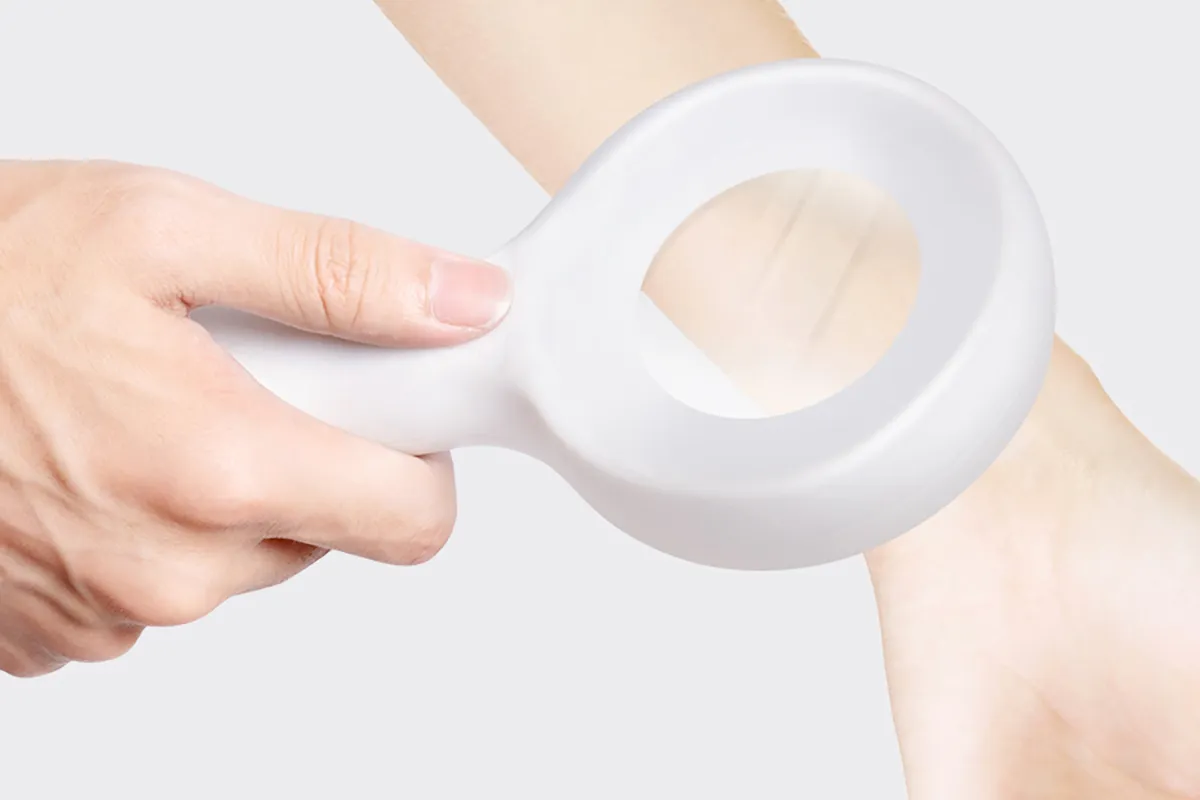
A dermatoscope is a handheld device that functions similarly to a magnifying glass but with greater magnification and an adjustable light source. A dermatoscope can magnify objects up to 10 times.

A dermatoscope can reveal subsurface details, while a magnifying glass or the naked eye can only show what's on the skin's surface. A dermatoscope uses an ultrasound gel to make the skin more translucent, allowing the examiner to assess the lesion's subsurface structure.

When used correctly, dermoscopes can reduce false positives and negatives. Dermoscopy is more accurate than visual inspection for identifying melanoma and excluding things that are not.

People May Ask

Otoscopes: Do They Enlarge?

Otoscopes with a handle and a head are the most widely used models. The head has a low-power magnifying lens that is usually about 8 diopters (3.00x Mag) and a light source.

Does An Otoscope Have A Magnifying Glass?

Unlike electronic otoscopes, the glass optical lens used in this device provides crisp, high-resolution magnification. You can see the ear very well thanks to the wide field of vision and magnification.

How Does Confocal Microscopy And Dermoscopy Differ from One Another?

The morphological structures of the skin, from the epidermis to the papillary dermis, can be seen by a procedure called dermatoscopy, which makes use of a magnifying glass. Reflectance confocal microscopy makes use of a laser to take cellular-level pictures of the skin's many layers.

What Is The Magnifying Glass Magnification Measurement Method?

Use the following formula to determine magnification: M (magnification) is equal to the object's height times the image's height, or ÷. To solve, enter your data into the formula. The image is magnified if your response is more than 1. The image is smaller than the object if your response falls between 0 and 1.

Does Mole Mapping Do Well?

An essential tool for melanoma early detection is melanoma mole mapping. Dermatologists may detect possible problems and offer prompt treatment by routinely checking your skin for changes.

How Come Dermoscopy Is Used?

Dermoscopy, also known as dermatoscopy or epiluminoscopy or epiluminescent microscopy, is the inspection of the skin by skin surface microscopy. The evaluation of pigmented skin lesions is the primary use of derm(at)oscopy. It may facilitate the diagnosis of melanoma in skilled hands.

How Much Does A Dermoscopy Magnify?

With the use of a handheld device called a dermoscopy, minute skin structures found in the papillary dermis and epidermis can be seen in vivo and on the face. The equipment has a magnification lens that is around 10×.

What Uses Does A Dermatoscope Serve?

A doctor or individual can inspect and diagnose skin lesions and disorders, including melanoma, using a dermatoscope, a hand-held visual assistance equipment. Examining the nails, hair, and scalp can also be facilitated by it. A dermatologist's practice typically has a dermatoscope.

What Distinguishes Dermatoscopy And Dermoscopy from One Another?

Dermoscopy is a non-invasive, in-vivo technique that has been traditionally helpful for the examination of suspected skin lesions. It is sometimes referred to as dermatoscopy, epiluminescence microscopy, or skin surface microscopy.

Do Doctors of Dermatology Use Magnifying Glasses?

The dermatologist will examine you from head to toe when you're ready for the screening, and he or she may use a magnifying lens to examine some areas more closely.

Dermatoscope Vs Magnifying Glass Products

China high quality dermatoscope vs magnifying glass manufacturer & factory

Scratch-resistant glass lens, large horizontal viewing area, and jumbo rectangular handheld magnifying glass (3X magnification)

China Professional dermatoscope vs magnifying glass

MagniPros 30X Illuminated Magnifying Glass - LED Illuminated Handheld Reading Magnifier with Three Light Modes - Ideal for Books, Detailed Inspection, Seniors, and Low Vision

China portable dermatoscope vs magnifying glass

Large Magnifier with Illuminating Glass A storage bag with four LED lights and a 5X/15X handheld illuminated magnifier Clean Cloth for Seniors to Read, Examine, and Explore (White)

China wholesale dermatoscope vs magnifying glass

PRECIOUS 4 U: 100x Portable Magnifying Glass Reading Lens with 3 LED Lights for Jewelry Loupe

China high quality dermatoscope vs magnifying glass

MOJINO 10X 30X Dual Glass Lens Handheld Illuminated Magnifying Glass Reading Magnifier for Seniors Read, Examine, Coins, Jewelry, and Exploration. Magnifying glass with 21 LED lights

China portable dermatoscope vs magnifying glass manufacturer & factory

Medokare Big Magnifying Glass Lens: 5X Portable Reading Magnifier for Elderly People, Adults, and Children, 4 Inch, 75 mm, Genuine Glass Magnifying Lens for Books, Newspapers, and Observation (Blue 75mm)

China affordable dermatoscope vs magnifying glass manufacturer & factory

Even illumination of the entire page viewing area is provided by the JUOIFIP 4X Large Magnifying Glass with 36 Adjustable LED Lights. Perfect for the Elderly and Those with Low Vision

China customized dermatoscope vs magnifying glass manufacturer & factory

With a 1.4-inch acrylic lens and an anti-counterfeiting UV black light, the MAGDEPO 10X Jewelers loupe SMD LED lit standing portable magnifying glass is ideal for seniors for reading, examination, coin collecting, and other hobbies.

China Professional dermatoscope vs magnifying glass manufacturer & factory

Handheld Magnifying Glass 30x, Lighted Magnifying Glass with 18 LEDs, Three Color Modes: Warm, Cold, and Magnifying Glass for Reading, Little Magnifier for Jewelers, Coins, and Coin-Exploring for Kids

China affordable dermatoscope vs magnifying glass

4X 大号攍大玻璃,带[防光和全可调光 LED] - 匀照明的姂看区域 - 昅读小字体、低视力老年人、组斑变性、磀查

Top Reviews

Robin Moore

This magnifying glass is the best one that I have ever had and I have purchased many hoping they would do the job I wanted. This one does.

Omri

I have a great deal of experience with magnifiers of all kinds because I work with insects and other little critters. The best magnifying glass I've ever used is this one; unlike most others, it genuinely allows me to see details in objects that are smaller than I can see on my own. For this kind of magnification, it is crucial that the illumination are good. Give it to a child who enjoys insects, plants, etc.; just make sure they take care not to break the glass lenses. Since there isn't a handheld magnifying glass, this cannot substitute a microscope. It's not portable either; I always have a jeweler's loupe with me in the field and in my purse. This size works well for handling living organisms; it's much more difficult to focus with a loupe or microscope on moving objects. Using this makes it much simpler to observe things like springtail colonies and my squirmy newborn lizards. Also useful for looking closely at little, but not tiny, items, like vintage jewelry.

Bobby G

I don't know why, but I've been having eye issues for a few years, mostly when it comes to attempting to read food product labels, so I'm not sure why I waited this long to acquire a magnifying glass. Within hours of using this product, my life has already been more pleasant. It definitely puts all of the fine print into reading focus, even though it is a little heavy—which is not an issue. The lighting is excellent. To be honest, I believe I will keep it on my nightstand to use as a flashlight. Fantastic product! I'm grateful.

0

Have questions on gear or your order?

Our Gear Guides are here to help! Get personal advice from pro creatives

Name
Subject
Email address
How can we help?

Instant Answers

How Can Dermoscopy Images Be Captured?

Dermoscopy images can be captured and stored in different ways, such as: • Using a smartphone or tablet with dermoscopic adapter, which consisted in the package.• Using a digital camera

Dermoscopy images can be captured and stored in different ways, such as:

• Using a smartphone or tablet with dermoscopic adapter, which consisted in the package.
• Using a digital camera with dermoscopic adapter, there’s 49mm screw size camera adapter available to order now.

Compatible phone/tablet models:
All iPhone models, 95% Android phones, 90% tablet. For phone/tablet size in 5.25-14mm

Compatible camera models:
All camera with built 49mm filter screw, such as Canon EOS 70D, 80D, 90D; Canon EOS R7, R10, R50, R100; Canon M100, M200, M50, Mark II; Canon G7X Mark III, Sony ZV-1

How Can I Connect My Phone to My Dermatoscope?

There’s universal phone adapter for all our dermoscopes. Please check the installation procedure bellow or watch operation guide. Smartphone Connector (1) Place phone adapter screw in the center of smartphone’s

There’s universal phone adapter for all our dermoscopes. Please check the installation procedure bellow or watch operation guide.

Smartphone Connector

(1) Place phone adapter screw in the center of smartphone’s main camera.
(2) Screw magnet attachment on phone adapter.
(3) Put dermoscope’s back ring and magnet attachment together

Take The Best Images

You need to adjust the focus ring after the dermoscpe connected on smartphone to get the best images.

How Can I Clean My Dermoscopy after Usage?

Cleaning your dermoscopy after usage is important to prevent cross-contamination and infection. The cleaning method may vary depending on the type and model of your dermoscopy, so you should always

Cleaning your dermoscopy after usage is important to prevent cross-contamination and infection. The cleaning method may vary depending on the type and model of your dermoscopy, so you should always follow the manufacturer’s instructions. However, some general steps are:

• Turn off and disconnect your dermoscopy from any power source or device.

• Wipe off any visible dirt or debris from the dermoscopy with a soft cloth or tissue.

• Disinfect the dermoscopy with an alcohol-based wipe or spray, or a disinfectant solution recommended by the manufacturer. Make sure to cover all surfaces, especially the lens and contact plate.

• Let the dermoscopy air dry completely before storing it in a clean and dry place.

• Do not use abrasive or corrosive cleaners, solvents, or detergents that may damage the dermoscopy.

• Do not immerse the dermoscopy in water or any liquid, unless it is waterproof and designed for immersion.

You should clean your dermoscopy after each use, or at least once a day if you use it frequently. You should also check your dermoscopy regularly for any signs of damage or malfunction, and contact the manufacturer or service provider if needed.

Polarized VS Non-Polarized Dermoscopy

A dermoscopy is a device that allows the examination of skin lesions with magnificationand illumination. By revealing subsurface structures and patterns that are not visible tothe naked eye. It can

A dermoscopy is a device that allows the examination of skin lesions with magnificationand illumination. By revealing subsurface structures and patterns that are not visible tothe naked eye. It can improve the diagnose accuracy of skin lesions, such as melanoma,basal cell carcinoma, seborrheic keratosis, etc.

There are two main types of dermoscopy: Non polarized and polarized dermoscopy.We’ve fitted most of our dermoscopys with polarized and non-polarized light. They canbe used in multiple skin structures.

Non-polarized contact Mode

In non-polarized mode, the instrument can provide information about the superficialskin structures, such as milia-like cysts, comedo-like openings, and pigment in theepidemis.

The dermoscopy requires applying a liquid such as mineral oil or alcohol to the skin andplacing the lens in contact with the skin. This reduces surface reflection and enhancesthe view of subsurface structures.

Image with non-polarized light (DE-3100)

Polarized contact Mode

In polarized mode, the instrument allows for visualization for deeper skin structures,such as blood vessels, collagen, and pigment in the dermis.

The dermoscopy does not need to be in contact with the skin or use any liquid. Theirpolarized light can help to eliminate surface reflection and allow visualization ofvascular structures.

Image with polarized light (DE-3100)

Polarized non-contact Mode

The dermoscopy can also use polarized light to examine the skin without direct contact.

In polarized non-contact mode, the instrument allows for examination infected areasand lesions that are painful for the patient, or the difficult to contact pigmented lesions,such as nails and narrow areas.

The contact plate should be removed in this mode, and it does not require applying aliquid to the skin. As it doesn’t require pressure or fluid application on the skin, it canalso avoid cross-contamination and infection risk.

Image in polarized non-contact mode (DE-3100)

How Effectiveness Is Dermoscopy

Compared with visual inspection, the dermoscopy can be used to capture and store skin lesion photos, which play an important role in early skin cancer examination. The dermoscopy allows the

Compared with visual inspection, the dermoscopy can be used to capture and store skin lesion photos, which play an important role in early skin cancer examination.

The dermoscopy allows the examination of skin lesions with magnification and illumination. This can be greatly avoiding the factors that cause interference to visual detection. Such as lighting, skin color, hair and cosmetics.

Several studies have demonstrated that dermoscopy is useful in the identification of melanoma, when used by a trained professional.

It may improve the accuracy of clinical diagnosis by up to 35%
It may reduce the number of harmless lesions that are removed
In primary care, it may increase the referral of more worrisome lesions and reduce the referral of more trivial ones

A 2018 Cochrane meta-analysis published the accuracy of dermoscopy in the detection.

Table 1. Accuracy of dermoscopy in the detection of melanoma in adults
Detection Method Sensitivity, % Specificity, % Positive Likelihood Ratio NegativeLikelihood Ratio
Visual inspection alone (in person) 76 75 3.04 0.32
Dermoscopy with visual inspection (in person) 92 95 18 0.08
Image-based visual inspection alone (not in person) 47 42 0.81 1.3
Dermoscopy with image-based visual inspection (not in person) 81 82 4.5 0.23
ROC—receiver operating characteristic. *Estimated sensitivity calculated on the summary ROC curve at a fixed specificity of 80%.

As we can see, the dermoscope can improve the accuracy of diagnosis of skin lesions, especially melanoma.

Table 1. Accuracy of dermoscopy in the detection of melanoma in adults
Detection Method Sensitivity, % Specificity, % Positive Likelihood Ratio NegativeLikelihood Ratio
Visual inspection alone (in person) 79 77 3.4 0.27
Dermoscopy with visual inspection (in person) 93 99 93 0.07
Image-based visual inspection alone (not in person) 85 87 6.5 0.17
Dermoscopy with image-based visual inspection (not in person) 93 96 23 0.07
ROC—receiver operating characteristic. *Estimated sensitivity calculated on the summary ROC curve at a fixed specificity of 80%.

Characteristics of the dermatoscopic structure of the skin lesions include:

• Symmetry or asymmetry
• Homogeny/uniformity (sameness) or heterogeny (structural differences across the lesion)
• Distribution of pigment: brown lines, dots, clods and structureless areas
• Skin surface keratin: small white cysts, crypts, fissures
• Vascular morphology and pattern: regular or irregular
• Border of the lesion: fading, sharply cut off or radial streaks
• Presence of ulceration

There are specific dermoscopic patterns that aid in the diagnosis of the following pigmented skin lesions:

• Melanoma
• Moles (benign melanocytic naevus)
• Freckles (lentigos)
• Atypical naevi
• Blue naevi
• Seborrhoeic keratosis
• Pigmented basal cell carcinoma
• Haemangioma

No products in the cart.

No products in the cart.

Hot Search Terms