


Case for iPhone – IBOOLO
Dermatoscope Comparison: A Guide to Finding The Best Skin Examination Tool
Navigate the market with confidence. Compare dermatoscopes and select the optimal device for your dermatological needs with our expert guide.
What Is A Dermatoscope?
A dermatoscope, or skin microscope, is a non-invasive diagnostic tool used to examine the skin's surface and sub-surface features with enhanced clarity. It's an essential instrument for dermatologists and anyone looking to monitor skin health closely.
Purpose of Dermatoscopes
- Diagnostic Tool: Dermatoscopes are invaluable for diagnosing skin conditions such as melanoma and other pigmented lesions.
- Educational Aid: They serve as a visual aid for dermatology students learning to identify various skin pathologies.
- Self-Monitoring: Individuals can use dermatoscopes for regular self-examinations to detect changes in moles or skin lesions.
How Do Dermatoscopes Work?
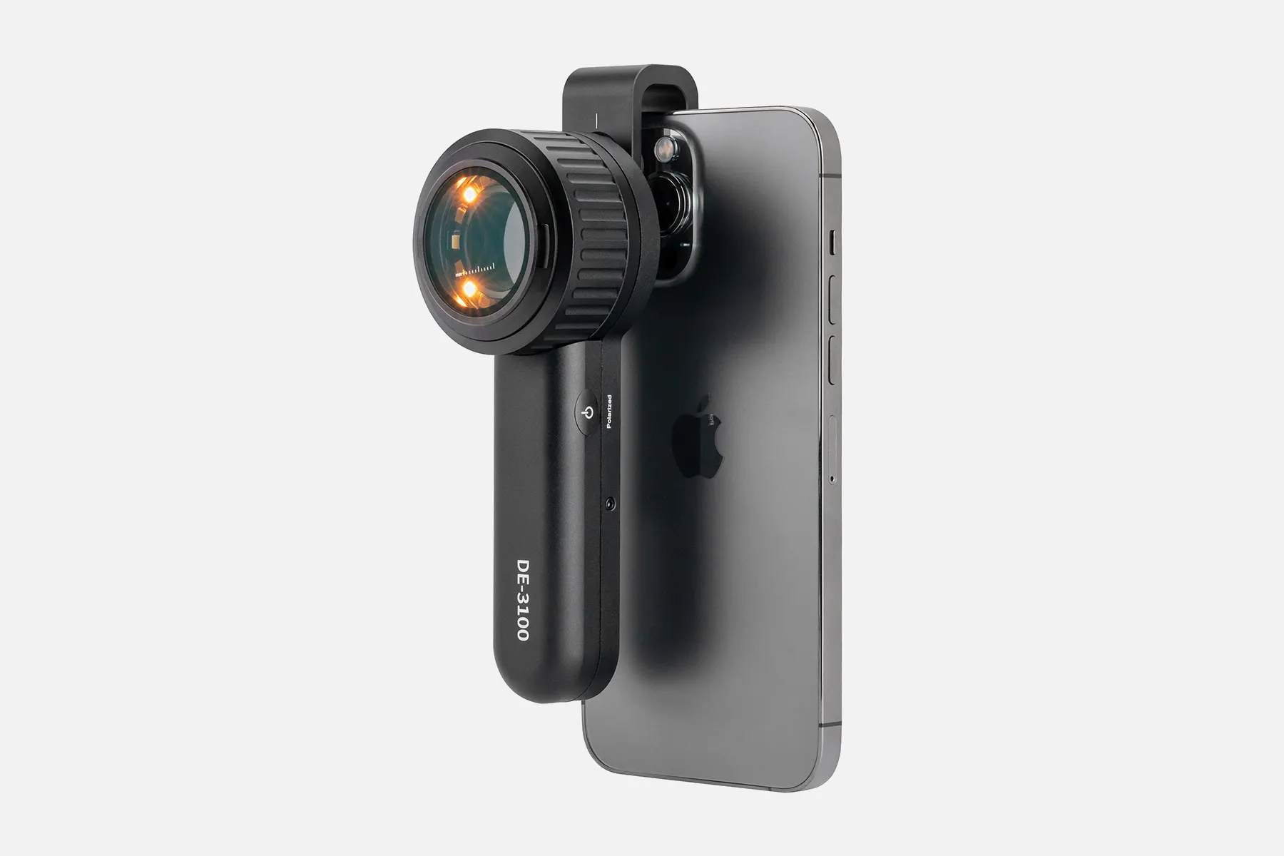
Dermatoscopes use a combination of polarized light and magnification to reveal the skin's structure. Here's a step-by-step look at their function:
1. Light Source: Dermatoscopes are equipped with a light source that illuminates the skin, reducing surface glare and enhancing the visibility of underlying structures.
2. Magnification: Users can adjust the level of magnification to inspect the skin in greater detail.
3. Polarized Light: This feature helps cancel out the reflection from the skin's surface, allowing for a clearer view of sub-surface details.
Dermatoscopio Compare: Key Comparison Factors
When comparing dermatoscopes, consider the following key factors to ensure you select the right device for your needs:
- Magnification Range: The extent of magnification available can impact the level of detail you can observe.
- Light Source Quality: The type and quality of the light source, including options for UV light, can affect the examination's accuracy.
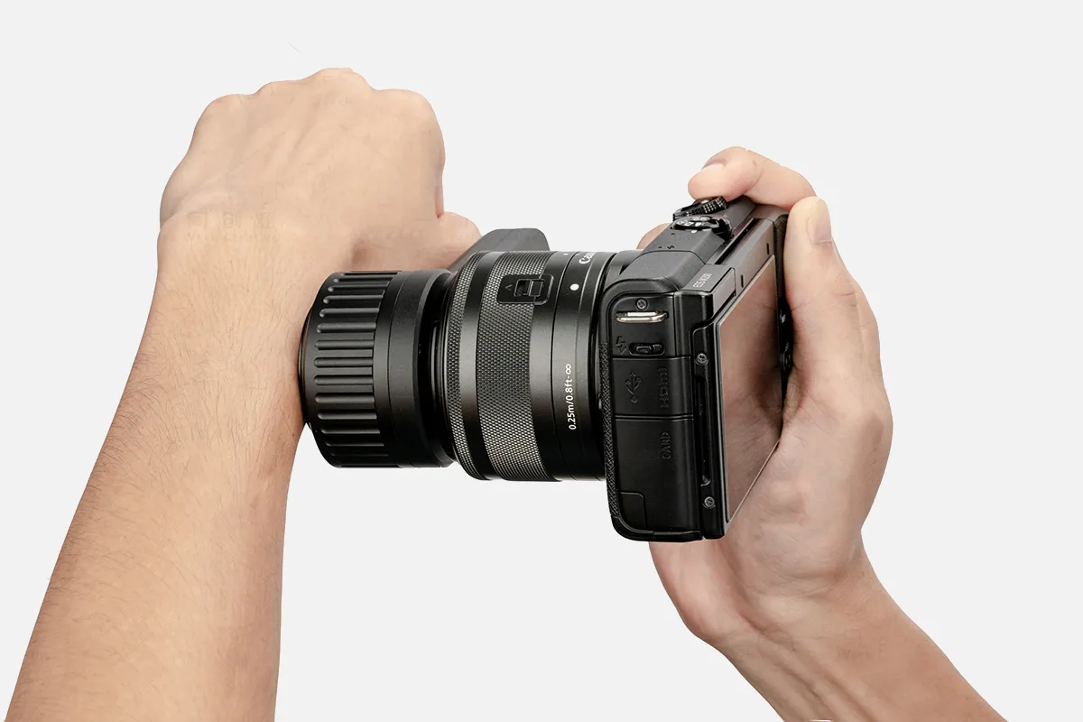
- Portability and Design: The size and weight of the dermatoscope, as well as its ergonomic design, can influence its ease of use.
- Additional Features: Consider any extra features such as digital imaging capabilities, connectivity with apps, or the ability to capture and store images.
Comparing dermatoscopes is a critical step in selecting the right device for your skin examination requirements. By evaluating magnification, light source quality, portability, and additional features, you can make an informed decision. Use our dermatoscopio compare guide to find the dermatoscope that meets your professional or personal skin health monitoring needs.
People May Ask
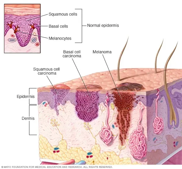
Cancerous cells in basal cellsThese cancers may manifest themselves as: regions that resemble cicatrices: flat, hard, pálida, or amarilla. Elévadas manchas rojizas que pueden provocar comezón. Enrojecidas o rosadas, translúcidas, brillosas y nacaradas protuberancias que pueden tener áreas de color negro, marrón o azul.
What symptoms are present in skin cancer?Instead,¿La mancha o el lunar tienen una forma irregular, with dos partes que se ven muy diferentes?Instead,¿Son los bordes dentados o irregulares?Instead,Color: ¿Cual es el color disparejo?Instead,Measuremente: ¿El lunar o la mancha is más amplio que una arveja?Additional things...
Indicative of the skin becoming more rough and irregular. This frequently happens when the skin scratches itself or becomes rascared. Liquefied skin may result from chronic irritation caused by such effects as an eczema. Typically, topical steroids and humectant cremas are used to treat liquefied skin.
It's a visible, decolorated, flat cutáneal area. Generally speaking, neither the texture nor the thickness of the skin are altered.
Pápula: a circumscribed, perceptible elevation of the skin that is at least one centimeter in diameter. A rise in the dermal or epidermal cell or stromal component is the cause. Mina placa se deno- mina cuando el diámetro supere 1 cm.
372.337)Elevated suelds are also required by dermatologists. Un dermatólogo in Estados Unidos recibe un salario medium de unos 372.337 dólares al año.
fifteen to thirty minutesEn nuestra oficina, el tiempo promedio de una consulta dermatológica es de 15 a 30 minutos, aunque la duración puede variar dependiendo del caso y de la complejidad.
The University of Guadalajara's Institution NumberTutorial Mode Presencial Constructed with Integrated Professional CompetenciesEl programa dura 3 años.Instead,En all, 640 hours were worked.Referendum I/2014/056 normativo
El tratamiento médico y quirúrgico de adultos y niños with trastornos y enfermedades de la piel, las membranas mucosas, el cabello y las uñas is un desigualdad de los dermatólogos del Mayo Clinic.
A non-invasive in vivo diagnostic technique called dermatoscopia was developed to study skin lesions. enhances early diagnosis of potentially malignant lesions, including melanoma, and improves the accuracy of diagnosis of hyperpigmented lesions.
Dermatoscopio Compare Products

Student-proof biological microscope for use in labs and homes: Compound Microscope EXM-150 with Monocular Head, 40–400X Magnification, Disc Diaphragm, and Cordless LED Illumination
YOCTOSUN Magnifying Glasses with 4 Detachable Lenses, 2 LED Lights, Storage Case, Head Strap, Hands-Free Headband Magnifier for Crafts, Jewelry, and Close Work
[1080P HD & Full Lighted View] TOMLOV DM1 Wireless Digital Microscope 50X-1000X Wireless Charging Mini USB Trichome Coin Microscope Camera Magnifier for iPhone, iPad, Android Phone, and Computer via WiFi

For iPhone, Android, and PC, Ninyoon 4K WiFi Microscope with Expert Stand, 50-1000X Digital USB Microscope Wireless Endoscope HD Camera Compatible with All Cellphones Windows Macintosh Tablet Android iPad Glimmer Windows
ZTEEERS Compound Binocular Microscope,40X-2500X Magnification with 10X/25X Eyepiece, LED Illumination, Abbe Condenser, Double-Layer Mechanical Stage,Microscope for Adults.

Windows/Mac OS/TV Compatible, 32GB TF Card Included, Dcorn 10 HDMI LCD Digital Microscope 1500X, Coin Microscope for Adults with 16MP Camera Sensor, Soldering Microscope with LED Lights Touch Control
For the 3GEN, Cameron Sino New 1150mAh Replacement Battery DL2 Dermatoscope, DL3 Dermatoscope, DermLite Pro, DermLite II, DermLite III, and so on
Portable Ear Light and Exam Kit for Home and Professional Use - Cynamed Mini Otoscope - 3X Magnifying Fiber Optic Scope with Extra Tips, Bulb, and Carrying Case - Pocket Diagnostic Tools (Maroon)
Hot Products


News & Blog

Top Reviews
Amy
With my DM9, I'm overjoyed! It easily fulfills the expectations. Tomlov's after-sale service is what makes them unique. I sent them an email with a query, and presto—they responded right away! Those days, who does that? Extremely pleased with the goods and the excellent relationship with their customer support
Robert M. Johnson
In order to determine whether it may be used on any of our PCB manufacturing, inspection, and rework lines for 0603 and 0805 SMDs (Surface Mount Devices), this was acquired. It's definitely worth it for the money. We intend to buy multiple additional units for each of our lines. Though it isn't intended to be, it isn't a replacement for high magnification scopes or trinoculars. It serves as an alternative to benchtop magnifying lenses and 4x and 10x magnifier headsets for quality assurance and inspection. On our lines, we will use photo capture, but we will not use video capture to MicroSD. Production staff also need very little training to learn the focus, which is sharp and clear. From our point of view, this is a tool, and a quite decent one at that, rather than a toy. A big thank you to the engineers that created this gadget!
Fred Malotte
The two side lights and the switch that lets you turn them on, off, or up are nice features. Comparing this to my previous digital version, it's better. Jewelry, coins, and other small objects will be examined using this.


