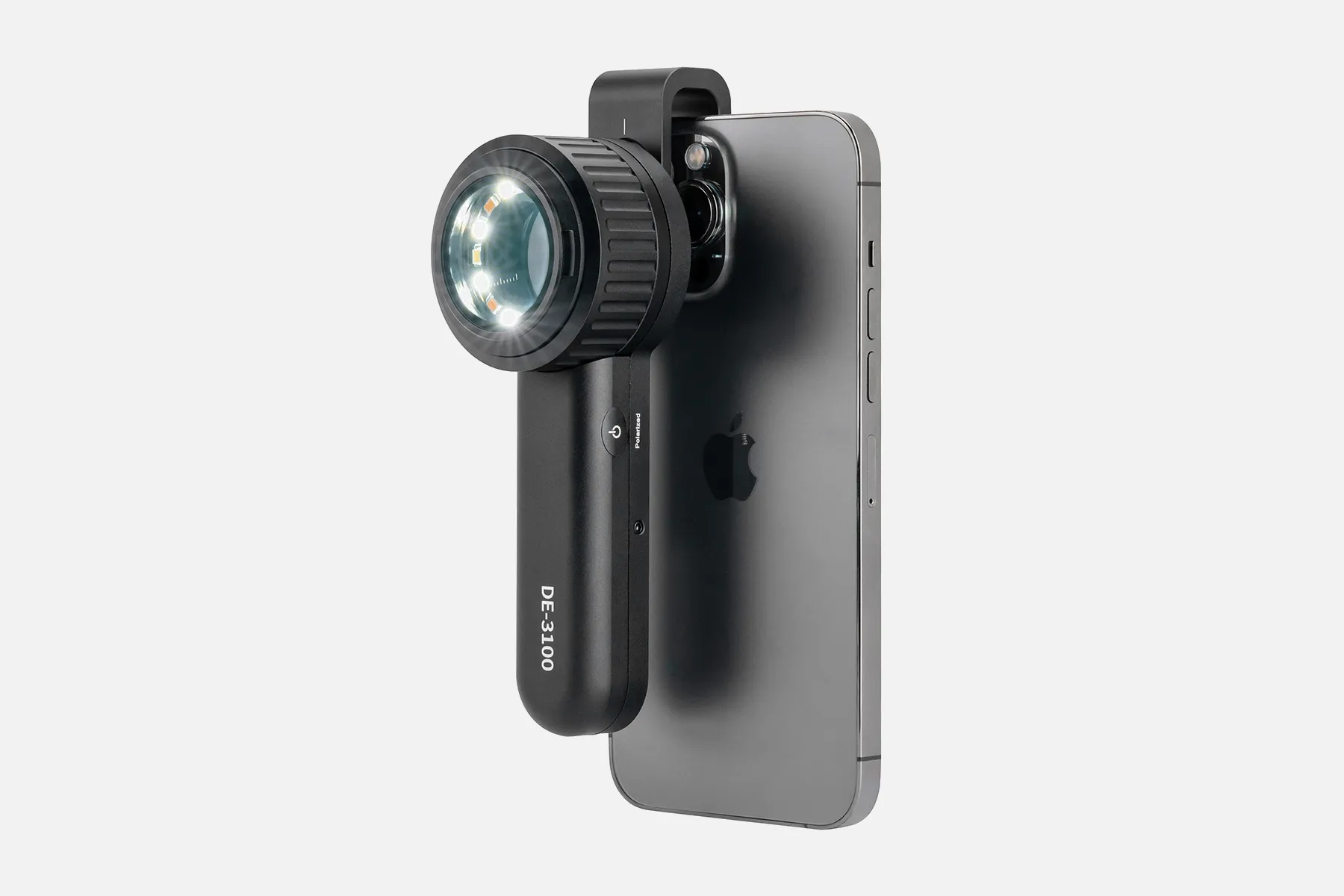
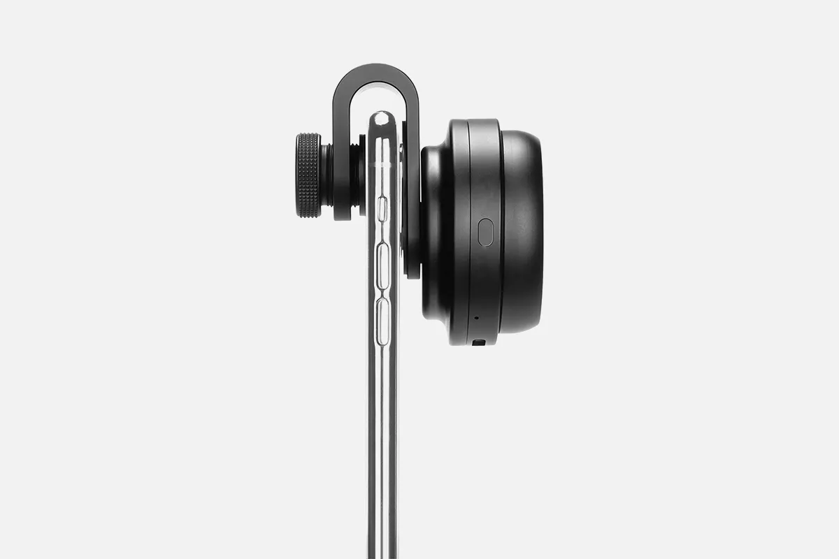
Exploring Dermoscopedia Resources

Dermoscopedia is an online resource for dermoscopy. Dermoscopy is a non-invasive diagnostic technique examining skin lesions with a dermatoscope. A dermatoscope is a handheld instrument that looks like a magnifying glass and can magnify up to 10 times. The technique also involves using a non-polarized light source, a transparent plate, and a liquid medium between the instrument and the skin.

Dermoscopy is also known as dermatoscopy, chemiluminescence, or skin surface microscopy. It's a cheap technique that can improve the accuracy of diagnosing non-pigmented skin tumors.

  • Dermoscopy can be used for:
  • Examining nail fold capillaries
  • Distinguishing skin conditions like lichen planus from others like eczema or psoriasis
  • Evaluating hair loss
  • Examining skin lesions

There are two types of dermoscopy:

  • Polarised dermoscopy: Doesn't require skin contact or fluid immersion
  • Nonpolarised dermoscopy: Always requires skin contact and fluid immersion

Understanding The Functional Utility of The Dermoscopedia Resource

Dermoscopy is a technique that utilizes a specialized medical device called a dermatoscope to examine skin lesions visually. Dermatoscopes have integrated optics to magnify subsurface structures and lighting to illuminate the skin.

During an examination, the gel is first applied to optimize light transmission from the device to the skin. The dermatoscope lens is then held firmly against the lesion while carefully scanning. This allows inspection of morphological patterns and blood vessel arrangements indicative of disease.

The dermatoscope's light and magnification enable the visualization of fine structures in various epidermal and dermal skin layers. Features normally invisible to the naked human eye become apparent. This facilitates improved detection accuracy for melanoma and other skin cancers compared to visual inspection alone, as confirmed by multiple clinical studies.

In summary, dermoscopes grant dermatologists an invaluable window into detailed skin anatomy below the surface. The enhanced magnification and lighting reveal key subsurface clues that can allow early identification of malignant lesions.

People May Ask

What Do The Dermoscopy's White Circles Represent?

A raised lesion may indicate invasive squamous cell carcinoma, while a flat lesion may indicate actinic keratosis. Both polarized and non-polarized dermatoscopy can detect dermatoscopic white circles.

Will There Be No Mole If Melanoma Develops?

A mole is not necessarily the first sign of melanoma. On skin that appears normal otherwise, it can also happen.

Is It Possible to Be Unaware of Melanoma for Two Years?

How long could you have melanoma without realizing it? It is contingent upon the kind of melanoma. Radial melanoma, on the other hand, might spread slowly over a decade, whereas nodular melanoma spreads quickly over a few weeks. Similar to a cavity, a melanoma can proliferate for years without showing any noticeable symptoms.

How Can A Mole's Malignant Status Be Determined?

pigment that has spread into the surrounding skin from the spot's edge. Redness or a fresh enlargement outside the mole's boundaries. Modification in feeling, including pain, sensitivity, or itching. A mole's surface may change, becoming scaly, leaking, bleeding, or developing a bulge or bump.

Dermatologists: How Accurate Are They?

Results of Classification. As can be seen in Table 1, the dermatologists' total mean sensitivity in the first phase of the trial was 59.4% (95% CI 53.3%-65.5%), their specificity was 70.6% (95% CI 62.3%-78.9%), and their accuracy was 65.0% (95% CI 62.3%-67.6%).

What Is The Best Biopsy for Skin?

Examine the punch biopsyThe most popular method for taking skin samples is this one.

Under Dermoscopy, How Does Melanoma Appear?

The pigmented lesion must lack pattern symmetry and color uniformity in addition to at least one of the following characteristics in order to be diagnosed as melanoma: numerous brown dots, pseudopods, radial streaming, scar-like depigmentation, peripheral block spots/globules, five to six colors, a blue-white veil,...

What Level of Specificity Does Dermoscopy Have?

In the naked-eye arm, referral sensitivity, specificity, positive and negative predictive values were 54.1%, 71.3%, 11.3%, and 95.8%, respectively, while in the dermoscopy arm, they were 79.2%, 71.8%, 16.1%, and 98.1%, respectively. Sensitivity and negative predictive value (P =) showed significant variations.

Does BCC Show up on A Dermoscopy?

Dermoscopy is a crucial diagnostic technique for BCC differential diagnosis. Its utility extends beyond identifying rather obvious lesions since it offers a dependable method of differentiating between various histologic subtypes and is capable of identifying very early lesions.

Why Would Someone Use A Dermatoscope?

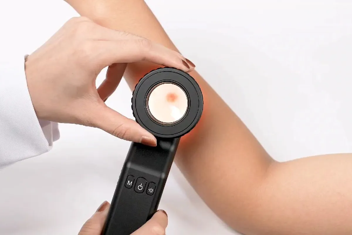
A handheld device known as a dermatoscope is used to perform dermoscopy. Subsurface skin structures in the epidermis, papillary dermis, and dermoepidermal junction-structures that are often invisible to the unaided eye-can be seen thanks to this method [2-4].

Dermoscopedia Products

China wholesale dermoscopedia

EverOne Superior Cardiology Stethoscope, 27-Inch Black Tube

China portable dermoscopedia products supply

Physicians and nurses can use the Kila Scopes Specialist Stethoscope, Professional Single Head Cardiology & Diagnostic Stethoscope with Accessories, K870 Orange.

China high quality dermoscopedia

Professional Dual Head Cardiology & Diagnostic Stethoscope for Physicians and Nurses - with Accessories, Kila Scopes Specialist Stethoscope, K751 Black

China affordable dermoscopedia products supply

Black, 27-inch EverOne Professional Style Cardiology Stethoscope

China affordable dermoscopedia

For nurses, emergency rooms, cardiologists, veterinarians, and fetal pediatrics, the Vive Precision Dualhead Stethoscope is a dual head diaphragm bell that doubles as a chestpiece device for doctors and students.

China high quality dermoscopedia products supply

Stethoscope Holder for Littmann - Genuine Leather Universal Stethoscope Holster with Velcro Closure and Padded Hip/Belt Clip

China wholesale dermoscopedia products supply

Classic Dual Head Paramed Stethoscope for Physicians, Nurses, Medical Students, Professionals in Pediatrics, Medicine, Cardiology, and Home Use with an Additional Diaphragm, Four Eartips, Accessory Case, and Name Tag - 29.5 Inch

China portable dermoscopedia

Nurses, men, women, and pediatric infants can use the 22-inch PARAMED Stethoscope, which is a classic single head cardiology device for medical and clinical use.

China Professional dermoscopedia

Universal Otoscope with Light Generation Earmuffs Infection Detector for Adults and Pediatrics Otoscope

China customized dermoscopedia products supply

Replacement Diaphragm and Silicone Stethoscope Parts for Adult and Pediatric Stethoscopes, BBTO 2 Sets Accessories for Stethoscope Ear Tips (Gray)

Top Reviews

rickyveljaec

My Littman Classic II stethoscope has been surpassed by this one. Compared to my prior stethoscope, which I had been using for the previous nine years, I can hear a lot more. I'm really happy with my purchase and would definitely buy from them again. I adore the vivid color selections.

Techno-Junkie

I purchased this equipment primarily to be able to monitor my bee colony's activity from outside the hives. Without being intrusive, I wanted to be able to assess the condition of my hives by opening each level, putting on my bee costume, and smoking. I can accomplish this with this stethoscope. I had to practice for a while before I could focus on the sound of the bees' beating wings as they went about their job. While the earpieces are fairly sensitive when flipped around, you can wear them and yet not hear anything in one direction. One of the ear piece tubes has an obvious tab big enough to write a brief word on, so I typed "Right," as that's the ear piece that goes to my right ear, not my left. What prevented them from labeling this during assembly? It's a minor gripe that doesn't detract from my evaluation. All in all, an excellent, reasonably priced stethoscope that can even detect heartbeats! I'm quite happy with it and would suggest it to anyone for the price.

jkb

As a dental hygienist, I purchased this for use in my line of work. It works really well for me, so far. I can hear the heartbeat more clearly than I can using the pricy stethoscopes that our office provides.

0

Have questions on gear or your order?

Our Gear Guides are here to help! Get personal advice from pro creatives

Name
Subject
Email address
How can we help?

Instant Answers

How Can Dermoscopy Images Be Captured?

Dermoscopy images can be captured and stored in different ways, such as: • Using a smartphone or tablet with dermoscopic adapter, which consisted in the package.• Using a digital camera

Dermoscopy images can be captured and stored in different ways, such as:

• Using a smartphone or tablet with dermoscopic adapter, which consisted in the package.
• Using a digital camera with dermoscopic adapter, there’s 49mm screw size camera adapter available to order now.

Compatible phone/tablet models:
All iPhone models, 95% Android phones, 90% tablet. For phone/tablet size in 5.25-14mm

Compatible camera models:
All camera with built 49mm filter screw, such as Canon EOS 70D, 80D, 90D; Canon EOS R7, R10, R50, R100; Canon M100, M200, M50, Mark II; Canon G7X Mark III, Sony ZV-1

How Can I Connect My Phone to My Dermatoscope?

There’s universal phone adapter for all our dermoscopes. Please check the installation procedure bellow or watch operation guide. Smartphone Connector (1) Place phone adapter screw in the center of smartphone’s

There’s universal phone adapter for all our dermoscopes. Please check the installation procedure bellow or watch operation guide.

Smartphone Connector

(1) Place phone adapter screw in the center of smartphone’s main camera.
(2) Screw magnet attachment on phone adapter.
(3) Put dermoscope’s back ring and magnet attachment together

Take The Best Images

You need to adjust the focus ring after the dermoscpe connected on smartphone to get the best images.

How Can I Clean My Dermoscopy after Usage?

Cleaning your dermoscopy after usage is important to prevent cross-contamination and infection. The cleaning method may vary depending on the type and model of your dermoscopy, so you should always

Cleaning your dermoscopy after usage is important to prevent cross-contamination and infection. The cleaning method may vary depending on the type and model of your dermoscopy, so you should always follow the manufacturer’s instructions. However, some general steps are:

• Turn off and disconnect your dermoscopy from any power source or device.

• Wipe off any visible dirt or debris from the dermoscopy with a soft cloth or tissue.

• Disinfect the dermoscopy with an alcohol-based wipe or spray, or a disinfectant solution recommended by the manufacturer. Make sure to cover all surfaces, especially the lens and contact plate.

• Let the dermoscopy air dry completely before storing it in a clean and dry place.

• Do not use abrasive or corrosive cleaners, solvents, or detergents that may damage the dermoscopy.

• Do not immerse the dermoscopy in water or any liquid, unless it is waterproof and designed for immersion.

You should clean your dermoscopy after each use, or at least once a day if you use it frequently. You should also check your dermoscopy regularly for any signs of damage or malfunction, and contact the manufacturer or service provider if needed.

Polarized VS Non-Polarized Dermoscopy

A dermoscopy is a device that allows the examination of skin lesions with magnificationand illumination. By revealing subsurface structures and patterns that are not visible tothe naked eye. It can

A dermoscopy is a device that allows the examination of skin lesions with magnificationand illumination. By revealing subsurface structures and patterns that are not visible tothe naked eye. It can improve the diagnose accuracy of skin lesions, such as melanoma,basal cell carcinoma, seborrheic keratosis, etc.

There are two main types of dermoscopy: Non polarized and polarized dermoscopy.We’ve fitted most of our dermoscopys with polarized and non-polarized light. They canbe used in multiple skin structures.

Non-polarized contact Mode

In non-polarized mode, the instrument can provide information about the superficialskin structures, such as milia-like cysts, comedo-like openings, and pigment in theepidemis.

The dermoscopy requires applying a liquid such as mineral oil or alcohol to the skin andplacing the lens in contact with the skin. This reduces surface reflection and enhancesthe view of subsurface structures.

Image with non-polarized light (DE-3100)

Polarized contact Mode

In polarized mode, the instrument allows for visualization for deeper skin structures,such as blood vessels, collagen, and pigment in the dermis.

The dermoscopy does not need to be in contact with the skin or use any liquid. Theirpolarized light can help to eliminate surface reflection and allow visualization ofvascular structures.

Image with polarized light (DE-3100)

Polarized non-contact Mode

The dermoscopy can also use polarized light to examine the skin without direct contact.

In polarized non-contact mode, the instrument allows for examination infected areasand lesions that are painful for the patient, or the difficult to contact pigmented lesions,such as nails and narrow areas.

The contact plate should be removed in this mode, and it does not require applying aliquid to the skin. As it doesn’t require pressure or fluid application on the skin, it canalso avoid cross-contamination and infection risk.

Image in polarized non-contact mode (DE-3100)

How Effectiveness Is Dermoscopy

Compared with visual inspection, the dermoscopy can be used to capture and store skin lesion photos, which play an important role in early skin cancer examination. The dermoscopy allows the

Compared with visual inspection, the dermoscopy can be used to capture and store skin lesion photos, which play an important role in early skin cancer examination.

The dermoscopy allows the examination of skin lesions with magnification and illumination. This can be greatly avoiding the factors that cause interference to visual detection. Such as lighting, skin color, hair and cosmetics.

Several studies have demonstrated that dermoscopy is useful in the identification of melanoma, when used by a trained professional.

It may improve the accuracy of clinical diagnosis by up to 35%
It may reduce the number of harmless lesions that are removed
In primary care, it may increase the referral of more worrisome lesions and reduce the referral of more trivial ones

A 2018 Cochrane meta-analysis published the accuracy of dermoscopy in the detection.

Table 1. Accuracy of dermoscopy in the detection of melanoma in adults
Detection Method Sensitivity, % Specificity, % Positive Likelihood Ratio NegativeLikelihood Ratio
Visual inspection alone (in person) 76 75 3.04 0.32
Dermoscopy with visual inspection (in person) 92 95 18 0.08
Image-based visual inspection alone (not in person) 47 42 0.81 1.3
Dermoscopy with image-based visual inspection (not in person) 81 82 4.5 0.23
ROC—receiver operating characteristic. *Estimated sensitivity calculated on the summary ROC curve at a fixed specificity of 80%.

As we can see, the dermoscope can improve the accuracy of diagnosis of skin lesions, especially melanoma.

Table 1. Accuracy of dermoscopy in the detection of melanoma in adults
Detection Method Sensitivity, % Specificity, % Positive Likelihood Ratio NegativeLikelihood Ratio
Visual inspection alone (in person) 79 77 3.4 0.27
Dermoscopy with visual inspection (in person) 93 99 93 0.07
Image-based visual inspection alone (not in person) 85 87 6.5 0.17
Dermoscopy with image-based visual inspection (not in person) 93 96 23 0.07
ROC—receiver operating characteristic. *Estimated sensitivity calculated on the summary ROC curve at a fixed specificity of 80%.

Characteristics of the dermatoscopic structure of the skin lesions include:

• Symmetry or asymmetry
• Homogeny/uniformity (sameness) or heterogeny (structural differences across the lesion)
• Distribution of pigment: brown lines, dots, clods and structureless areas
• Skin surface keratin: small white cysts, crypts, fissures
• Vascular morphology and pattern: regular or irregular
• Border of the lesion: fading, sharply cut off or radial streaks
• Presence of ulceration

There are specific dermoscopic patterns that aid in the diagnosis of the following pigmented skin lesions:

• Melanoma
• Moles (benign melanocytic naevus)
• Freckles (lentigos)
• Atypical naevi
• Blue naevi
• Seborrhoeic keratosis
• Pigmented basal cell carcinoma
• Haemangioma

No products in the cart.

No products in the cart.

Hot Search Terms