


DE-400 Dermatoscope – IBOOLO
Explanation of What An Electronic Dermatoscope Is
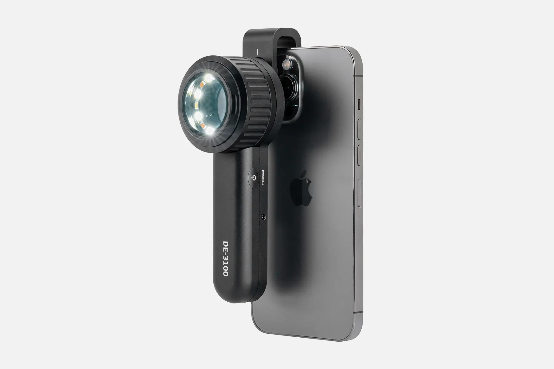
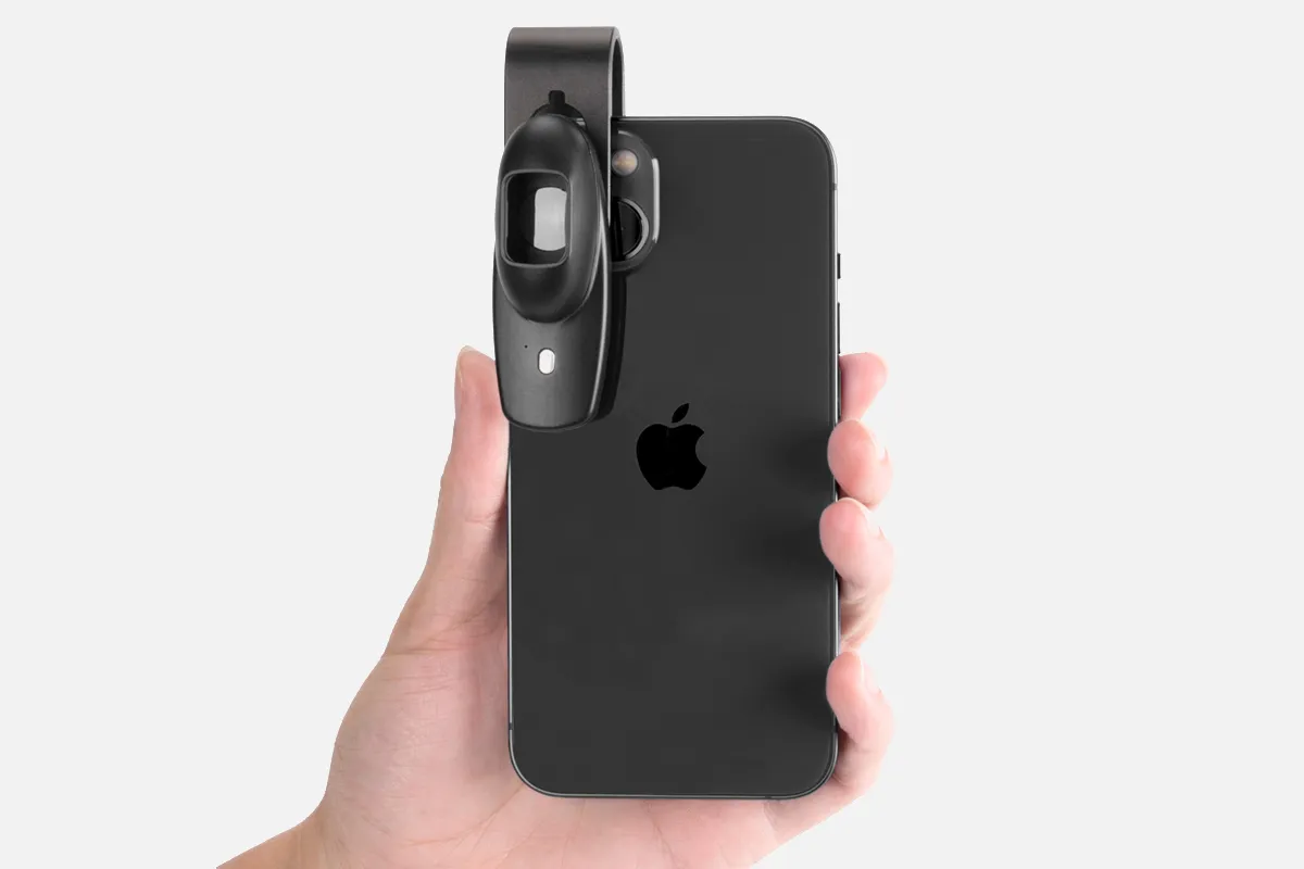
An electronic dermatoscope is a digitally-enabled dermatoscope used to evaluate skin. While standard dermatoscopes utilize optics and lighting to magnify lesions, electronic versions incorporate advanced digital capabilities.
These devices contain sensitive CMOS/CCD imaging sensors to capture clinical-grade dermoscopic photos and HD video. Some models integrate multi-spectral lighting modes like polarized, non-polarized, and UV to accentuate specific skin characteristics. The visual data aids in tracking changes over time.
Embedded processors and memory enable onboard storage, analysis features like side-by-side image comparisons and data transfer connectivity. Wireless and wired options allow linking to external mobile devices, web platforms, and electronic medical record systems.
Electronic dermatoscopes transform traditional manual inspection into sophisticated digital imaging suites. Quantifiable visual documentation combined with software-enhanced workflow aim to improve clinical monitoring, coordination, and diagnoses - especially for detecting melanoma and managing patient cases over longer durations.
How Does An Electronic Dermatoscope?
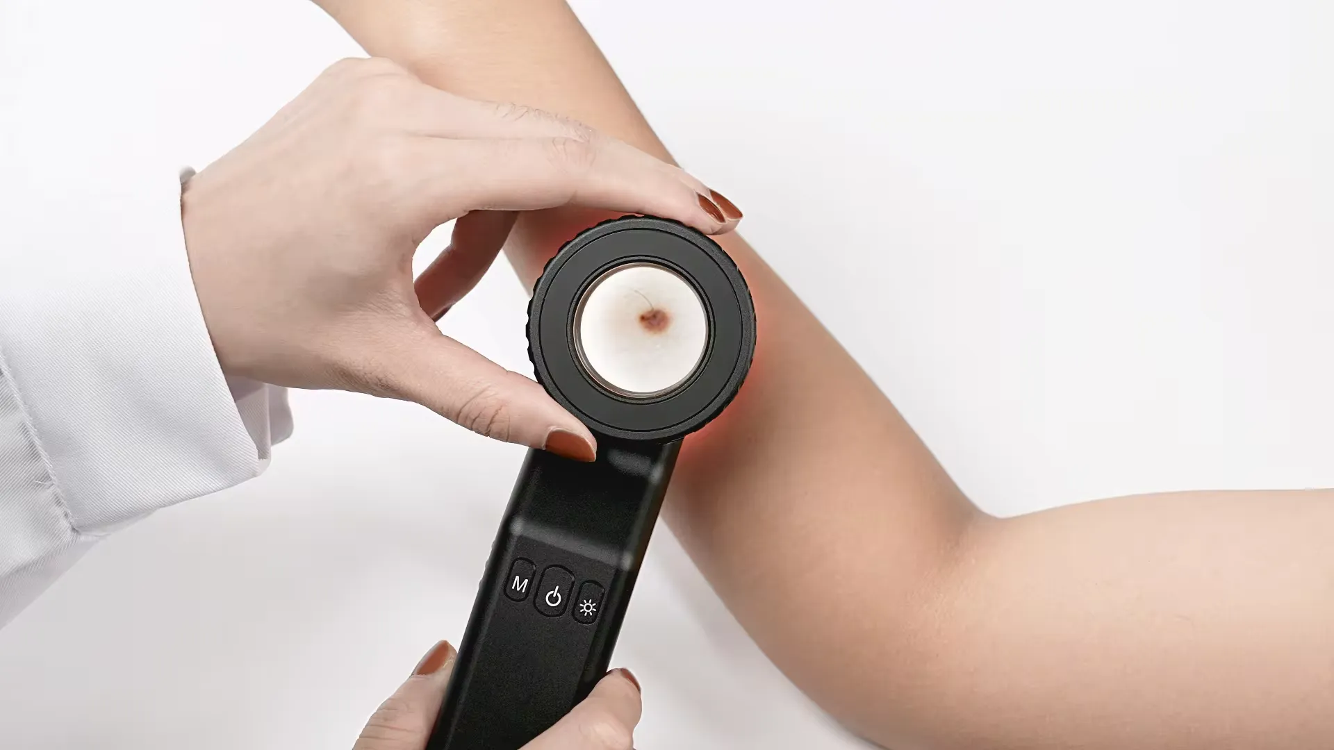
A dermatoscope is a tool similar to a camera used to examine skin lesions. It has a magnifier, a light source, and a transparent plate. The basic principle of dermoscopy is to transilluminate a lesion to study it with high magnification.
Here's how a dermatoscope works:
1. A clinician applies an ultrasound gel or oil to the skin to improve image clarity.
2. The clinician gently presses the dermatoscope into the skin.
3. The dermatoscope transilluminates the lesion.
4. The dermatoscope records images for future comparison.
Some things to consider when choosing a dermatoscope include:
- Compatibility with smartphones and cameras
- Polarization, such as polarized, linear-polarized, unpolarised, or variable
- Contact or non-contact viewing
- Magnification, such as 10x, 16x, or 40x
Some dermatoscopes have built-in magnets to connect and disconnect contact plates or compatible electronic devices, such as smartphones or tablets.
People May Ask
Capture images of your worries. Send them in for an electronic version of the Virtual Skin Cancer Spot Check. Within three business days, a dermatological specialist will reply with recommendations for further care. It really is that easy.
When determining if a mole should be removed because it may be a melanoma, dermatologists frequently utilize a dermatoscope.
Pigment localization is shown by dots and globules. The site is identified by the color: blue or grey dots are caused by melanophages in the dermis, brown globules are caused by junctional nests of melanocytes, and black dots are caused by free melanin in the stratum corneum. Central black spots and globules are typical features of benign lesions.
Dermoscopy dates back to the middle of the modern era, with significant contributions by Ernst Karl Abbe, Unna, Muller, Saphier, and others, as well as Borel's discovery (1655–1656) that lay the groundwork.
Only potentially cancerous moles are seen visually on your skin. It is not able to confirm for you that you have it. A biopsy test is the sole method available for diagnosing the illness. Should your physician determine that a mole is a concern, they will administer a numbing injection and remove as much of the mole as feasible using a scrape.
Visiting a dermatologist might provide you with piece of mind if you discover a worrisome area. Unlike any other medical professional, dermatologists specialize on skin care and have the greatest experience in identifying skin cancer. Go to Find a dermatologist to locate a dermatologist in your area.
Your physician applies a gel or oil to your skin. After that, they place the dermatoscope against your skin to take a close look at the affected area. Your skin is unaffected or damaged by this.
An otoscope, sometimes known as an auriscope, is a medical instrument used for ear examination. Otoscopes are used by medical professionals to explore ear problems and to test for sickness during routine check-ups. The ear canal, tympanic membrane, and eardrum may be visible with an otoscope.
With a dermatoscope, features can be evaluated down to the reticular dermis, and pictures can be taken for further analysis. Transillumination of a lesion for high magnification examination and visualization of minor details is the fundamental idea behind dermoscopy.
How does digital dermatoscopy work? Digital dermatoscopy is one of the most advanced and precise techniques available for identifying alterations in pigmented skin lesions. Melanoma can result from pigmented skin lesions like nevi or moles.
Electronic Dermatoscope Products

Optelec Farview Portable Low Vision Magnifier

Tofficu Pocket Microscope with Camera for Phone and Pocket Microscope Microscope with Digital Camera Handy Phone Microscope Mini Microscope Handheld Microscope Phone Cellphones for Lens White

100X Mini LED Magnifier Microscope, Handheld Microscope, Pen Microscope with Adjustable Focus and Integrated LED Light

TEHAUX Pocket Microscope: A Portable Hand-Powered Magnifying Glass Microscope Tiny Microscope Dermatoscope with Micro Camera Pocket White Miniature Clip-on Camera Phone
Sanko DINOAM2101 Electron Microscope, Dino-Lite Basic E, Basic Function, First Model, Magnification: 10-230 Times

With a universal clip and LEDs, the PUSOKEI 100X phone microscope is a compact and lightweight USB micro magnifier suitable for children, adults, and trichome coins.

Convenient LED pocket microscope with movable focus that can magnify objects up to 100 times
Kids Pocket Microscope Pen with 100X Magnification and Built-in LED Light, Jewelers Loupe, 100X Mini LED Magnifier Microscope
One-piece, 100x zoom mini led magnifier microscope with UV and LED light, jewelers loupe magnifying glass for mobile phone, Othmro 1Pcs Mini 100X Microscope.

Battery Replacement for 3GEN CS DL2 Dermatoscope, DL3 Dermatoscope, DermLite Pro, DermLite II, DermLite III, and so on
Hot Products


News & Blog


Top Reviews
Linda
For my mother, who has macular degeneration, this has been incredibly beneficial. She can operate it rather easily, albeit it is a little tricky to move the object beneath the magnifier.
J. Socrates
While the Eschenbach device works perfectly, two out of the three light panels are no longer functional. The device's spoken voice says something extremely quickly. The sound is a little weak, but it seems to be saying "Battery low." So perhaps there's a problem with the lights' charging. I will investigate that. If this is the case, then another example of Eschenbach's clever design is to only turn on the main light when the battery becomes low rather than leaving the user in the dark. I'll follow up.
Hookedon
In the world of work, tools such as these are essential for someone who is legally blind and has low vision. One issue is that they can be quite expensive. I was looking for something that was lightweight and easy to carry. It's fantastic. I was worried about the screen breaking after watching YouTube videos and reading the IBOOLO description, so I thought I would have to buy a case. Its inclusion of a hard case astounded me. There are tons of features. It's simple to use the menus. Everybody talks. It has a 22X zoom. I usually use 12X for it. It's convenient that I can use the touch screen or the zoom buttons. The photos have excellent quality. clear and concise The picture on my other video magnifier, a 7-inch Optelec that is meant to be color, appears washed out. This gadget is quite excellent. I therefore adore this video magnifier. will utilize it daily at work. But there are two problems. I don't mean to be critical, but these wouldn't be a deal-breaker. I do, however, want the company to see this.


