What Is A Dermatoscopy Phone Attachment?

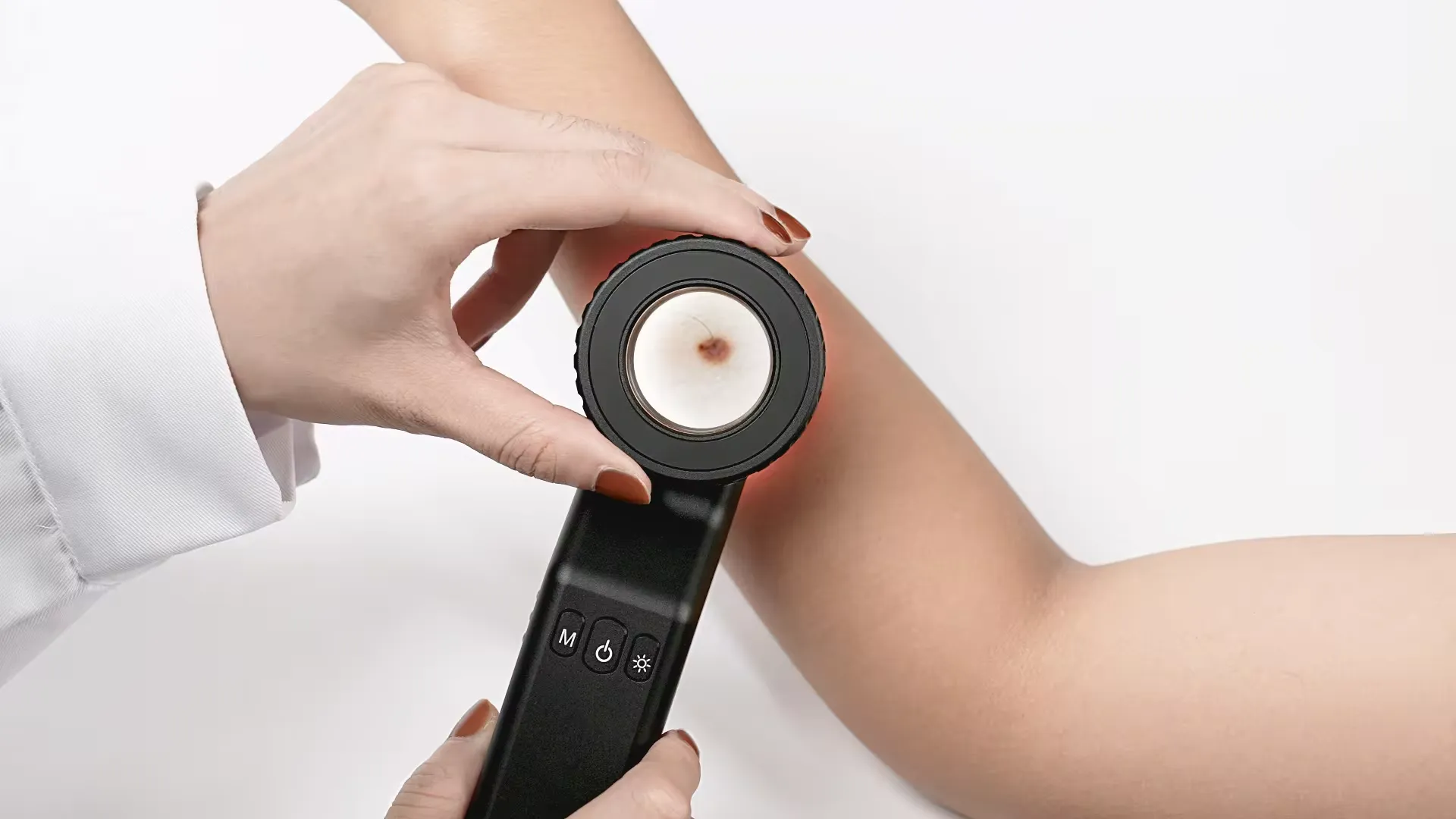
A dermatoscope is a specialized magnifying device used to inspect skin lesions, revealing subsurface visual details impossible to see with the naked eye alone. A dermatoscope phone attachment is an accessory that securely connects a dermatoscope to a smartphone such as an iPhone or Android device.

When selecting a dermatoscopy attachment for smartphone-based skin photography and tracking, key factors to evaluate include:

  • Device Compatibility - Ensure the mount correctly fits your phone model
  • Polarizing Filter - polarization reduces skin surface reflections and glare
  • Magnification power - 10x is standard but higher magnification shows finer structures
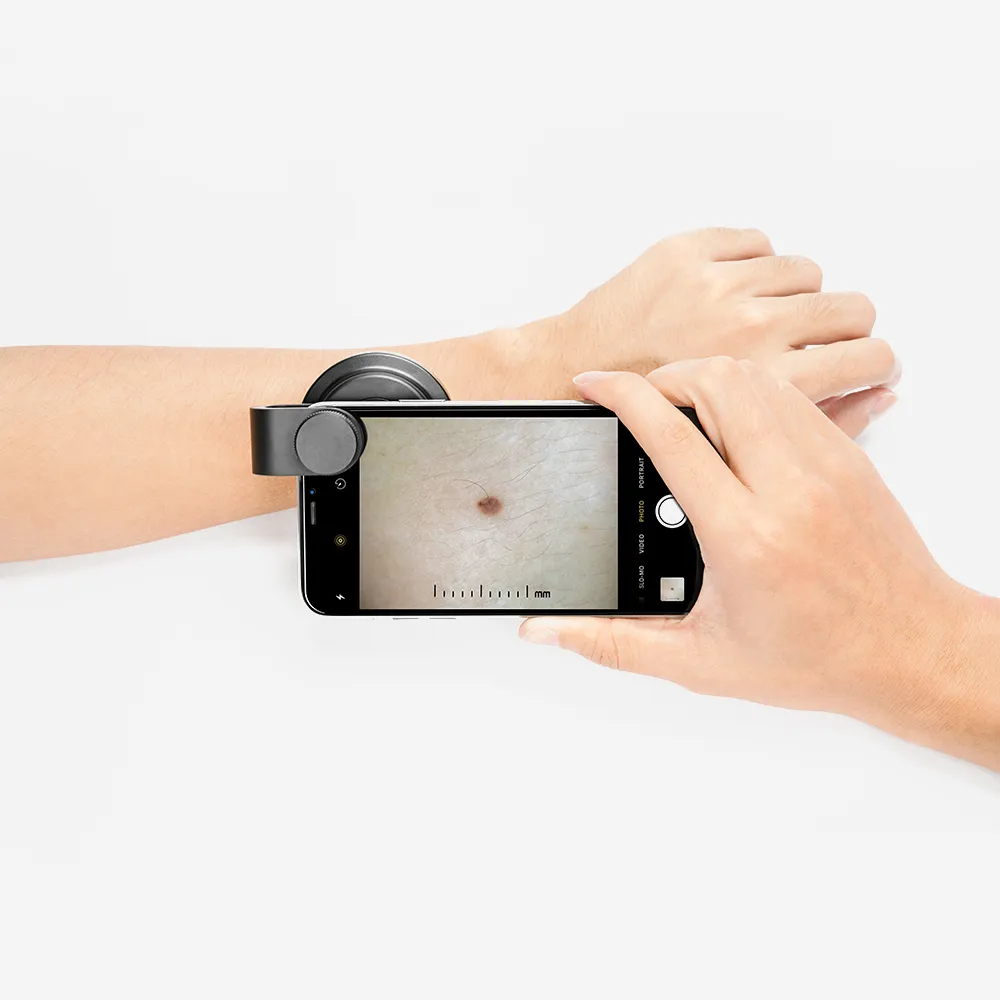
  • Image Quality - Should capture fine details and allow proper focus
  • Ergonomics - The phone should be stable and easy to position on lesions

The main benefit of interfacing a smartphone with a dermatoscope is documenting clinical images to monitor suspicious moles over time. Photos can also be shared remotely with specialists. Choosing an optimal attachment facilitates mobile skin surface microscopy.

How to Attach A Dermatoscope to A Smartphone

There are various methods to securely mount a dermatoscopy onto a smartphone to conduct hands-free skin examinations and documentation:

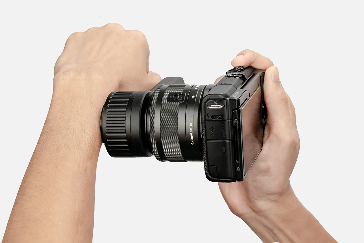
Sklip Design - The Sklip model slides over and straddles the smartphone directly over its rear camera lens.

DermLite Range - DermLite units feature proprietary magnetic connector cables (MCC) to interface with iPhone, Android, and tablet devices.

Universal phone Adaptor - Adjustable screw widths allow these mounts to grip diverse phone sizes, typically incorporating overhanging cradles to position phones.

Phone Case Method - Special smartphone cases with integrated magnets or fittings aim to stabilize specific iPhone and Android models for skin imaging.

In practice, properly centering the phone’s camera under the mounted dermatoscope helps accurately capture magnified clinical images and subtle skin details. Applying a drop of oil often improves visibility. Taking multiple photos while applying mild pressure facilitates inspecting the lesion from different angles. Proper mounting and positioning are key for quality visualization.

People May Ask

Is A Mole Necessary for Melanoma to Exist?

Although some melanomas are pink, red, purple, or skin-colored, the majority are black or brown in hue. While the majority of melanomas start in normal skin, about 30% do so in pre-existing moles. Since most melanomas do not begin as moles, it is extremely crucial to monitor any changes in your skin.June 21, 2021....
The symptoms, stages, diagnosis, treatment, and prevention of melanomaOhioHealthCare.orgwww.clevelandclinic.org/my/health/14391-melanoma

Is Magnification of 100X Beneficial?

100x - This provides an excellent all-around view of Jupiter, allowing you to see all four of its moons in the field of view and to observe cloud detail on the planet. A little orange dot on the planet (if it is on the side facing Earth) and the Great Red Spot can also be seen.

Is It Possible to Determine An Image's Magnification?

The size of the image divided by the object's size is commonly used to calculate magnification. Magnification can be computed using the following straightforward formula: size of the image / size of object.

Under A Microscope, Is Melanoma Visible?

A melanoma can be measured using a specialized tool and examined under a microscope to find its thickness. The treatment plan that your care team chooses is influenced by the thickness of the melanoma. Surgery may not be necessary for thinner melanomas if the malignancy and some surrounding healthy tissue can be removed.

When Utilizing An Otoscope, What Safety Measures Should Be Followed?

Make sure the patient is seated comfortably, at eye level, and that privacy is respected. 2. Inspect the surrounding scalp, pinna, and outer meatus. Examine the area for scars from prior surgery, infection, discharge, edema, and indications of skin abnormalities or lesions.

Is Holding Your Phone up to Your Ear Safe?

Put your phone on [speaker phone" or hold it away from your ear if you're not using a hands-free gadget. Switch sides when conversing on the phone. Since cell phones generate radiation even when they are not in use, avoid keeping your phone in your pocket, on your belt, or anyplace near your body.

Does An Otoscope Allow You to See An Ear Infection?

How Ear Infections Are Diagnosed by Physicians. The only surefire way to find out if your child has one is for a medical professional to use an otoscope, a tiny flashlight with a magnifying lens, to examine inside her ear.

Does An Otoscope Provide for Ear Infection Detection?

Reflectometry using acoustics. By measuring the amount of sound that is reflected back from the eardrum, this test provides an indirect indicator of middle ear fluid levels. The majority of sound is normally absorbed by the eardrum. The eardrum will reflect more sound, nevertheless, the more pressure there is from the middle ear fluid.

Do Earphones Have The Potential to Infect Ears?

They have the ability to hold earwax in place and push it upward. This could lead to an accumulation of earwax and an ear infection. In the ears, earbuds can also retain fluid and moisture. The bacteria that cause an ear infection may begin to flourish in this warm, moist environment.

How Does A Smartphone Otoscope Work?

The TYM smartphone otoscope is not like other ordinary otoscopes since it can take pictures and videos, save them in the patient's file, and share them safely with other medical experts.

Dermatoscope Phone Attachment Products

China Professional dermatoscope phone attachment

Digital Microscope: Wireless, Handheld, USB-Powered Pocket Microscope with 50x-1000x Zoom Fixed Focus HD Magnifier and LED Light; Compatible with iPhone, Android Phone, MacBook, and Windows PC (Black); Inspection Camera

China Professional dermatoscope phone attachment products supply

Compact 200X Phone Mini Pocket Microscope with LED Light and Universal Clip, 99% Smartphone Compatible Portable Digital Microscope Camera Attachments, Microworld for Children and Adults (Black, 200X Without CPL)

China affordable dermatoscope phone attachment

COHEALI 3 pieces Miniature Microscope Camera Microscope Miniaturized Phone Dermatoscope Electronic Microscope Cellular Microscope Carrying Handheld Compact Cell Phone White with Pocket Magnifying Glass and Pocket Camera

China customized dermatoscope phone attachment

Small Microscope, Telescopic Design, 60x Portable Handheld USB Microscope Camera, Broad Irradiation Range (9882(RD)2 black)

China customized dermatoscope phone attachment products supply

The LED-lit, compact clip-on miniature mobile phone microscope by Akozon is a 9595W, 60X magnifying glass microscope.

China wholesale dermatoscope phone attachment products supply

Phone Attachment for EXCEART Microscope Handy Phone Microscope Small Microscope for Pocket Microscope Electronic Microscope Microscope Microscope for Cell Phone Hand with White Lens and Lights

China portable dermatoscope phone attachment products supply

ABOUFAN Triple-Lens Cell Phone LED Lens Phone Microscope, Black Phone Microscope with Universal Clip Microscope Magnification Lens for All Phones, Clip-on Magnifier

China high quality dermatoscope phone attachment products supply

APEXEL Microphone for Android/iPhone, 100x Magnification, LED Light, CPL Handheld Pocket, Suitable for Accessories for Smartphones Macro Focus Glass as a Present.

China affordable dermatoscope phone attachment products supply

APEXEL Portable Microscope, Small Microscope for iPhone Camera Lens Attachment, 100X Microscopes with Universal Clip Compatible with All Smartphones, Transparent Micro Loupe Lens for Children and Adults, Trichome Coin

China wholesale dermatoscope phone attachment

Three pieces of NUOBESTY s tiny, portable, magnifying glass phone attachments for dermatoscopes, mobile phones, and hand-held, external white digital microscopes

Top Reviews

SC Dad

I asked it to examine the trichomes on my plants. It functions flawlessly, is simple to use, and I have no issues!

john Meyers

As a child, I enjoy disassembling things. I had some fun and ease taking pictures of the insides of several antique electronics. I used this lens to take a close-up photo of a dead fly today, and it worked well! I'm happy that I got this phone purchase.

Martin Hamblin

These pictures are of my hair and a crystal onyx jewelry that I own. I really enjoyed looking at various objects, including my hands, using this lens. For the lens to work well, you do need to be relatively close to the thing; picture it like a scientific microscope. With this lens, I can now see everything around me in a minuscule detail! Seeing how all these commonplace items appear up close is fascinating, awesome, and enjoyable. Excellent for youth and young adults. The crystal is shown in two different photos: one with the integrated led lights off and the other with them on. I have an LG velvet and an iPhone 11, and this fits both phones. It's also incredibly simple to put on and take off. You can move the slider to align the lens so that it screws on in front of your camera.

0

Have questions on gear or your order?

Our Gear Guides are here to help! Get personal advice from pro creatives

Name
Subject
Email address
How can we help?

Instant Answers

How Can Dermoscopy Images Be Captured?

Dermoscopy images can be captured and stored in different ways, such as: • Using a smartphone or tablet with dermoscopic adapter, which consisted in the package.• Using a digital camera

Dermoscopy images can be captured and stored in different ways, such as:

• Using a smartphone or tablet with dermoscopic adapter, which consisted in the package.
• Using a digital camera with dermoscopic adapter, there’s 49mm screw size camera adapter available to order now.

Compatible phone/tablet models:
All iPhone models, 95% Android phones, 90% tablet. For phone/tablet size in 5.25-14mm

Compatible camera models:
All camera with built 49mm filter screw, such as Canon EOS 70D, 80D, 90D; Canon EOS R7, R10, R50, R100; Canon M100, M200, M50, Mark II; Canon G7X Mark III, Sony ZV-1

How Can I Connect My Phone to My Dermatoscope?

There’s universal phone adapter for all our dermoscopes. Please check the installation procedure bellow or watch operation guide. Smartphone Connector (1) Place phone adapter screw in the center of smartphone’s

There’s universal phone adapter for all our dermoscopes. Please check the installation procedure bellow or watch operation guide.

Smartphone Connector

(1) Place phone adapter screw in the center of smartphone’s main camera.
(2) Screw magnet attachment on phone adapter.
(3) Put dermoscope’s back ring and magnet attachment together

Take The Best Images

You need to adjust the focus ring after the dermoscpe connected on smartphone to get the best images.

How Can I Clean My Dermoscopy after Usage?

Cleaning your dermoscopy after usage is important to prevent cross-contamination and infection. The cleaning method may vary depending on the type and model of your dermoscopy, so you should always

Cleaning your dermoscopy after usage is important to prevent cross-contamination and infection. The cleaning method may vary depending on the type and model of your dermoscopy, so you should always follow the manufacturer’s instructions. However, some general steps are:

• Turn off and disconnect your dermoscopy from any power source or device.

• Wipe off any visible dirt or debris from the dermoscopy with a soft cloth or tissue.

• Disinfect the dermoscopy with an alcohol-based wipe or spray, or a disinfectant solution recommended by the manufacturer. Make sure to cover all surfaces, especially the lens and contact plate.

• Let the dermoscopy air dry completely before storing it in a clean and dry place.

• Do not use abrasive or corrosive cleaners, solvents, or detergents that may damage the dermoscopy.

• Do not immerse the dermoscopy in water or any liquid, unless it is waterproof and designed for immersion.

You should clean your dermoscopy after each use, or at least once a day if you use it frequently. You should also check your dermoscopy regularly for any signs of damage or malfunction, and contact the manufacturer or service provider if needed.

Polarized VS Non-Polarized Dermoscopy

A dermoscopy is a device that allows the examination of skin lesions with magnificationand illumination. By revealing subsurface structures and patterns that are not visible tothe naked eye. It can

A dermoscopy is a device that allows the examination of skin lesions with magnificationand illumination. By revealing subsurface structures and patterns that are not visible tothe naked eye. It can improve the diagnose accuracy of skin lesions, such as melanoma,basal cell carcinoma, seborrheic keratosis, etc.

There are two main types of dermoscopy: Non polarized and polarized dermoscopy.We’ve fitted most of our dermoscopys with polarized and non-polarized light. They canbe used in multiple skin structures.

Non-polarized contact Mode

In non-polarized mode, the instrument can provide information about the superficialskin structures, such as milia-like cysts, comedo-like openings, and pigment in theepidemis.

The dermoscopy requires applying a liquid such as mineral oil or alcohol to the skin andplacing the lens in contact with the skin. This reduces surface reflection and enhancesthe view of subsurface structures.

Image with non-polarized light (DE-3100)

Polarized contact Mode

In polarized mode, the instrument allows for visualization for deeper skin structures,such as blood vessels, collagen, and pigment in the dermis.

The dermoscopy does not need to be in contact with the skin or use any liquid. Theirpolarized light can help to eliminate surface reflection and allow visualization ofvascular structures.

Image with polarized light (DE-3100)

Polarized non-contact Mode

The dermoscopy can also use polarized light to examine the skin without direct contact.

In polarized non-contact mode, the instrument allows for examination infected areasand lesions that are painful for the patient, or the difficult to contact pigmented lesions,such as nails and narrow areas.

The contact plate should be removed in this mode, and it does not require applying aliquid to the skin. As it doesn’t require pressure or fluid application on the skin, it canalso avoid cross-contamination and infection risk.

Image in polarized non-contact mode (DE-3100)

How Effectiveness Is Dermoscopy

Compared with visual inspection, the dermoscopy can be used to capture and store skin lesion photos, which play an important role in early skin cancer examination. The dermoscopy allows the

Compared with visual inspection, the dermoscopy can be used to capture and store skin lesion photos, which play an important role in early skin cancer examination.

The dermoscopy allows the examination of skin lesions with magnification and illumination. This can be greatly avoiding the factors that cause interference to visual detection. Such as lighting, skin color, hair and cosmetics.

Several studies have demonstrated that dermoscopy is useful in the identification of melanoma, when used by a trained professional.

It may improve the accuracy of clinical diagnosis by up to 35%
It may reduce the number of harmless lesions that are removed
In primary care, it may increase the referral of more worrisome lesions and reduce the referral of more trivial ones

A 2018 Cochrane meta-analysis published the accuracy of dermoscopy in the detection.

Table 1. Accuracy of dermoscopy in the detection of melanoma in adults
Detection Method Sensitivity, % Specificity, % Positive Likelihood Ratio NegativeLikelihood Ratio
Visual inspection alone (in person) 76 75 3.04 0.32
Dermoscopy with visual inspection (in person) 92 95 18 0.08
Image-based visual inspection alone (not in person) 47 42 0.81 1.3
Dermoscopy with image-based visual inspection (not in person) 81 82 4.5 0.23
ROC—receiver operating characteristic. *Estimated sensitivity calculated on the summary ROC curve at a fixed specificity of 80%.

As we can see, the dermoscope can improve the accuracy of diagnosis of skin lesions, especially melanoma.

Table 1. Accuracy of dermoscopy in the detection of melanoma in adults
Detection Method Sensitivity, % Specificity, % Positive Likelihood Ratio NegativeLikelihood Ratio
Visual inspection alone (in person) 79 77 3.4 0.27
Dermoscopy with visual inspection (in person) 93 99 93 0.07
Image-based visual inspection alone (not in person) 85 87 6.5 0.17
Dermoscopy with image-based visual inspection (not in person) 93 96 23 0.07
ROC—receiver operating characteristic. *Estimated sensitivity calculated on the summary ROC curve at a fixed specificity of 80%.

Characteristics of the dermatoscopic structure of the skin lesions include:

• Symmetry or asymmetry
• Homogeny/uniformity (sameness) or heterogeny (structural differences across the lesion)
• Distribution of pigment: brown lines, dots, clods and structureless areas
• Skin surface keratin: small white cysts, crypts, fissures
• Vascular morphology and pattern: regular or irregular
• Border of the lesion: fading, sharply cut off or radial streaks
• Presence of ulceration

There are specific dermoscopic patterns that aid in the diagnosis of the following pigmented skin lesions:

• Melanoma
• Moles (benign melanocytic naevus)
• Freckles (lentigos)
• Atypical naevi
• Blue naevi
• Seborrhoeic keratosis
• Pigmented basal cell carcinoma
• Haemangioma

No products in the cart.

No products in the cart.

Hot Search Terms