


DE-400 Dermatoscope – IBOOLO
Digital Dermatoscope: A Comprehensive Guide to Technology, Applications, And Maintenance
In the realm of skin health assessment, the digital dermatoscope is revolutionizing traditional observation methods. This advanced tool integrates cutting-edge optical technology with digital imaging, enabling clear visualization of skin microstructures and providing intelligent image management for professional medical evaluations. This article offers an in-depth exploration of the digital dermatoscope, covering its technical principles, diverse clinical applications, and practical guidelines for operation and maintenance. Importantly, as an auxiliary observation tool, the digital dermatoscope supports professional medical judgment, with all findings requiring comprehensive evaluation by trained healthcare professionals.
What Is A Digital Dermatoscope And How It Transforms Skin Assessment
A digital dermatoscope is a modern diagnostic aid that combines traditional dermoscopy optics with digital imaging capabilities. Unlike conventional dermatoscopes, this device digitizes magnified images, transforming skin observation into a more precise and traceable process.
Key Technological Advancements
The digital dermatoscope achieves three major breakthroughs:
1. Image Digitization: High-resolution sensors capture detailed skin microstructures.
2. Real-Time Recording: Images can be instantly stored and reviewed.
3. Intelligent Analysis: Some models feature basic image-processing algorithms for enhanced diagnostics.
Impact on Skin Assessment
These advancements have fundamentally changed skin evaluation:
- Transition from instantaneous judgment to traceable records.
- Enable multi-timepoint image comparisons.
- Facilitate remote consultations and collaborative discussions.
- Provide comprehensive data for professional medical assessments.
It is critical to note that the digital dermatoscope serves as an auxiliary tool. All observations must be interpreted by qualified medical professionals in conjunction with clinical expertise.
Why The Digital Dermatoscope Is A Game-Changer in Dermatology
The digital dermatoscope is hailed as a transformative tool in dermatology due to its unique advantages in clinical practice. Below, we explore its core benefits.
Core Advantages
1. Workflow Optimization:
- Reduces examination time by approximately 40%.
- Automates image archiving.
- Standardizes report generation.
2. Enhanced Observation Capabilities:
- Supports variable magnification (20-100x).
- Incorporates multispectral imaging.
- Offers 3D reconstruction technology.
3. Improved Collaboration:
- Cloud storage enables multi-device access.
- Standardized image formats ensure compatibility.
- Supports remote consultations.
Real-World Impact
Clinical studies indicate a 35% improvement in observation consistency with the digital dermatoscope. Additionally, 90% of professional users report enhanced workflow efficiency, while teaching hospitals note significant improvements in educational demonstrations. However, these benefits rely on proper operation and an understanding of the device’s role as an auxiliary tool requiring professional validation.
Unveiling The Magic of The Digital Dermatoscope
For first-time users, the digital dermatoscope captivates with its advanced technological features. Let’s dive into what makes this high-tech tool stand out.
Technological Highlights
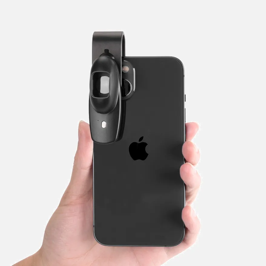
- Miniaturized HD Camera: An 8mm-diameter camera delivering 4K resolution.
- Smart Illumination System: Automatically adjusts brightness and color temperature.
- Wireless Connectivity: Enables real-time image sharing.
- Ergonomic Design: Allows one-hour fatigue-free operation.
Enhanced User Experience
Simplified Operation:
- One-touch autofocus.
- Touchscreen controls.
- Voice annotation support.
Superior Image Quality:
- Color accuracy >98%.
- 14-bit dynamic range.
- Noise levels <0.5%.
Versatile Features:
- Interchangeable filters.
- External display compatibility.
- Integration with major medical systems.
These innovations make the digital dermatoscope a powerful tool for professional skin observation, though its outputs must always be evaluated by medical experts.
Inside The Digital Dermatoscope: A Peek at Its High-Tech Core
The digital dermatoscope owes its precision to a meticulously engineered internal structure. Let’s break down its core components.
Key Components
1. Optical System:
- Multi-coated lens assembly.
- Polarized light filters.
- Anti-reflective coatings.
2. Imaging System:
- 1/2.3-inch CMOS sensor.
- Digital signal processor.
- USB 3.0 data interface.
3. Illumination System:
- Ring-shaped LED array.
- Constant-current driver circuit.
- Advanced heat dissipation.
Technical Specifications
- Optical resolution: 228 lp/mm.
- Distortion control: <1.5%.
- Working distance: Adjustable 2-5 cm.
- Power consumption: <3.5W.
Manufacturing Excellence
- Military-grade sealing.
- Nanoscale mirror polishing.
- Medical-grade materials.
- Electromagnetic compatibility design.
These design elements ensure the digital dermatoscope delivers reliable performance, providing high-quality data for professional assessments.
How The Digital Dermatoscope Achieves Precision Skin Observation
The digital dermatoscope’s ability to deliver precise observations stems from its sophisticated technical processes.
Operational Workflow
1. Optical Acquisition:
- Uniformly illuminates the target area.
- Eliminates surface reflections.
- Captures deep-layer scattered light.
2. Signal Conversion:
- Performs photoelectric conversion.
- Executes analog-to-digital conversion.
- Applies digital noise reduction.
3. Image Processing:
- Automatic white balance.
- Edge enhancement.
- Color correction.
Precision Assurance
- Daily auto-calibration.
- Temperature compensation algorithms.
- Illumination uniformity checks.
- Periodic professional validation.
Operational Guidelines
- Maintain standard working distance.
- Ensure clean skin surfaces.
- Select appropriate magnification.
- Record environmental parameters.
These processes ensure the digital dermatoscope provides stable, reliable data, but all findings must be professionally evaluated.
Imaging Technology: Unlocking Skin Microstructure Clarity
The digital dermatoscope’s ability to reveal skin microstructures relies on its advanced imaging technology.
Key Imaging Components
1. Multispectral Illumination:
- Uses LED arrays (400-1000 nm).
- Switchable polarized/non-polarized modes.
- Adaptive brightness for varying skin tones.
2. High-Precision Optics:
- Apochromatic lenses to eliminate chromatic aberration.
- 20-layer anti-reflective coatings.
- Patented light-guiding structures.
3. Digital Imaging Processing:
- 14-megapixel CMOS sensor.
- Real-time noise reduction.
- Multi-frame synthesis.
Technical Highlights
- Resolution: 4.2 μm/pixel.
- Depth of field: 0.8-1.2 mm.
- Color depth: 16-bit.
- Dynamic range: 90 dB.
This synergy of technologies ensures the digital dermatoscope delivers clear, detailed images, though final interpretations require professional expertise.
How to Use A Digital Dermatoscope Correctly
To maximize the digital dermatoscope’s performance, follow these standardized operating procedures:
Preparation
1. Device Check:
- Ensure battery level >80%.
- Verify lens cleanliness.
- Confirm available storage space.
2. Environmental Setup:
- Maintain ambient light at 50-100 lux.
- Control room temperature (18-26°C).
- Prepare coupling gel if needed.
Operation Steps
1. Skin Preparation:
- Clean the observation area.
- Remove surface oils and cosmetics.
- Keep skin in its natural state.
2. Device Configuration:
- Select magnification (start at 10x).
- Adjust light intensity (auto/manual).
- Choose imaging mode (polarized/non-polarized).
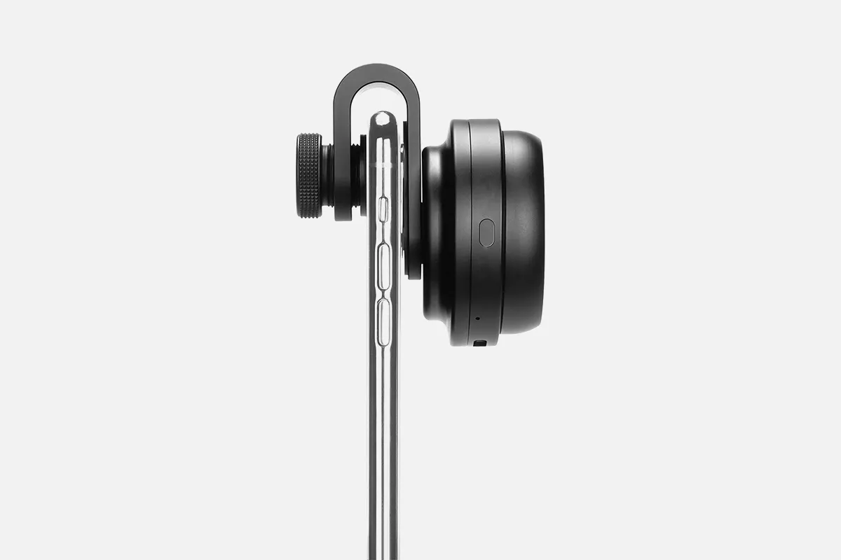
3. Image Acquisition:
- Maintain 1.5-2 cm distance.
- Hold steady for 3 seconds.
- Capture from multiple angles (minimum 3).
Precautions
- Clean contact areas after each use.
- Avoid direct lens exposure to strong light. ascended.
- Perform regular white balance calibration.
- All findings require professional medical confirmation.
Applications of The Digital Dermatoscope: from Clinics to Spas
The digital dermatoscope is widely used across various professional settings due to its versatility.
Medical Applications
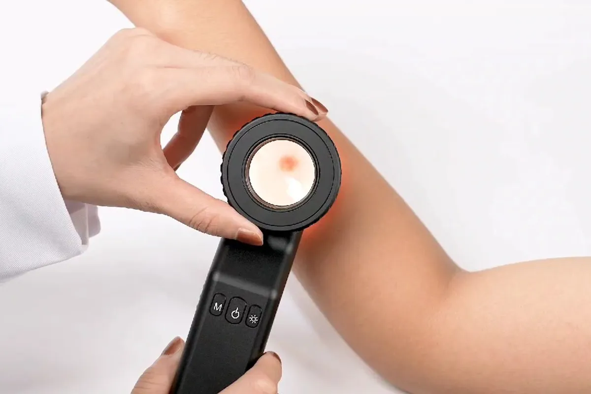
1. Dermatology Clinics:
- Routine skin examinations.
- Lesion observation and documentation.
- Treatment progress tracking.
2. Health Screening Centers:
- Skin health profiling.
- High-risk patient monitoring.
- Baseline image creation.
Aesthetic Applications
- Skin condition assessments.
- Treatment plan development.
- Before-and-after treatment comparisons.
Other Uses
- Medical education: Teaching demonstrations.
- Research: Data collection.
- Telemedicine: Remote image sharing.
- Pharmacies: Basic skin checks.
In all scenarios, the digital dermatoscope serves as an auxiliary tool, with professional medical evaluation remaining essential.
Maintenance Tips for Prolonging Digital Dermatoscope Lifespan
Proper maintenance ensures the longevity of a digital dermatoscope.
Daily Maintenance
- Wipe down after use with specialized lens cleaners.
- Avoid harsh solvents.
- Store in a moisture-proof case.
Regular Checks
- Monthly performance tests.
- Quarterly professional calibration.
- Annual comprehensive servicing.
Storage Guidelines
- Use a dedicated protective case.
- Avoid extreme temperatures.
- Store battery at 50% charge.
- Organize accessories systematically.
These practices ensure the digital dermatoscope remains a reliable tool for professional use.
The digital dermatoscope represents a significant advancement in skin health assessment, offering high-definition imaging, intelligent features, and efficient data management. From its technical principles to its wide-ranging applications in clinical and aesthetic settings, the digital dermatoscope enhances professional observation capabilities. However, it remains an auxiliary tool, and its findings must always be validated by trained medical professionals. By adhering to proper operation and maintenance protocols, users can maximize the digital dermatoscope’s potential as a vital tool in skin health evaluation.
People May Ask
melanoma. Although most melanomas are brown or black in color, some might have pink, tan, or even white hues. Not all melanomas are spherical like regular moles; some have colored sections. They could proliferate swiftly or even penetrate the skin around them.
With a dermatoscope, features can be evaluated down to the reticular dermis, and pictures can be taken for further analysis. Transillumination of a lesion for high magnification examination and visualization of minor details is the fundamental idea behind dermoscopy.
As they expand, melanomas may begin as flat patches. 4. If you can feel it, you should get it checked out, even if some moles can also be raised. Occasionally, when assessing melanoma, the "E" in the ABCDE guidance refers to "evolving." This is a result of the gradual changes in melanomas' size, shape, and color.
Thick pigmented lines around appendageal apertures, commonly referred to as rhomboidal structures, are the hallmarks of lentigo maligna's dermoscopic features.Unevenly colored follicular apertures.Instead,globules and dots of slate grey.
Is melanoma possible from a common mole? Melanoma, the most dangerous kind of skin cancer, is extremely uncommon to develop from a common mole. Those with numerous little or multiple large moles are more likely to acquire melanoma, even though common moles are not malignant (1).
Not all moles with scabs are malignant. But sometimes, scabby moles are malignant. If you are unable to link the scabbing to a recognized skin injury, it is crucial that you have them examined.
Initial clinical symptoms and misdiagnoses. In our study, the initial physician visit resulted in an erroneous diagnosis for 30% of the melanomas. This aligns with the outcomes of previous cohorts. Fortin and colleagues discovered a 25% rate of initial misdiagnosis, but Bristow and Acland reported a 33% rate of wrong diagnosis.
pigment that has spread into the surrounding skin from the spot's edge. Redness or a fresh enlargement outside the mole's boundaries. Modification in feeling, including pain, sensitivity, or itching. A mole's surface may change, becoming scaly, leaking, bleeding, or developing a bulge or bump.
An essential tool for melanoma early detection is melanoma mole mapping. Dermatologists may detect possible problems and offer prompt treatment by routinely checking your skin for changes.
With a dermatoscope, features can be evaluated down to the reticular dermis, and pictures can be taken for further analysis. Transillumination of a lesion for high magnification examination and visualization of minor details is the fundamental idea behind dermoscopy.
Digital Dermatoscope Products

Anykit Digital Otoscope with a gyroscope, 4.5-inch screen, 3.9mm ear scope camera with six lights, 32GB card, and an ear wax removal tool Allows for the capture of photos and videos
Dino-Lite AM3113T USB Digital Microscope with 0.3 MP, 10x - 50x, 230x Optical Magnification, Microtouch, Measurement, and Discontinued
SupereyesB005+1~200X Portable USB Digital Microscope, Endoscope Loupe, Otoscope Magnifier with 11mm Health Kit Tripod Diameter

Digital video otoscope/earscope Firefly DE550 Wireless

USB-C and Lightning Compatible Digital Microscope

The VMS-007-DX2 is a USB digital microscope with 5 Megapixels, x300 magnification, the ability to capture and record photos and videos, up to 2592 x 1944 resolution, 8 LEDs, and an adjustable stand.
The adult 4.3-inch IPS coin microscope with 1000X magnification is made by Hayve.Eight programmable LED coin collection suppliesCompatible with WindowsTF Card, 32GB

Digital Portable Microscope with 8 LED Magnifier, Manually Adjustable for Computer Phone and Tablet, 50X-500X 0.3MP USB Magnification

手持式 USB 数码显微敜带金属支枴,便携式高清 1000 倍放大检查摄像头,带 8 个 LED 灯,怂用于 Android Mac Windows 电脑
Vividia Digital Borescope Microscope with 12mm Diameter OTG Android and USB PC Compatible with a Professional Multi-Functional Metal Stand
Hot Products


News & Blog


Top Reviews
Edmond D.
Fantastic watch, excellent numbers are visible at night, and Italian is a language that is easy to imitate. I really like this watch.
Rhisian
The lampshade really highlights the nursery motif. It's really gorgeous and precisely what we were looking for—the green is a little bit lighter than it appears in the photo, more like a light sage.
Mrs K Frampton
Though they appear smaller in the photo, these are quite adorable and a really affordable way to alter the appearance of a regular lightbulb. To add a special touch, I painted a couple of these as well???????


