


DE-400 Dermatoscope – IBOOLO
Dermatoscopio Precio: Unveiling The Costs And Benefits
The dermatoscopic has become an essential tool in dermatological diagnosis, allowing physicians to examine the skin's surface and subsurface layers with precision. However, the array of options available on the market can be overwhelming, especially when it comes to pricing. This article demystifies the factors influencing dermatoscopio prices and provides a guide for making an informed purchasing decision.
When Should I Get A Dermatoscopic Examination?
Dermatoscopio precio is an essential, non-invasive diagnostic tool for monitoring skin lesions, aiding in early cancer detection, diagnosing unidentified spots, and assessing inflammatory and infectious skin conditions. It's invaluable for surgical planning, evaluating treatments, and educational purposes, offering a quick, painless way to enhance diagnostic accuracy and inform treatment decisions in dermatology.
The Dermatoscopio Basics
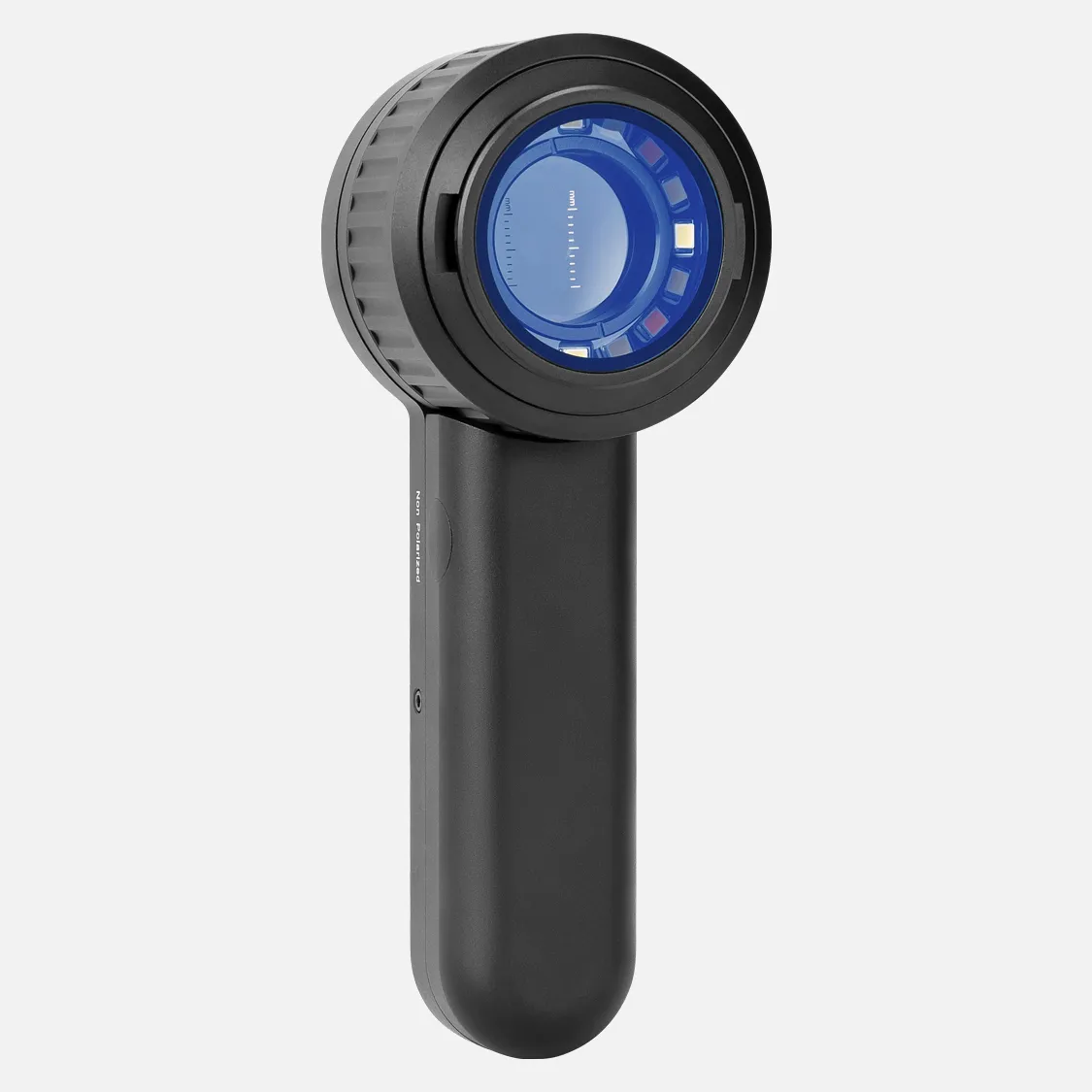
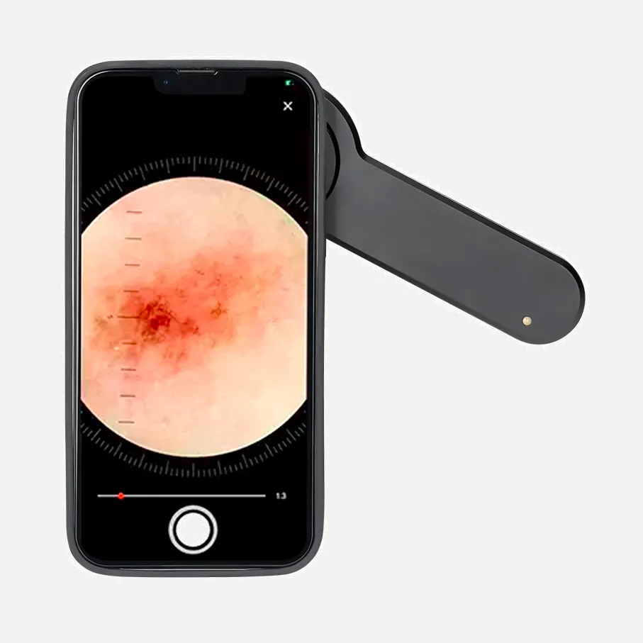
Before delving into prices, it's crucial to understand what a dermatoscopio is and how it functions. A dermatoscopio is an amplification device used to examine skin lesions and detect cellular changes. Its design has evolved from the early magnifying glasses to modern models with digital imaging and polarization technology.
Factors Affecting Price
1. Type of Illumination: Dermatoscopio with built-in LED light sources tends to be more expensive.
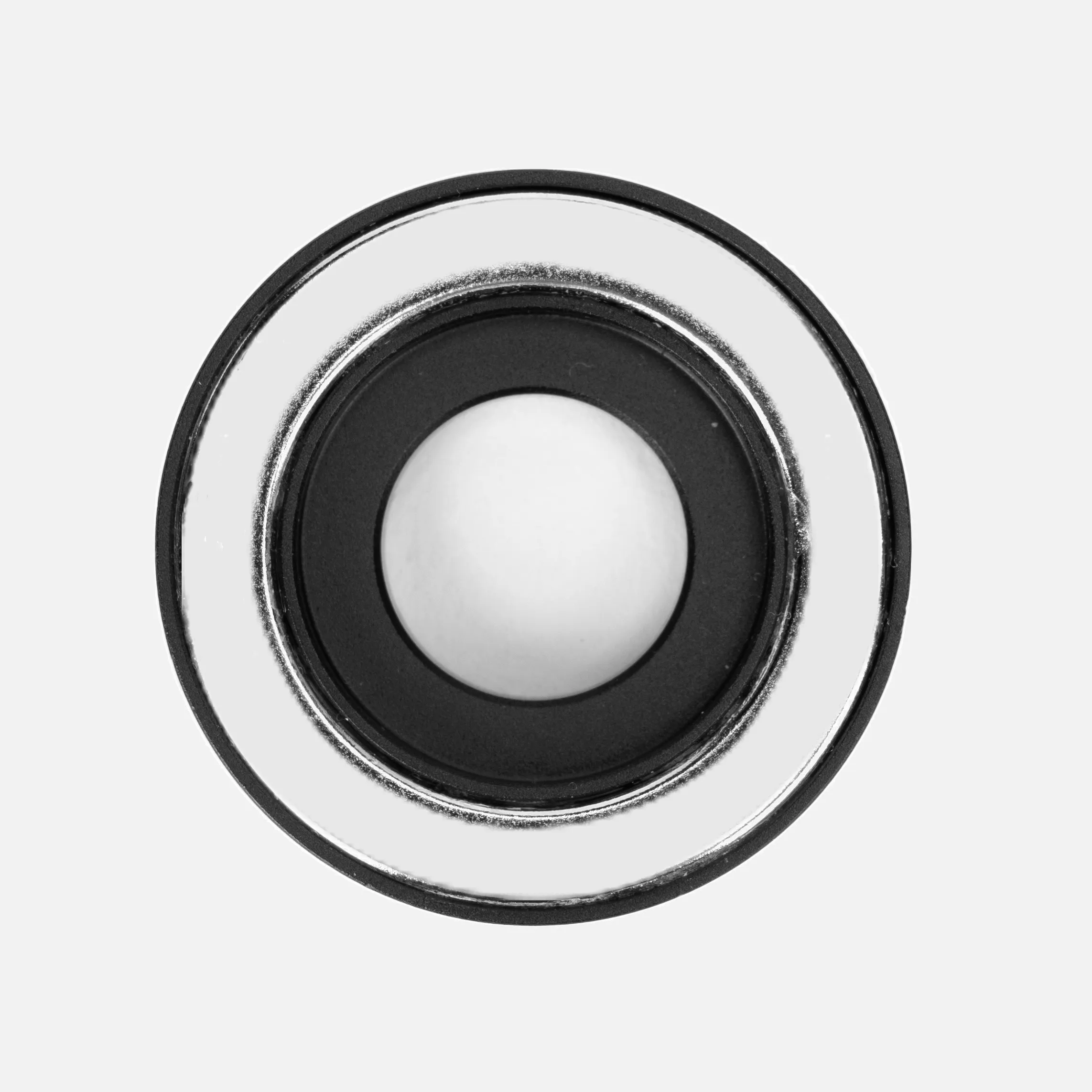
2. Magnification: Dermatoscopio with adjustable magnification levels often cost more.
3. Digital Technology: Dermatoscopio precio with built-in cameras and digital storage capabilities generally have a higher price tag.
4. Advanced Features: Cross-polarization, spectroscopy, and image analysis software are features that can increase the price.
Price Range
Dermatoscopio can be found across a wide price spectrum, ranging from basic models for around $50 to cutting-edge systems that can cost over $2,000. Considerations for purchase: When choosing a dermatoscopio, consider, Your budget and needs. The frequency of use and the type of patients you will attend to. The ease of use and training capacity. Warranty and post-sale support.
Brands And Prices
Prized for its precision in dermatology, iboolo dermatoscopic spans a spectrum of models, each tailored to specific needs and priced according to its advanced features. High magnification, advanced LED lighting, digital capabilities, polarization technology, portable designs, analytical software, robust warranties, and strong market reputation all contribute to the cost, yet each reflects iboolo's dedication to innovation and quality.
Where to Buy Iboolo Dermatoscopios
IBOOLO Official Website: The most direct source is the iboolo official website(https://www.iboolo.com/), where customers can find the full range of dermatoscopios, accessories, and often the best pricing and exclusive offers.
Professional Conferences and Trade Shows: Cibolo may have a presence at professional medical conferences and trade shows, where attendees can get hands-on demonstrations and special event pricing.
When purchasing an iboolo dermatoscopic, it's essential to ensure that you are buying from a reputable source to guarantee the authenticity of the product and to receive the full manufacturer's warranty and support.
Call to Action
The price of a dermatoscopio is an investment in the quality of healthcare provided to patients. By carefully considering features, the needs of your practice, and reviews from other users, you can find the dermatoscopic that best fits your budget and requirements. Visit our website for more information on available dermatoscopios and to contact us directly with any questions or to request a quote.
People May Ask
Tiny bump that is raised and firm on the skin, with a distinct border that is easily visible. El color de las pápulas is rojiza, morada, marrone o rosada.
A medical doctor with a license who later became a dermatologist is known as a dermatólogo. Esta educación le brinda acceso a vastos conocimientos acerca del cuerpo humano y las razones que pueden generar daños o afecciones en la piel.
When the moon that appears on your skin or body is not harmful, it can be removed with a CO2 laser. This effective vaporization technique completely removes the moon without leaving any scars behind.
El retículo pigmentado negativo (RPN) is una malla o red que presenta huecos oscuros junto con líneas claras. It's a controversial and frequently occurring dermoscópic sign. There is agreement to acknowledge its existence in certain Spitz6–9 nevi.
Several techniques, such as tumor biopsies and dermatoscopy, are used to diagnose skin cancer. To determine the best course of treatment and establish a prognosis in each case, a diagnosis of cutaneous cancer and the kind of tumor is essential.
Usted o su asesor médico verá si existen lunares, marcas de nacimiento u áreas con un color, tamaño, forma o textura anormales para realizar un examen de cáncer de piel. Should a portion of your skin appear abnormal, you may need to conduct tests to determine whether it is cancerous.
La examinación cutánea is un componente esencial del examen físico estándar. All of the body's skin is meticulously examined as part of this examination. The assessment focuses on identifying abnormal signs in the skin, such as the cabelludo, orificios, uñas, and mucosal surfaces.
The 30- to 40-minute digital dermatoscopy is a non-invasive test that doesn't require much prior preparation.
Los LEDs deben estar direcionados in la dirección de la lesión que será examinada para ser usados con un dermatoscopio. It should be placed about 12 mm from the skin. To maintain an ideal space or a piece that is occasionally included for such purpose, one can use their hands.
A non-invasive in vivo diagnostic technique called dermatoscopia was developed to study skin lesions. enhances early diagnosis of potentially malignant lesions, including melanoma, and improves the accuracy of diagnosis of hyperpigmented lesions.
Dermatoscopio Precio Products

MDF ProCardial Cardiology Stethoscope, Adult, Dual Head, Black Tube, Gold Chestpiece-Headset, MDF797K11, made of stainless steel
USB Charging Headband Magnification Glasses for Jewelry Craft Watch Repair Hobby 5 Replaceable Lenses Hands-Free Head Mount Magnifying Glasses with LED Light 1.5X, 2.0X, 2.5X, 3.5X, and 1.0X (White)

Adjustable Magnifying Glass with Light for Crafts, Reading, and Close Work - Black - Brightech LightView Pro Magnifying Desk Lamp, 2.25x Light Magnifier with Clamp

ELITRA Heavy Duty Scrubber Sponge, Odorless, Blue, with Smell Resistant Hydrophilic Foam Technology, 24 Pack
Made from cellulose and coconut fibers, AIRNEX Biodegradable Compressed Cellulose Sponges and Scrubbing Pads for Dishes are non-scratchy and come in a set of 24.

Kila Scopes 专业听诊器 - 帓业单头心脏病和诊断听诊器 适用于医生和护士-带配件,K971 酒红
This product is a portable digital microscope with a 4 LCD screen, a 1500X pocket microscope for kids with LED lights, and a 32GB memory card for adults.Has a Camera and Video Function

The Toyofmine USB digital microscope is a portable handheld electronic coin magnifier that can magnify objects from 10x to 200x. It also has a welder camera and an otoscope.
With eight programmable LED lights, an LCD digital microscope with 1000x magnification, PC view, and Windows compatibility, the Elikliv EDM4 4.3-inch coin microscope
MioEco Natural Reusable Unsponge Kitchen Sponges - Washable & Organic Dish Towels, Non-Scratch Organic Sponge for Dishes, Zero Waste Sponge, Set of 4
Hot Products


News & Blog


Top Reviews
Mary Trent
I've had this stethoscope since I started working in the intensive care unit, and I really like it! Even though I've always used a Littmann, I wasn't willing to pay the price they were asking for a Littmann when I started looking for a cardiology-style stethoscope. I'm happy I chose to go with this Kila after finding it and reading excellent reviews!
Kristina Davidson
Great sound for listening in the back of an ambulance. The only problem is head is a little heavy, so you need to make sure you do not hit your head or the patients as you are leaning over. Overall the quality seems very good and the stethoscope is comfortable in the ears and around the neck. I have also bought the refurb kit from Kila Labs and their equipment inproved the ultrascope that I had been using.
Becky
is somewhat lengthy, but it provides a better sound quality than my Lithman cardiac stethoscope.


