


DE-400 Dermatoscope – IBOOLO
What Is A Smartphone Dermatoscope?
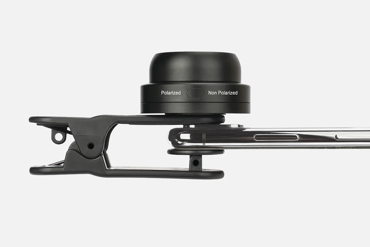
A smartphone dermatoscope refers to a type of dermatoscope designed to interface with smartphone cameras to capture clinical-grade dermoscopic images and video.
A compatible mounting device securely aligns a smartphone’s rear camera with the eyepiece of a standard contact dermatoscope. This enables the smartphone to visualize and document the enhanced magnified perspective of skin lesions under evaluation.
By leveraging smartphone computational power, camera sensors, display capabilities, connectivity, apps, and storage capacity - smartphone dermatoscopes can transform mobile devices into versatile point-of-care screening and tracking tools. Users can build a shareable record of worrisome moles, coordinate care, and support remote diagnoses.
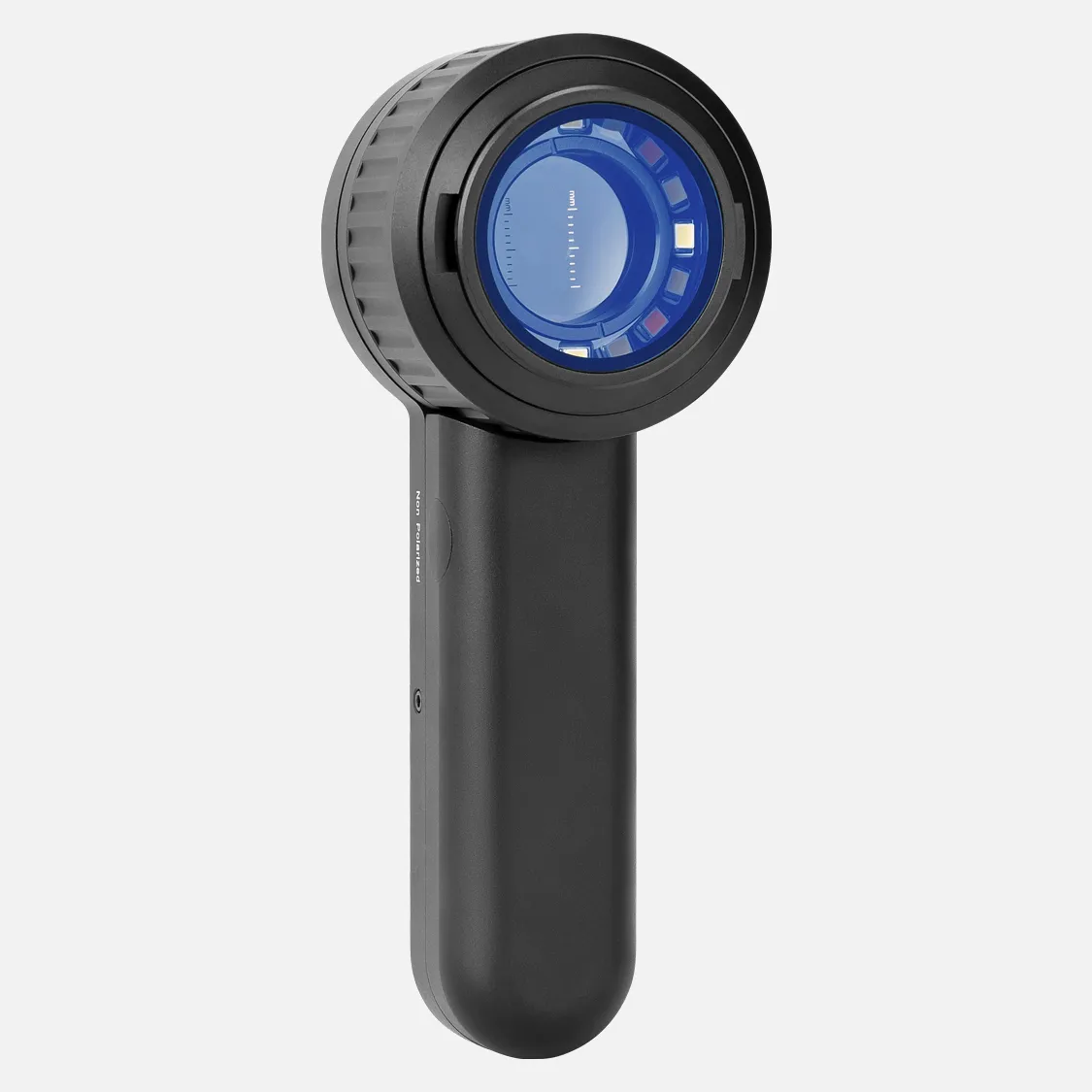
Key factors in smartphone models include adjustable magnification level, lighting technique (polarized vs. non-polarized), ergonomic handling, intuitive controls/display, and seamless image capture workflows for health monitoring and telemedicine. Further testing aims to validate applications delivering clinical equity.
Smartphone dermatoscopes provide an integrated digitized skin assessment platform combining precision optics with mobile functionality - promising expanded access to precision screening.
How Does A Smartphone Dermatoscope?
A dermatoscope uses a smartphone's camera and optical systems to take images of skin lesions. These images can be used as a form of preventive screening for cancer, especially when professional assistance is not available.
Here's how to perform a dermoscopy at home using a smartphone:
1. Put one drop of oil on the mole
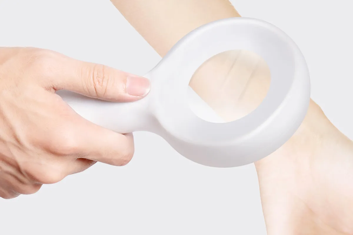
2. Place the dermatoscope on the skin and apply gentle downward pressure
3. Take a picture with the smartphone
4. Touch the center of the screen to focus the image
5. Store clear images
Mobile dermoscopes can also connect to dermatology software to streamline workflows. For example, a loupe can be taped to the back of a smartphone with a lens similar to the smartphone's camera lens. Alcohol gel can be used as the dermoscopy fluid, with the lesions viewed through the fluid.
Dermoscopy can help identify and differentiate between melanocytic lesions and non-melanoma skin cancers.
People May Ask
Common warts typically manifest in a clinic setting as papillomatous hyperkeratotic dome-shaped papules with bleeding, linear, hairpin-like, and dotted capillaries, among other dermoscopic symptoms.
Not because they are unable of identifying melanoma, but rather because they deny themselves the opportunity to observe and investigate it, is probably the reason why doctors fail to detect it.
A handheld device known as a dermatoscope is used to perform dermoscopy. Subsurface skin structures in the epidermis, papillary dermis, and dermoepidermal junction-structures that are often invisible to the unaided eye-can be seen thanks to this method [2-4].
Surface melanoma is characterized by its asymmetrical, irregular shape and structure. One or more of the following dermoscopic characteristics are typically present in superficial melanomas: Blue-white veil. Numerous brown spots.
The free mobile app UMSkinCheck (available for iOS and Android) is designed for skin cancer self-examination and surveillance. It lets users take and save a whole body photo collection, monitor identified lesions and moles, download educational materials and videos, and find a skin cancer specialist.
Cancer of the Basal CellDermoscopic parameters linked to BCCs include the lack of a pigment network and the presence of certain characteristics such as ulceration, arborizing vessels, enormous ovoid nests of blue-gray, multiple blue-gray globules, leaf-like areas, and spoke wheel areas.
Dermoscopy can help diagnose melanoma more accurately, but it cannot take the place of a histopathologic examination.
Conclusions. Dermascopy is seen as having a significant role in enhancing skin examinations in primary care by both GPs who use it and those who do not. It was noted that a major obstacle to its broader adoption is the requirement for sufficient training in dermoscopy and dermatology more broadly.
Dermoscopy is a non-invasive, in-vivo technique that has been traditionally helpful for the examination of suspected skin lesions. It is sometimes referred to as dermatoscopy, epiluminescence microscopy, or skin surface microscopy.
On the mole that bothers you, apply one drop of oil. Directly touch the skin with the dermatoscope, press down gently, and use your smartphone to take a picture. Touching the screen's center may be necessary to concentrate the mole's image. Take and preserve clear dermoscopic pictures.
Smartphone Dermatoscope Products

Coin magnifier with light, 2K LCD Digital Microscope 1200X, Dcorn 7 24MP HDMI Microscope, Soldering Coin Microscope with Lights, Extension Tube & 32GB Card Included

With 30 adjustable LED lights, a parfocal lens, wireless remote control, and PC compatibility, the SZJMS 10.1 Digital Microscope 1600X features an IPS Touch Screen Coin Microscope and a 1080P 12MP Soldering Microscope.
10x–220x Magnification Dino Lite USB Handheld Digital Microscope True Resolution: 0.3MP/1.3MP/5.0MP; Software for Windows, Mac, iOS, and Android Included; Supports PC, Tablet, and Mobile Devices

Shipenophy 100X Pocket Microscope: Handy, Handheld Magnification Microscope Pen with Adjustable Focusing LED Magnifier for Home and Laboratory Use
Handheld Fiber Optical Microscope, Ashta 200x 400x Examining fiber terminations under coaxial illumination using a small, portable microscope equipped with 1.25mm and 2.5mm adapters
Nonslip Digital Microscope, 50X1600X Magnifying Endoscope Camera for 7 Full View

LED Pocket Size Handheld Microscope/Computer Magnifier with 50X–500X Magnification, 0.3MP USB Digital Microscope

50X Subminiature Portable Microscope Magnifier for Small Microscope Magnifiers for Jewelry Appraisal: Compact Pocket Microscope with LED Currency Detecting Light

Dino-Lite USB Digital Microscope AM3113 - 0.3 MP, 10 - 50 x 230 x Optical Magnification, Measurement Compatible
Record nature and the world with the GOSKY Smartphone Adapter Mount Regular Size, which fits nearly all smartphones on the market and is compatible with binoculars, monoculars, spotting scopes, telescopes, and microscopes.
Hot Products


News & Blog


Top Reviews
Stephen Hiatt
Excellently crafted, with a clean, clear quality. It was ideal for checking the stylus rake angle on my cartridge, which is exactly what I did with it. Although I had trouble understanding some of the help sections, the software that came with it is efficient. I saw a few instructional videos on YouTube that showed me how to set up the program and use the drawing feature. simple as pie.
D. Banici
I was first a little let down by the resolution because I was used to DSLRs and films. But I quickly discovered that it's plenty, and you really don't need any more information than that. You zoom in and concentrate to see more. Two microscopes hooked in and recording at the same time can be supported by the provided software, which functions flawlessly with Windows 7 and Windows 8.
Customer
I still own the Dino-lite AM3111 that I originally purchased. Although I'm in love with it, I wish I had purchased the optional micro-touch to capture object photos. I could have traded up to the AM3111T, but instead I just bought it so I would have two in case I needed them. They truly are incredible.


