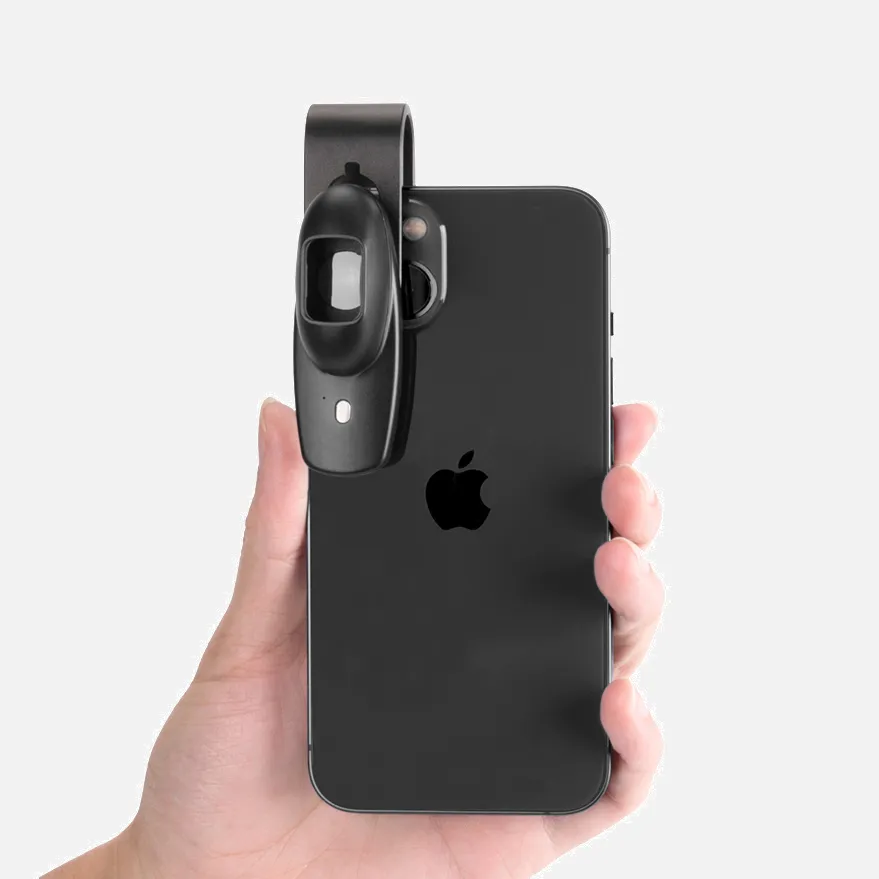
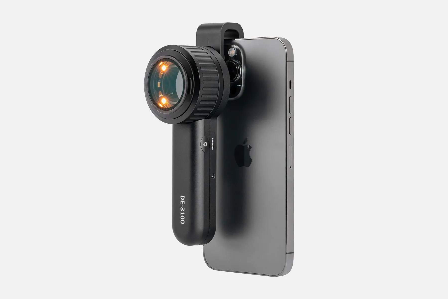
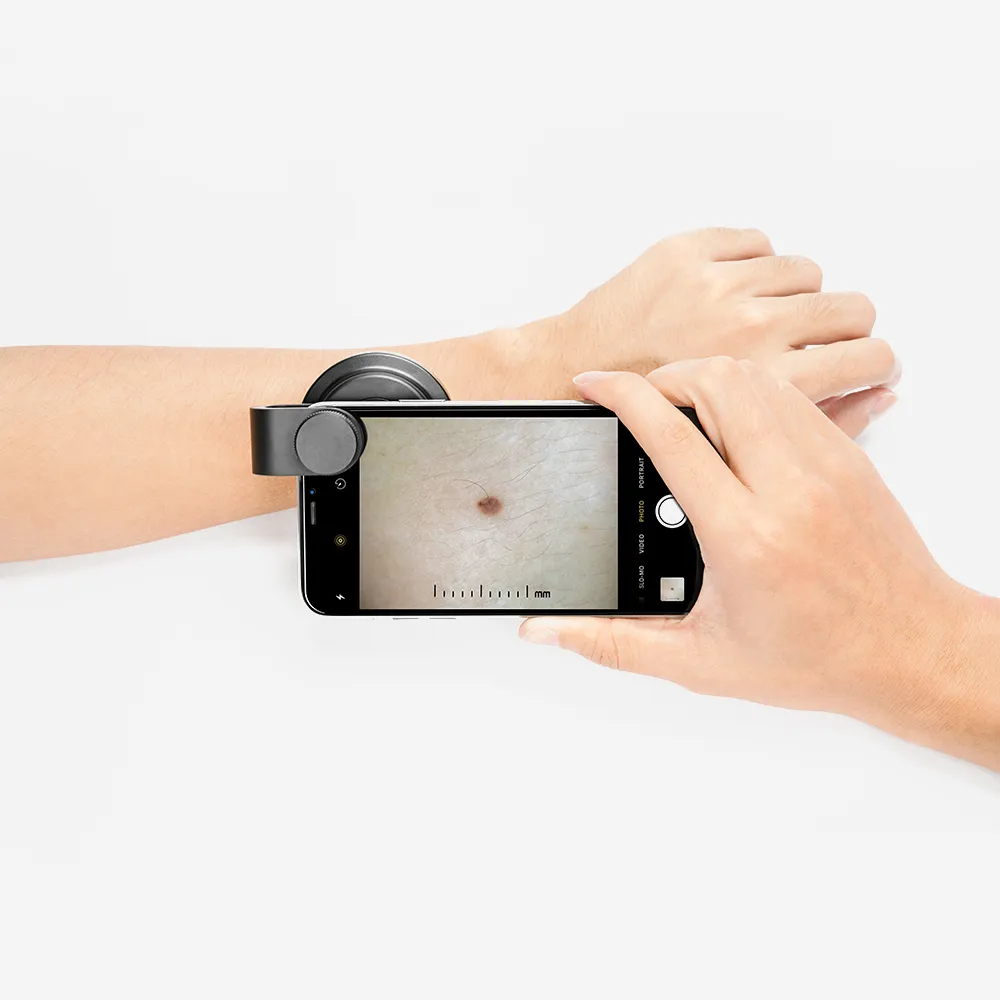
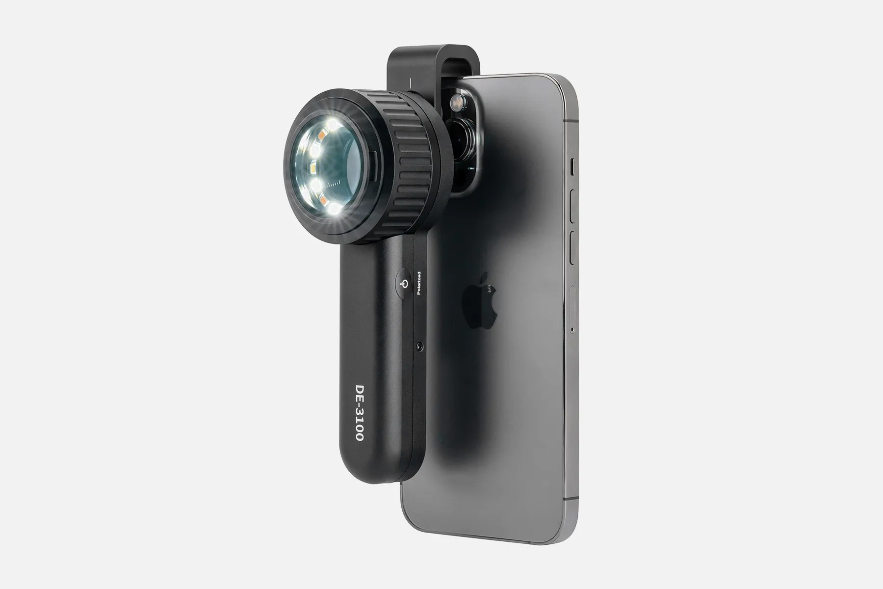
What Is Polarized Dermoscopy?

Polarized dermoscopy is the dermoscopic technique of using polarized lighting to perform clinical skin evaluations. Unlike standard non-polarized dermoscopes, which emit single-plane light, polarized dermoscopes leverage dual-plane polarized illumination to suppress surface reflections that can obscure subsurface details.

Cross-polarization uses a polarizing filter on the incident light emitted into the skin and a second orthogonally-aligned polarizing filter on the return pathway to effectively block reflections off the stratum corneum. This improves visualization of deeper morphological structures without requiring gel or skin contact with the lens.

By reducing shine and glare, polarized dermoscopes reveal vital subsurface clues in the epidermal, dermal-epidermal junction, and papillary dermis layers. This magnified perspective of pigmented networks, vessels, and structural formations facilitates enhanced screening, inspection, and clinical decisions regarding potential biopsy of lesions.

Polarized dermoscopy opens a clearer window into skin architecture for superior anatomical assessment and diagnostics. The technology offers advanced illumination techniques to serve precision inspection needs beyond standard non-polarized devices.

How Does Polarized Dermoscopy?

Polarized dermoscopy (PD) is a technique that can help visualize deeper layers of the skin. PD works by blocking light that reflects off the surface more effectively than non-polarized dermoscopy (NpD). This allows pD to show better deep structures, such as the dermo-epidermal junction and superficial dermis.

pD uses light-emitting diodes to provide illumination. Light passing through the source polarizer is unidirectional and will be rejected by the detector polarizer.

Dermoscopy works by transilluminating a lesion to be studied with high magnification to visualize subtle features. The principle of dermoscopy is based on the understanding that the stratum corneum reflects light, which reduces the ability to see structures under the skin.

People May Ask

What Do Dermoscopy Crystalline Formations Mean?

structural crystalsThese are caused by an overabundance of collagen and can be observed in melanoma, dermatofibroma, scar, squamous cell carcinoma, basal cell carcinoma with fibroplasia, and Spitz naevi. Chrysalis structure was a misnomer when it was first used.

What Distinguishes Polarized Light?

If this electric field's direction varies arbitrarily over time, light is said to be unpolarized. Unpolarized light is produced by halogen lighting, incandescent bulbs, LED spotlights, sunlight, and many other common light sources. Polarized light is characterized as having a well-defined direction of the electric field.

How Is It Possible to Distinguish between Light That Is Polarized And Unpolarized?

polarized light Non-polarized illuminationInstead,When light is polarized, the electric field only oscillates in one direction. Its electric field is oscillating in all directions.2. Light that is polarized is coherent by nature. Unpolarized light cannot be coherent in the natural world.

How Does Polarized Light Vary from Non-Polarized Light?

Non-polarized light is defined as light waves with several directions of reflection. Conversely, polarized light waves are ones that only reflect in one direction or location. Both horizontal and vertical paths are taken by non-polarized light.

Why Do We Use Light That Is Polarized?

To lessen glare, sunglasses with polarization are utilized. Stress analysis experiments are conducted in the plastics industry using Polaroid filters. Polarization is used in the creation and presentation of three-dimensional films. Transverse and longitudinal waves can be distinguished using polarization.

How Come Polarized Light Is Superior?

There are several imaging applications where polarization control might be helpful. To reduce hot spots from reflecting surfaces, boost contrast, and remove glare from light dispersion, polarizers are put over a light source, lens, or both.

What Varieties Exist for Dermoscopes?

Dermatoscopes come in two primary varieties: handheld, portable, and stationary mounted. A magnifying optic with a magnification of at least ten times makes up a hand-held dermatoscope, together with a transilluminating light source.

What May Be Observed Using Polarized Dermoscopy on Chrysalis Structures?

Shiny, brilliant white, orthogonal linear streaks-which we have named "chrysalis structures"-are frequently visible in skin lesions with elevated collagen levels. Neither the unassisted eye nor nonpolarized dermoscopy can make these structures visible.

How Does Polarized And Nonpolarized Dermoscopy Explain The Observed Variations?

The superficial dermis and dermo-epidermal junction are two deeper layers that can be seen with polarized dermoscopy. To see superficial layers, nonpolarized dermoscopy is employed (the superficial epidermis to the dermo-epidermal junction).

For What Purposes Is A Polarized Dermoscopy Preferable?

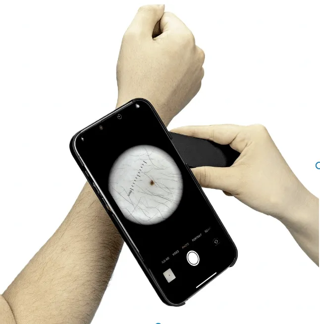
Contactless polarized dermatoscopyFurthermore, because this mode is non-contact, no pressure is applied to the areas of skin that need to be checked, making it especially appropriate for checking lesions and infected areas that cause pain for the patient.

Polarized Dermoscopy Products

Revo Sunglasses Harbor: Metal Navigator Frame, UV Filtering Polarized Lens

Women s Bloomoak Polarized Trendy Hexagonal Night Driving Glasses with UV 400 Protection and Anti-Glare Technology

China handheld polarized dermoscopy manufacturer & factory

3Gen Hybrid Polarized Dermallite DL200 Dermatoscope

China Professional polarized dermoscopy

MEDCASE Radiance Otoscope with Light German Fiber Optic Otoscopes: Excellent for Professional Use - Ear Scope with LED Light and Speculum for Ear Examination and Diagnosis

China Professional polarized dermoscopy manufacturer & factory

MEDCASE Radiance Otoscope - Professional Grade Ear Exam and Diagnosis Instrument with LED Light and Speculum - Pink - Lifetime Warranty

polarized dermoscopy manufacturer & factory

Coyote Eyewear UV Protection Bifocal Polarized Sunglasses BP-13 Sunglasses Reader

China wholesale polarized dermoscopy manufacturer & factory

DUCO Luminous Eyewear Night Driving Anti-glare Polarized Glasses 2181Y

China customized polarized dermoscopy manufacturer & factory

UV Protection Sunglasses N44 with a Flip-up Polarized Lens for Prescription Glasses by RuiJinGen

Top Reviews

sach509

This is intended for anyone working in the field of dermatology. I purchased and returned several dermatoscopes before deciding to purchase this one, and I have no regrets about it. Plus you'll only find at this price here

Jaye E. Benjamin

Strong craftsmanship in contrast to other plastic scopes I've owned. The LEDs have good brightness and optics. I'm not sure what advantage the option to adjust the illumination's color temperature offers. Although this instrument costs more than other dermatoscopes, it is obviously made to last.

supermom

This light is excellent for my dental loupes. The battery lasts all day, which is really great.

0

Have questions on gear or your order?

Our Gear Guides are here to help! Get personal advice from pro creatives

Name
Subject
Email address
How can we help?

Instant Answers

How Can Dermoscopy Images Be Captured?

Dermoscopy images can be captured and stored in different ways, such as: • Using a smartphone or tablet with dermoscopic adapter, which consisted in the package.• Using a digital camera

Dermoscopy images can be captured and stored in different ways, such as:

• Using a smartphone or tablet with dermoscopic adapter, which consisted in the package.
• Using a digital camera with dermoscopic adapter, there’s 49mm screw size camera adapter available to order now.

Compatible phone/tablet models:
All iPhone models, 95% Android phones, 90% tablet. For phone/tablet size in 5.25-14mm

Compatible camera models:
All camera with built 49mm filter screw, such as Canon EOS 70D, 80D, 90D; Canon EOS R7, R10, R50, R100; Canon M100, M200, M50, Mark II; Canon G7X Mark III, Sony ZV-1

How Can I Connect My Phone to My Dermatoscope?

There’s universal phone adapter for all our dermoscopes. Please check the installation procedure bellow or watch operation guide. Smartphone Connector (1) Place phone adapter screw in the center of smartphone’s

There’s universal phone adapter for all our dermoscopes. Please check the installation procedure bellow or watch operation guide.

Smartphone Connector

(1) Place phone adapter screw in the center of smartphone’s main camera.
(2) Screw magnet attachment on phone adapter.
(3) Put dermoscope’s back ring and magnet attachment together

Take The Best Images

You need to adjust the focus ring after the dermoscpe connected on smartphone to get the best images.

How Can I Clean My Dermoscopy after Usage?

Cleaning your dermoscopy after usage is important to prevent cross-contamination and infection. The cleaning method may vary depending on the type and model of your dermoscopy, so you should always

Cleaning your dermoscopy after usage is important to prevent cross-contamination and infection. The cleaning method may vary depending on the type and model of your dermoscopy, so you should always follow the manufacturer’s instructions. However, some general steps are:

• Turn off and disconnect your dermoscopy from any power source or device.

• Wipe off any visible dirt or debris from the dermoscopy with a soft cloth or tissue.

• Disinfect the dermoscopy with an alcohol-based wipe or spray, or a disinfectant solution recommended by the manufacturer. Make sure to cover all surfaces, especially the lens and contact plate.

• Let the dermoscopy air dry completely before storing it in a clean and dry place.

• Do not use abrasive or corrosive cleaners, solvents, or detergents that may damage the dermoscopy.

• Do not immerse the dermoscopy in water or any liquid, unless it is waterproof and designed for immersion.

You should clean your dermoscopy after each use, or at least once a day if you use it frequently. You should also check your dermoscopy regularly for any signs of damage or malfunction, and contact the manufacturer or service provider if needed.

Polarized VS Non-Polarized Dermoscopy

A dermoscopy is a device that allows the examination of skin lesions with magnificationand illumination. By revealing subsurface structures and patterns that are not visible tothe naked eye. It can

A dermoscopy is a device that allows the examination of skin lesions with magnificationand illumination. By revealing subsurface structures and patterns that are not visible tothe naked eye. It can improve the diagnose accuracy of skin lesions, such as melanoma,basal cell carcinoma, seborrheic keratosis, etc.

There are two main types of dermoscopy: Non polarized and polarized dermoscopy.We’ve fitted most of our dermoscopys with polarized and non-polarized light. They canbe used in multiple skin structures.

Non-polarized contact Mode

In non-polarized mode, the instrument can provide information about the superficialskin structures, such as milia-like cysts, comedo-like openings, and pigment in theepidemis.

The dermoscopy requires applying a liquid such as mineral oil or alcohol to the skin andplacing the lens in contact with the skin. This reduces surface reflection and enhancesthe view of subsurface structures.

Image with non-polarized light (DE-3100)

Polarized contact Mode

In polarized mode, the instrument allows for visualization for deeper skin structures,such as blood vessels, collagen, and pigment in the dermis.

The dermoscopy does not need to be in contact with the skin or use any liquid. Theirpolarized light can help to eliminate surface reflection and allow visualization ofvascular structures.

Image with polarized light (DE-3100)

Polarized non-contact Mode

The dermoscopy can also use polarized light to examine the skin without direct contact.

In polarized non-contact mode, the instrument allows for examination infected areasand lesions that are painful for the patient, or the difficult to contact pigmented lesions,such as nails and narrow areas.

The contact plate should be removed in this mode, and it does not require applying aliquid to the skin. As it doesn’t require pressure or fluid application on the skin, it canalso avoid cross-contamination and infection risk.

Image in polarized non-contact mode (DE-3100)

How Effectiveness Is Dermoscopy

Compared with visual inspection, the dermoscopy can be used to capture and store skin lesion photos, which play an important role in early skin cancer examination. The dermoscopy allows the

Compared with visual inspection, the dermoscopy can be used to capture and store skin lesion photos, which play an important role in early skin cancer examination.

The dermoscopy allows the examination of skin lesions with magnification and illumination. This can be greatly avoiding the factors that cause interference to visual detection. Such as lighting, skin color, hair and cosmetics.

Several studies have demonstrated that dermoscopy is useful in the identification of melanoma, when used by a trained professional.

It may improve the accuracy of clinical diagnosis by up to 35%
It may reduce the number of harmless lesions that are removed
In primary care, it may increase the referral of more worrisome lesions and reduce the referral of more trivial ones

A 2018 Cochrane meta-analysis published the accuracy of dermoscopy in the detection.

Table 1. Accuracy of dermoscopy in the detection of melanoma in adults
Detection Method Sensitivity, % Specificity, % Positive Likelihood Ratio NegativeLikelihood Ratio
Visual inspection alone (in person) 76 75 3.04 0.32
Dermoscopy with visual inspection (in person) 92 95 18 0.08
Image-based visual inspection alone (not in person) 47 42 0.81 1.3
Dermoscopy with image-based visual inspection (not in person) 81 82 4.5 0.23
ROC—receiver operating characteristic. *Estimated sensitivity calculated on the summary ROC curve at a fixed specificity of 80%.

As we can see, the dermoscope can improve the accuracy of diagnosis of skin lesions, especially melanoma.

Table 1. Accuracy of dermoscopy in the detection of melanoma in adults
Detection Method Sensitivity, % Specificity, % Positive Likelihood Ratio NegativeLikelihood Ratio
Visual inspection alone (in person) 79 77 3.4 0.27
Dermoscopy with visual inspection (in person) 93 99 93 0.07
Image-based visual inspection alone (not in person) 85 87 6.5 0.17
Dermoscopy with image-based visual inspection (not in person) 93 96 23 0.07
ROC—receiver operating characteristic. *Estimated sensitivity calculated on the summary ROC curve at a fixed specificity of 80%.

Characteristics of the dermatoscopic structure of the skin lesions include:

• Symmetry or asymmetry
• Homogeny/uniformity (sameness) or heterogeny (structural differences across the lesion)
• Distribution of pigment: brown lines, dots, clods and structureless areas
• Skin surface keratin: small white cysts, crypts, fissures
• Vascular morphology and pattern: regular or irregular
• Border of the lesion: fading, sharply cut off or radial streaks
• Presence of ulceration

There are specific dermoscopic patterns that aid in the diagnosis of the following pigmented skin lesions:

• Melanoma
• Moles (benign melanocytic naevus)
• Freckles (lentigos)
• Atypical naevi
• Blue naevi
• Seborrhoeic keratosis
• Pigmented basal cell carcinoma
• Haemangioma

No products in the cart.

No products in the cart.

Hot Search Terms