


DE-400 Dermatoscope – IBOOLO
Revolutionizing Dermatology: A Glimpse into The Future of Ddermoscope Innovation
The dermoscope, a pivotal tool in dermatological examinations, has transcended its humble beginnings as a simple magnifying glass to become an indispensable device in the early detection and diagnosis of skin conditions. This article delves into the evolution of dermoscope, its current applications, and the promising horizons on the cusp of technological breakthroughs.
The Dermoscope Journey: A Historical Perspective
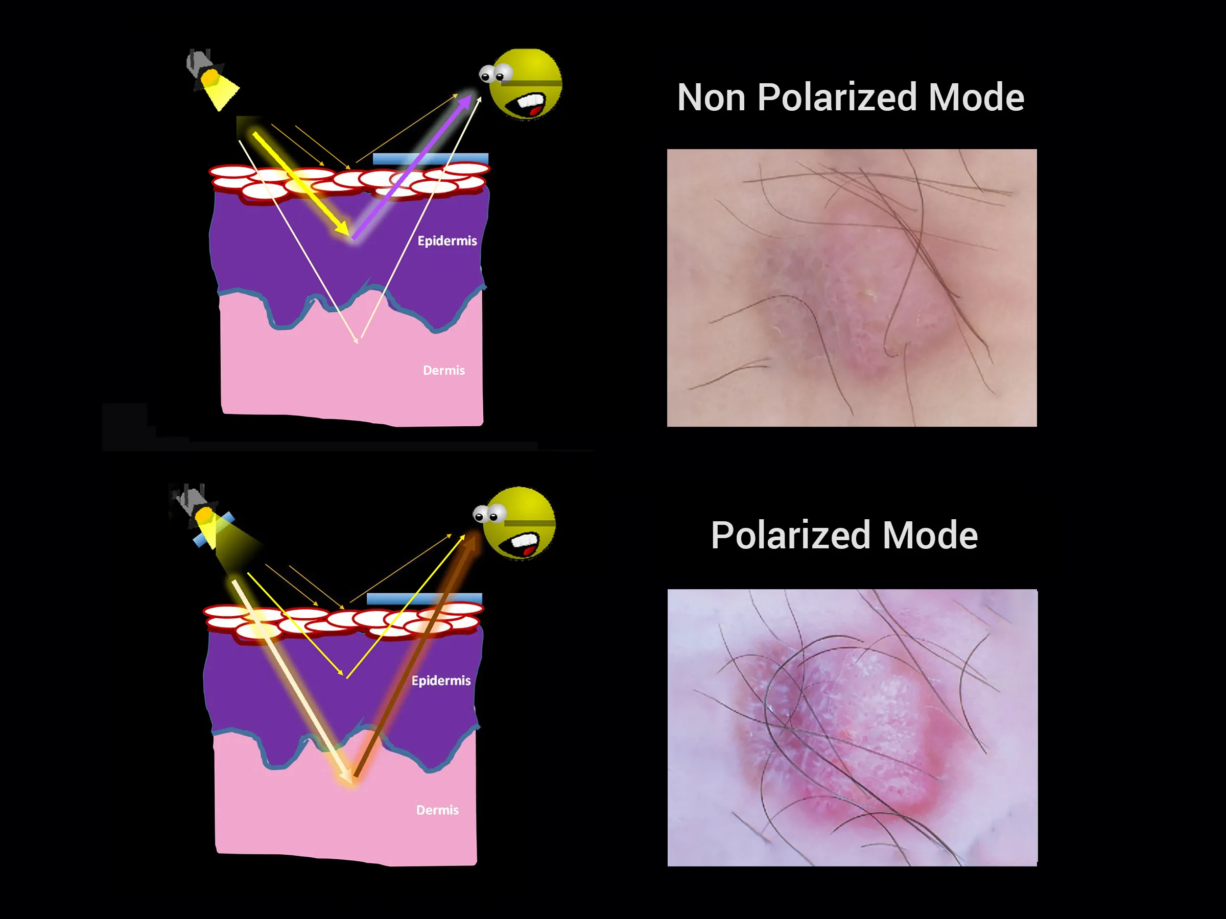
In the early 1900s, a dermoscope was born out of necessity—a basic magnifying glass to scrutinize skin lesions. Fast forward to the mid-20th century, and the introduction of polarized light technology marked a significant leap, allowing for clearer visualization of the skin's subsurface. The late 20th century witnessed the integration of digital imaging, giving rise to the modern dermoscope capable of capturing and storing images for detailed analysis.
The Digital Revolution: Dermoscopes in The 21st Century
The dawn of the 21st century brought rapid advancements in digital technology, transforming dermoscopes into sophisticated instruments. These now boast connectivity with computers for real-time imaging, storage, and analysis. The incorporation of advanced features like spectral analysis, skin pattern recognition, and 3D imaging has further elevated the capabilities of dermoscopes. The integration of AI has been a game-changer, adjuvant treatment and analysis that enhances diagnostic precision.
The Versatility of Dermoscopes
Dermoscopes serve as a multifaceted tool in dermatology, adept at identifying and distinguishing a myriad of skin lesions, from benign moles to malignant melanomas. They unveil the intricate pigment network and vascular patterns beneath the skin's surface, providing critical insights into conditions that might otherwise go undetected. Moreover, dermoscopes facilitate the monitoring of known lesions, ensuring timely intervention and optimal patient outcomes.
Reliability And The Role of Dermoscopes
The reliability of dermoscopes in dermatological practice is well-established, with studies affirming their heightened accuracy in diagnosing skin cancers compared to the naked eye. However, the reliability is contingent upon standardized classifications, operational proficiency, and objective image interpretation. The advent of quality control standards in dermoscopy has been pivotal in mitigating variability and enhancing diagnostic consistency.
Beyond The Magnifying Glass
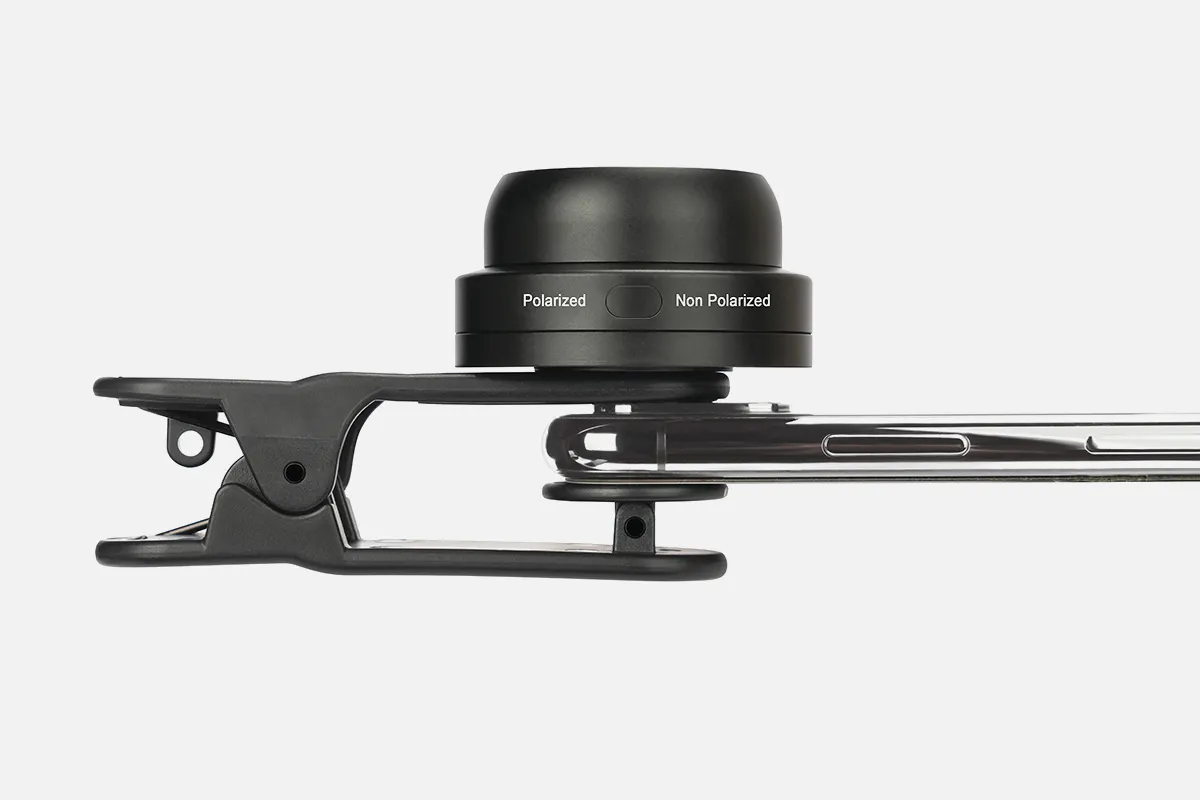
The dermoscope is far more than an ordinary magnifying glass. It is a testament to the confluence of magnification, optical technology, and digital innovation. With magnification capabilities ranging from 10x to 40x, dermoscopes offer a detailed glimpse into the skin. Polarized light is a distinguishing feature that minimizes surface glare, revealing deeper structures with clarity.
The Dermatoscope Vs. Dermoscope: Terminological Nuances
While "dermatoscope" and "dermoscope" are frequently used interchangeably, the latter is often associated with the more advanced models prevalent in contemporary practice. The distinction, though subtle, reflects the evolution from basic handheld magnifiers to cutting-edge diagnostic devices.
The Future of Dermoscopic Technology
As we peer into the future, dermoscopes are poised to integrate even more sophisticated features, heralding a new era of skin health management. Anticipated advancements include real-time lesion analysis, remote diagnostics, and personalized skin health monitoring. The trajectory of dermoscope technology mirrors the broader strides in medical innovation, promising a future where skin conditions are detected earlier, managed more effectively, and where prevention is within reach.
The journey of the dermoscope from a rudimentary magnifying glass to a cornerstone of modern dermatology is a narrative of human ingenuity. As technology progresses, the future of dermoscopy is set to transform how we understand, diagnose, and treat skin conditions, ensuring that the health of our largest organ is in capable hands.
People May Ask
How long could you have melanoma without realizing it? It is contingent upon the kind of melanoma. Radial melanoma, on the other hand, might spread slowly over a decade, whereas nodular melanoma spreads quickly over a few weeks. Similar to a cavity, a melanoma can proliferate for years without showing any noticeable symptoms.
pigment that has spread into the surrounding skin from the spot's edge. Redness or a fresh enlargement outside the mole's boundaries. Modification in feeling, including pain, sensitivity, or itching. A mole's surface may change, becoming scaly, leaking, bleeding, or developing a bulge or bump.
The pigmented lesion must lack pattern symmetry and color uniformity in addition to at least one of the following characteristics in order to be diagnosed as melanoma: numerous brown dots, pseudopods, radial streaming, scar-like depigmentation, peripheral block spots/globules, five to six colors, a blue-white veil,...
In the naked-eye arm, referral sensitivity, specificity, positive and negative predictive values were 54.1%, 71.3%, 11.3%, and 95.8%, respectively, while in the dermoscopy arm, they were 79.2%, 71.8%, 16.1%, and 98.1%, respectively. Sensitivity and negative predictive value (P =) showed significant variations.
You can be sure that a board-certified dermatologist has the necessary expertise to correctly diagnose and treat any health issues pertaining to your skin, hair, and nails, so as a patient, you can feel more at ease and confidence in their treatment.
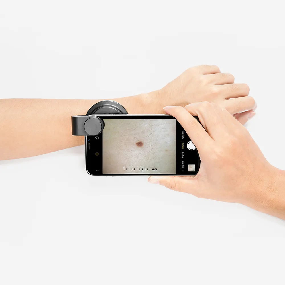
A noninvasive in vivo method called dermoscopy is mainly employed to examine skin lesions [1]. Skin-surface microscopy, incident light microscopy, dermatoscopy, and epiluminescence microscopy are synonyms. A handheld device known as a dermatoscope is used to perform dermoscopy.
Cancer of the Basal CellDermoscopic parameters linked to BCCs include the lack of a pigment network and the presence of certain characteristics such as ulceration, arborizing vessels, enormous ovoid nests of blue-gray, multiple blue-gray globules, leaf-like areas, and spoke wheel areas.
The pigmented lesion must lack pattern symmetry and color uniformity in addition to at least one of the following characteristics in order to be diagnosed as melanoma: numerous brown dots, pseudopods, radial streaming, scar-like depigmentation, peripheral block spots/globules, five to six colors, a blue-white veil,...
According to reports, dermoscopy's sensitivity can vary from 60% to 100%, depending on a number of variables including the examiners' level of experience and the lesions' diagnostic complexity. Dermoscopy can help diagnose melanoma more accurately, but it cannot take the place of a histopathologic examination.
Dermoscopy was shown to be more accurate than visual examination alone in both comparisons, according to the meta-analysis, with RDORs of (a) 4.7 (95% CI 3.0 to 7.5; P
Dermoscope Products
Replacement Diaphragm and Silicone Stethoscope Parts for Adult and Pediatric Stethoscopes, BBTO 2 Sets Accessories for Stethoscope Ear Tips (Gray)
Nurses, men, women, and pediatric infants can use the 22-inch PARAMED Stethoscope, which is a classic single head cardiology device for medical and clinical use.

Classic Dual Head Paramed Stethoscope for Physicians, Nurses, Medical Students, Professionals in Pediatrics, Medicine, Cardiology, and Home Use with an Additional Diaphragm, Four Eartips, Accessory Case, and Name Tag - 29.5 Inch

Stethoscope Holder for Littmann - Genuine Leather Universal Stethoscope Holster with Velcro Closure and Padded Hip/Belt Clip

For nurses, emergency rooms, cardiologists, veterinarians, and fetal pediatrics, the Vive Precision Dualhead Stethoscope is a dual head diaphragm bell that doubles as a chestpiece device for doctors and students.

Black, 27-inch EverOne Professional Style Cardiology Stethoscope
Professional Dual Head Cardiology & Diagnostic Stethoscope for Physicians and Nurses - with Accessories, Kila Scopes Specialist Stethoscope, K751 Black

Physicians and nurses can use the Kila Scopes Specialist Stethoscope, Professional Single Head Cardiology & Diagnostic Stethoscope with Accessories, K870 Orange.
EverOne Superior Cardiology Stethoscope, 27-Inch Black Tube
ZetaLife 2-in-1 Otoscope Set by Zyrev with 50 Additional Disposable Tips
Hot Products


News & Blog


Top Reviews
Tiffany
In exchange for a fantastic promo, I've written this review! The timing couldn't have been better when I was looking for a new stethoscope for my new position! The traditional single-head cardiology stethoscope is incredibly lightweight. You don't have to be concerned about it becoming heavy or falling off, like a double-headed stethoscope sometimes does. The quality of the sound is good. Long-term benefits include the additional bell and ear pieces that were included. Looking forward to using this vintage single-head cardiology stethoscope!
rickyveljaec
My Littman Classic II stethoscope has been surpassed by this one. Compared to my prior stethoscope, which I had been using for the previous nine years, I can hear a lot more. I'm really happy with my purchase and would definitely buy from them again. I adore the vivid color selections.
Techno-Junkie
I purchased this equipment primarily to be able to monitor my bee colony's activity from outside the hives. Without being intrusive, I wanted to be able to assess the condition of my hives by opening each level, putting on my bee costume, and smoking. I can accomplish this with this stethoscope. I had to practice for a while before I could focus on the sound of the bees' beating wings as they went about their job. While the earpieces are fairly sensitive when flipped around, you can wear them and yet not hear anything in one direction. One of the ear piece tubes has an obvious tab big enough to write a brief word on, so I typed "Right," as that's the ear piece that goes to my right ear, not my left. What prevented them from labeling this during assembly? It's a minor gripe that doesn't detract from my evaluation. All in all, an excellent, reasonably priced stethoscope that can even detect heartbeats! I'm quite happy with it and would suggest it to anyone for the price.


