


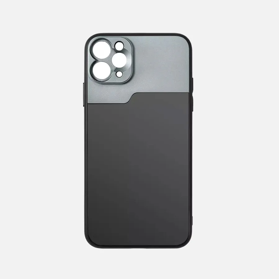
DE-400 Dermatoscope – IBOOLO
Comprehensive Analysis of Dermatoscopio Dermlite: Core Advantages, Proper Usage, And Maintenance
In the realm of modern dermatological assessment, the dermatoscopio DermLite has emerged as a leading tool for healthcare professionals due to its superior optical performance and user-centric design. This article provides a detailed, professional analysis of the DermLite dermatoscope, exploring its key advantages, including patented polarized light technology, modular design, and exceptional portability. We will outline the correct operational procedures, from basic setup to advanced applications, and offer practical maintenance tips to ensure long-term performance. While the dermatoscopio DermLite delivers high-quality imaging for skin observation, it is critical to note that all findings must be interpreted by qualified medical professionals. This article aims to equip users with a thorough understanding of the DermLite dermatoscope’s functionality and proper usage protocols.
What Is The DermLite Dermatoscope?
The dermatoscopio DermLite is a specialized medical device designed to assist in the detailed examination of skin surfaces and subsurface structures. Utilizing advanced optical magnification and specialized lighting, the DermLite dermatoscope enables healthcare professionals to observe intricate skin details with enhanced clarity. It serves as an essential tool in dermatological assessments but is strictly an observational aid, requiring professional medical evaluation for any conclusions.
Compared to traditional visual inspections, the dermatoscopio DermLite offers significantly clearer imaging of skin structures. Models like the DermLite DL3 incorporate polarized light technology to minimize surface glare, revealing subsurface details with precision. Its portable, ergonomic design makes the DermLite dermatoscope ideal for use in various settings, including clinical examinations and health screenings. The DermLite series includes a range of models, from entry-level to advanced, catering to diverse professional needs. These devices typically feature high-quality optical lenses, LED lighting systems, and ergonomic casings, with some models offering digital imaging capabilities for documentation. However, all observations made with the dermatoscopio DermLite must be interpreted in conjunction with comprehensive medical assessments.
How Does The DermLite Dermatoscope Work?
The dermatoscopio DermLite operates through a combination of advanced optical and lighting technologies to provide detailed visual information about the skin. The operational process can be broken down into the following key components:
1. Illumination System
- Equipped with specialized LED light sources for uniform illumination.
- Select models, such as the dermatoscopio DermLite DL4, use cross-polarized technology to reduce surface glare.
- Optional use of coupling agents enhances image clarity for specific observations.
2. Optical Magnification
- High-quality optical lenses provide 10x to 20x magnification.
- Multi-coated lenses ensure superior image sharpness and color accuracy.
3. Image Presentation
- Enables clear visualization of epidermal and superficial dermal structures.
- Distinct tissue types display specific color and morphological characteristics.
To use the DermLite dermatoscope effectively, follow these steps:
1. Clean the area of skin to be examined.
2. Select the appropriate observation mode (dry or wet).
3. Maintain optimal distance and angle for observation.
4. Systematically document findings for professional review.
It is critical to emphasize that results from the dermatoscopio DermLite require interpretation by trained medical professionals, as the device does not provide diagnostic conclusions.
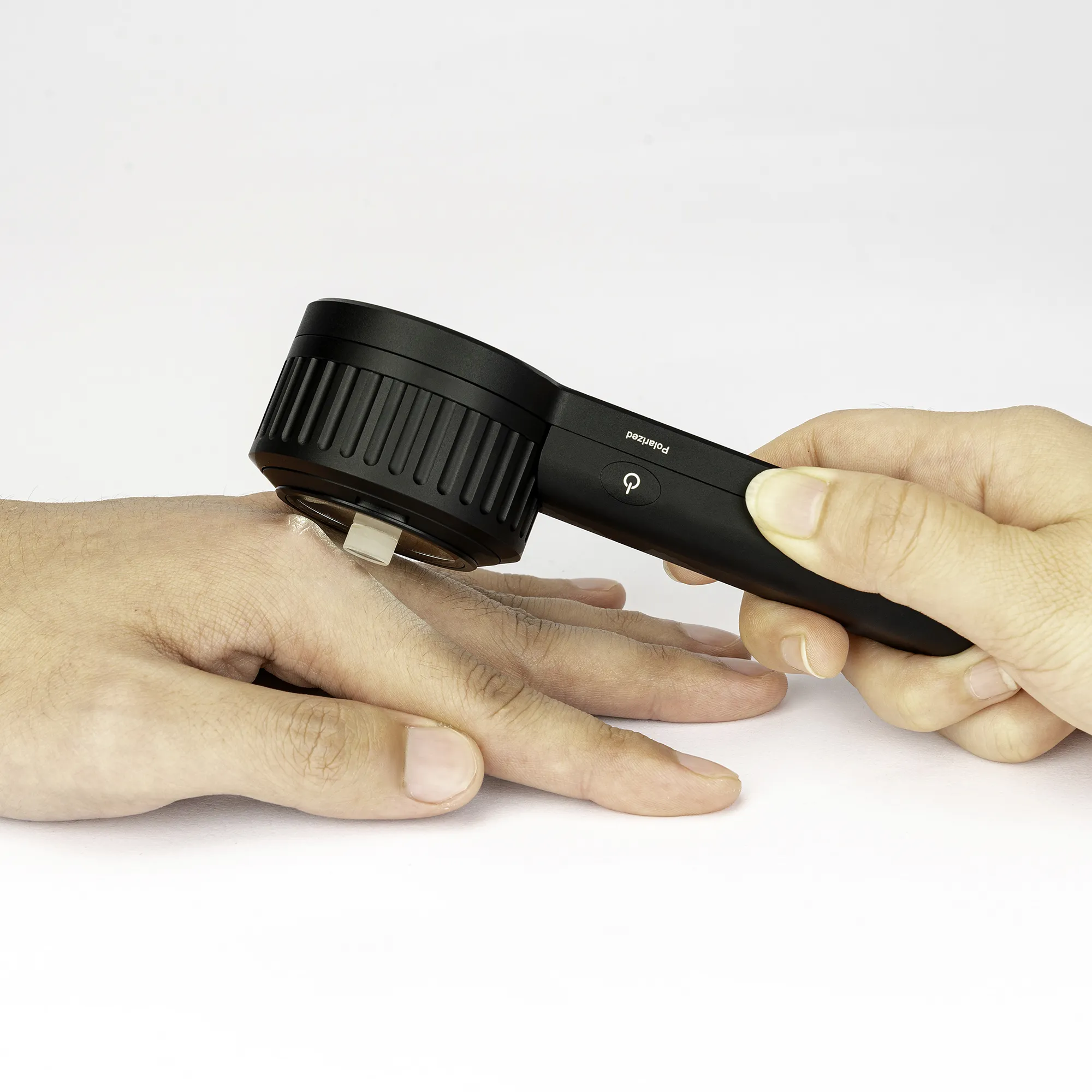
Why Choose The DermLite Dermatoscope? Product Structure And Workflow
The dermatoscopio DermLite is designed with practical usability in mind, incorporating features that enhance its functionality:
- Modular Design: Interchangeable lens components for versatile applications.
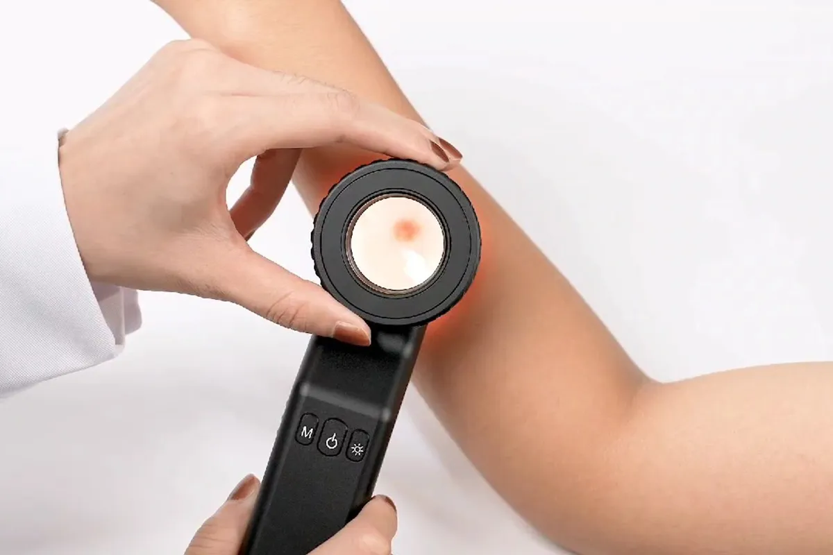
- Ergonomic Grip: Reduces fatigue during extended use.
- Multi-Mode Illumination: Adapts to various observation needs.
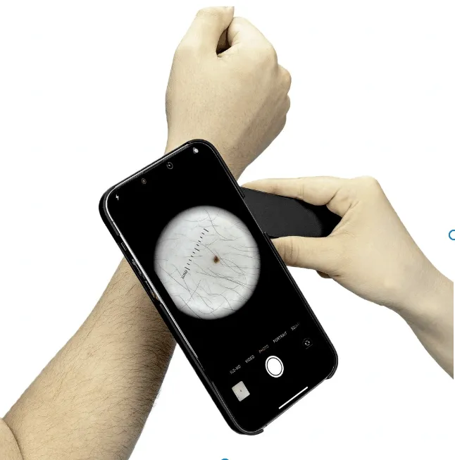
- Portability: Compact enough to fit in a pocket, ideal for mobile professionals.
A typical workflow for the DermLite dermatoscope includes:
1. Preparation: Verify battery charge and clean the observation window.
2. Mode Selection: Adjust settings based on the observation objective.
3. Observation: Maintain stability and observe from multiple angles.
4. Documentation: Use compatible apps or accessories to archive images.
The dermatoscopio DermLite’s technical specifications, such as light intensity and magnification, are rigorously calibrated for consistent performance. However, all observations must be evaluated alongside other clinical data.
Can The DermLite Dermatoscope Aid in Skin Examination? Application Scope
The dermatoscopio DermLite is a valuable tool across various dermatological applications, including:
1. Skin Feature Observation
- Detailed visualization of epidermal textures and pigment distribution.
- Clear imaging of superficial vascular patterns.
2. Health Monitoring Support
- Tracks changes in skin features over time.
- Provides comparable images for follow-up assessments.
3. Professional Assessment Reference
- Offers detailed visual data for medical professionals.
- Enhances standardization in health screening processes.
Limitations to Note:
- The DermLite dermatoscope has limited depth penetration and cannot visualize deep tissue structures.
- Observation outcomes may vary by skin type.
- All findings require comprehensive clinical evaluation by professionals.
Proper use of the dermatoscopio DermLite enhances observation efficiency, but it remains an auxiliary tool requiring professional oversight.
What Makes The DermLite Dermatoscope Stand out? Key Product Advantages
The dermatoscopio DermLite distinguishes itself through its optical excellence, user-friendly design, and expandability:
Optical Performance:
- Patented polarized light technology eliminates surface reflections, enhancing subsurface visibility.
- Models like the DermLite dermatoscope DL4 feature adjustable LED intensity for consistent illumination in diverse lighting conditions.
- Specialized optical systems ensure true color reproduction, minimizing image distortion.
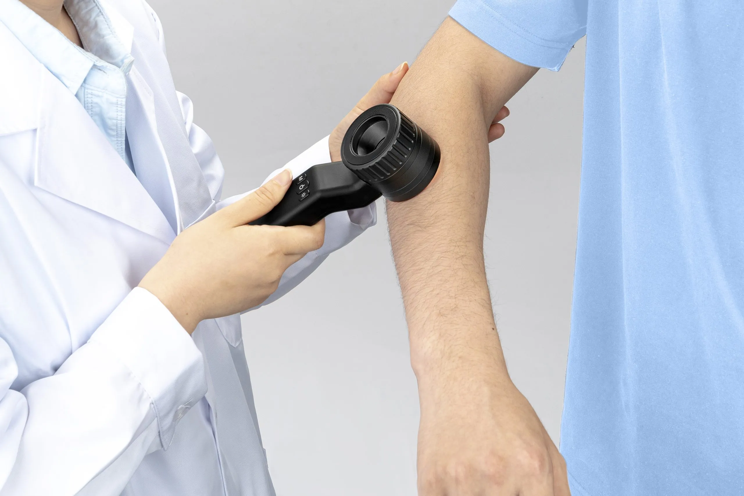
User-Friendly Design:
- Lightweight construction (most models <200g) for easy handling.
- Intuitive single-hand operation with ergonomic buttons.
- Fast-charging technology (1-hour charge for 8 hours of use).
- Anti-slip grip for enhanced operational stability.
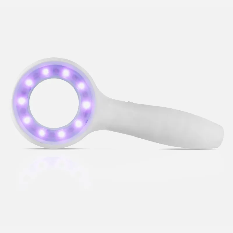
Expandability:
- Interchangeable lenses (10x/20x).
- UV adapters for specialized observations.
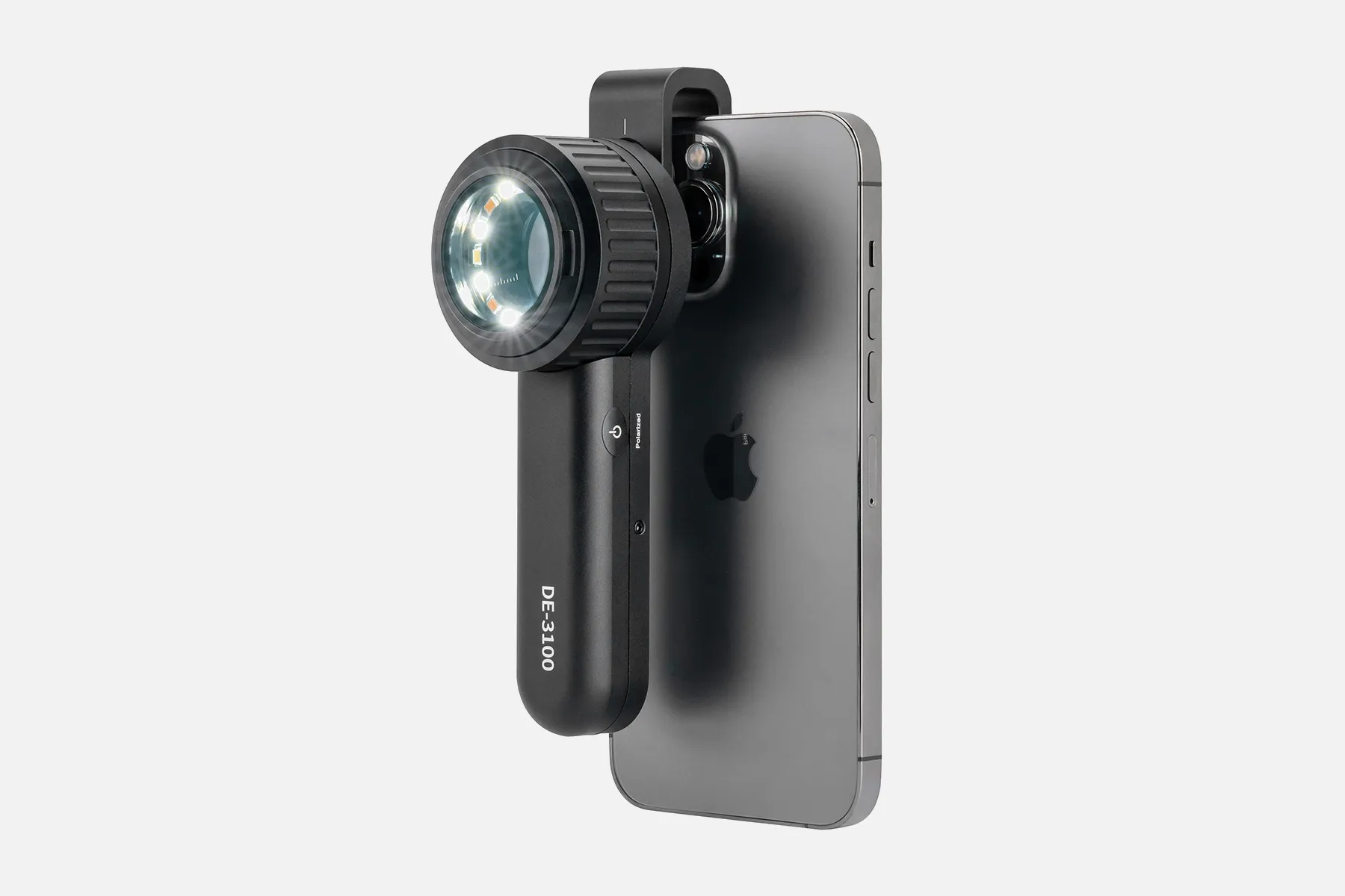
- Smartphone connectivity for seamless image documentation.
- Protective cases for safe transport.
While the dermatoscopio DermLite delivers high-quality imaging, all observations must be interpreted by medical professionals, as the device serves solely as an observational aid.
How to Properly Use The DermLite Dermatoscope? Step-by-Step Guide
Correct usage of the dermatoscopio DermLite is essential for optimal performance. Follow these steps:
Preparation:
1. Device Check: Ensure sufficient battery charge and clean lenses.
2. Environment Setup: Use appropriate lighting, avoiding direct sunlight.
3. Skin Preparation: Clean the observation area to remove oils or cosmetics.
Operational Steps:
1. Mode Selection:
- Dry mode for quick screenings.
- Wet mode with coupling agents for enhanced detail.
- Polarized mode for subsurface structure observation.
2. Observation Techniques:
- Maintain a 1-2 cm distance from the skin.
- Use multiple angles for comprehensive visualization.
- Switch to higher magnification for detailed analysis.
3. Image Documentation:
- Save images via compatible apps.
- Add annotations for clarity.
- Archive images for future reference.
Precautions:
- Clean the device after each use.
- Avoid excessive vibration or drops.
- Schedule regular professional calibration.
- All findings must be reviewed by medical professionals.
Where Can The DermLite Dermatoscope Be Applied? Practical Use Cases
The dermatoscopio DermLite is versatile, supporting various professional scenarios:
Clinical Settings:
- Routine examinations in dermatology clinics.
- Skin health assessments in wellness centers.
- Pre- and post-treatment evaluations in cosmetic dermatology.
Specialized Populations:
- Monitoring skin changes in elderly patients.
- Assessing occupational skin conditions.
- Screening high-risk groups for skin health issues.
Telemedicine Applications:
- Image capture for remote consultations.
- Documentation for home visits by healthcare providers.
- Longitudinal tracking for health management.
Educational And Research Use:
- Demonstrations in medical education.
- Hands-on training for dermatology students.
- Image-based data collection for research.
In all cases, the DermLite dermatoscope serves as an auxiliary tool, with professional medical evaluation required for all observations.
How to Configure And Optimize DermLite Dermatoscope Settings?
Optimizing the dermatoscopio DermLite settings enhances image quality. Key configuration tips include:
1. Light Intensity Adjustment:
- Start at 50% brightness.
- Reduce for lighter skin, increase to 70-80% for darker skin.
- Adjust for mucosal areas as needed.
- Avoid prolonged use at maximum brightness.
2. Magnification Selection:
- Use 10x for general observation, 20x for detailed analysis.
- Begin with low magnification to locate areas of interest, then switch to higher magnification.
3. Polarization Mode:
- Non-polarized mode for surface features.
- Polarized mode for deeper structures.
- Allow 3 seconds for mode transitions.
4. Image Settings:
- Set contrast to 60-70%.
- Maintain default sharpness, adjusting minimally if needed.
- Calibrate white balance before each session.
Note: Settings should be tailored to specific observation needs, ideally under professional guidance. The DermLite dermatoscope provides reference images only, not diagnostic conclusions.
How to Maintain And Care for Your DermLite Dermatoscope?
Proper maintenance ensures the longevity of the dermatoscopio DermLite:
Daily Cleaning:
- Wipe the device with a dedicated cleaning cloth after use.
- Clean lenses with specialized lens paper.
- Disinfect the body with recommended agents.
- Remove dust from interfaces regularly.
Periodic Maintenance:
- Check light intensity monthly.
- Calibrate white balance quarterly.
- Schedule professional inspections biannually.
- Conduct comprehensive maintenance annually.
Storage Requirements:
- Store at 5-40°C with <80% humidity.
- Use a protective case to prevent damage.
- Avoid direct sunlight exposure.
Troubleshooting:
- Blurry lenses: Use approved cleaning solutions.
- Power issues: Inspect charging ports.
- Connectivity problems: Restart the device.
- Image abnormalities: Contact authorized service providers.
Tip: Maintain a maintenance log to track care activities. Professional servicing by authorized providers ensures consistent performance. The dermatoscopio DermLite remains an auxiliary tool, with all findings requiring medical evaluation.
The dermatoscopio DermLite stands out as a premier tool in dermatological observation, offering unmatched optical clarity, ergonomic design, and versatile functionality. Its patented polarized light technology, modular components, and portability make it a valuable asset for healthcare professionals. However, the DermLite dermatoscope is strictly an observational aid, and all findings must be interpreted by qualified medical personnel. By adhering to proper usage protocols and maintenance practices, users can maximize the dermatoscopio DermLite’s performance, ensuring reliable support in skin health assessments.
How Does Dermatoscopio Dermlite?
The DermLite DL100 from 3Gen is an advanced digital dermatoscope that utilizes innovative optics and software for skin evaluation. It features a no-oil, non-contact platform with 10x magnification and high-intensity LED lighting with cross-polarizers.
This dermatoscope enables detailed visualization of subsurface skin structures without requiring direct contact or immersion fluid. The specialized optical system produces color-corrected images to reveal information about skin lesions' color, size, shape, and morphology.
Embedded image processing software provides additional functionality like side-by-side comparisons for tracking changes over time. Dermatoscopy can be integrated with smartphones or PCs for image capture, storage, and analysis.
The 3Gen DermLite DL100 is a versatile, non-contact dermatoscope leveraging advanced optics and digital technology. It provides dermatologists with an enhanced platform for evaluating pigmented and non-pigmented skin abnormalities. Detailed dermoscopic images facilitate remote consultations and assist in the early detection of skin cancers.
People May Ask
Identify Your Skin SpotsSkin cancer comes in three different forms: melanoma, basal cell carcinoma, and squamous cell carcinoma. The ability to recognize every kind of skin cancer is a skill taught to dermatologists. Make an appointment with the dermatologist over the phone if you find anything strange during your self-examination.
The diameter of a scabies mite is 0.2–0.5 mm. It burrows a track in the stratum corneum of the epidermis to deposit its eggs. With a dermatoscope that has an x 10 magnification, both mites and burrows are plainly visible.
Compared to not using dermoscopy, the diagnosis accuracy for melanoma was significantly greater (log odds ratio 4.0 [95% CI 3.0 to 5.1] versus 2.7 [1.9 to 3.4]; an improvement of 49%, p = 0.001). The level of experience of the examiners had a substantial impact on the accuracy of the dermoscopy diagnosis.
Do dermatoscopes have accuracy? A 2018 Cochrane study found that when used by a qualified practitioner, dermatoscopes are more accurate than the human eye alone in the diagnosis of melanomas. This is important since it can save someone time and possibly avoid the needless operation.
sanitization and cleaning...
Isopropyl alcohol (70% vol.) can be used to wipe out and sanitize the DermLite body. In the optical sections of the device, avoid using alcohol or disinfectants.
Similar to a magnifying glass, a dermatoscope is a portable device. It has the ability to magnify objects up to ten times. Your physician applies a gel or oil to your skin. After that, they place the dermatoscope against your skin to take a close look at the affected area.
With a dermatoscope, features can be evaluated down to the reticular dermis, and pictures can be taken for further analysis. Transillumination of a lesion for high magnification examination and visualization of minor details is the fundamental idea behind dermoscopy.
Handheld dermatoscope including changeable polarization, dermoscopic white and UV, torch LED, and 10x magnification. desktop charging base with auxiliary USB output and IceCap storage. 2 meter USB-C to USB-USB cable.
0:00 > 3:02It always appears polarized. In the middle. The pigment boost works similarly; all you have to do is tap that more
DermLite and Prompt Identification of Skin CancerWith the use of the DermLite product range, an expert eye can identify skin cancer and other skin diseases at an early stage. Each DermLite has an LED light source, a magnification lens, and the majority of them feature polarizing filters to reduce glare.
Dermatoscopio Dermlite Products
Featuring WF10x and WF20x eyepieces, 20X/40X/80X magnification, 2X and 4X objectives, upper and lower LED lighting, a reversible black/white stage plate, a pillar stand, and 120V or battery power, the AmScope SE306R-PZ-LED forward-mounted binocular stereo
A complete science kit that includes 10 microscope slide specimens, 5 blank slides, a 16-page user guide, and a phone adapter is the perfect choice for students studying science with a 40x–1000x microscope for adults.

GUVOP 50X-1000X Magnification WiFi Portable Handheld USB Microscopes with 8 LED & Stand: Wireless Digital Microscope Camera Compatible with iPhone, Android, Electronic Microscope for Kids and Adults - Blue
BEBANG Portable Microscope for Children Students Adult Microbiology Observation Laboratory Learning, Educational Set, 60x–120x Handheld Mini Microscope with LED Light and 5 Slide Samples
20x/50x/100x Magnification, 17mm Field of View, Pen Light Included in AmScope H2510 Handheld Stand Measuring Microscope
Arsir 7.3*4.4 Large Folding Lighted Magnifier with 48 LED Lights (3 Modes), Rectangular Handhold, Full-Page 5X Magnifying Glass for Reading Black Magnifying Lens Gifts for Seniors Who Read Books and Prints
Radio Frequency Face Machine for Residential Use High Frequency Face Massager with EMS, Light Therapy for Wrinkles Lifting and Anti-Aging Skin Tightening and Rejuvenation
Skin Tightening, Wrinkle Reduction, Hair Care: YourMate PhotoTherapy Device: High Frequency Facial Wand Machine with Argon Tubes for Face, Neck, and Hair

For kids and adults, a 3.9mm 720PHD WiFi ear scope with 6 LED lights that is compatible with both Android and iPhone is available with a wireless otoscope ear camera with dual view.

USSERA Medicube Age-R (Device alone)
Hot Products


News & Blog


Top Reviews
Lane Nelson
Works flawlessly on both Android and iPhone. Excellent for keeping an eye on your child's ears to determine whether the problem is ongoing or just transient. Helped me avoid a third round of antibiotics and surgical procedures recommended by a pushy specialist, after I showed a series of photos to his pediatrician, that showed the ears clear themselves. My sister used it to identify a tick that looked like a tiny black dot on a skin.
Barbara Sanchez
This ottoscope has performed admirably. Simple to operate and link to a phone. You can record videos and take photos, and you can store those as well. It also has a good battery life. I've only charged it once in the roughly five months I've had it.
Cindy M.
You "see" what this small lightweight endoscope picks up on its camera on your cell phone via an app. You don't actually look in the scope like your doctor does when examining you. This means you can easily look in your own ears and see a large image on your phone which is an added benefit. This unit came with a small card with two bar codes, one for downloading the app on your phone and a second to turn off ads on the app. Once the app is installed on your phone, you push the button to turn on the endoscope and find it via phone wifi settings to connect to it. Once you tell the app what kind of device it is, you are good to go. Clean a cone with alcohol, put it on the scope, and look in your ear (or wherever you need to look). You can capture photos with the touch of a button that you could easily send to your doctor if needed. The scope came fully charges, has a charger cord, little tools for use in your ear, and four starter alcohol swabs for cleaning before and after use. For the price, this will be a val


