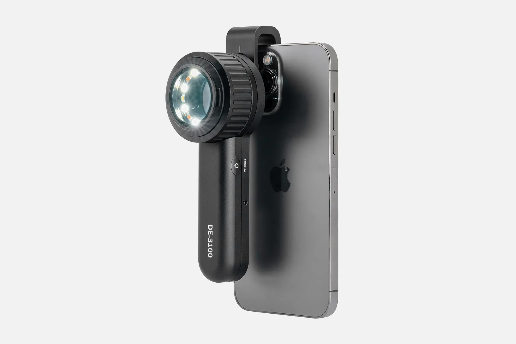
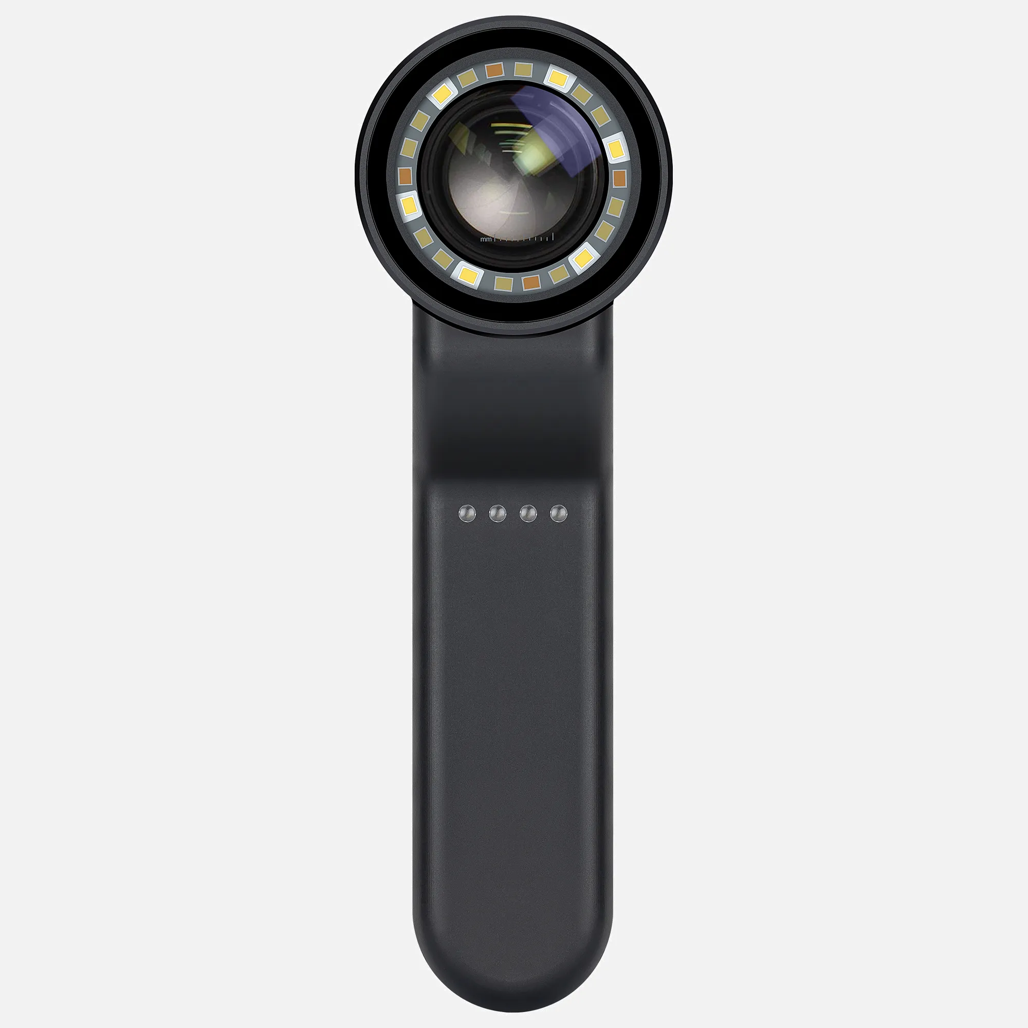
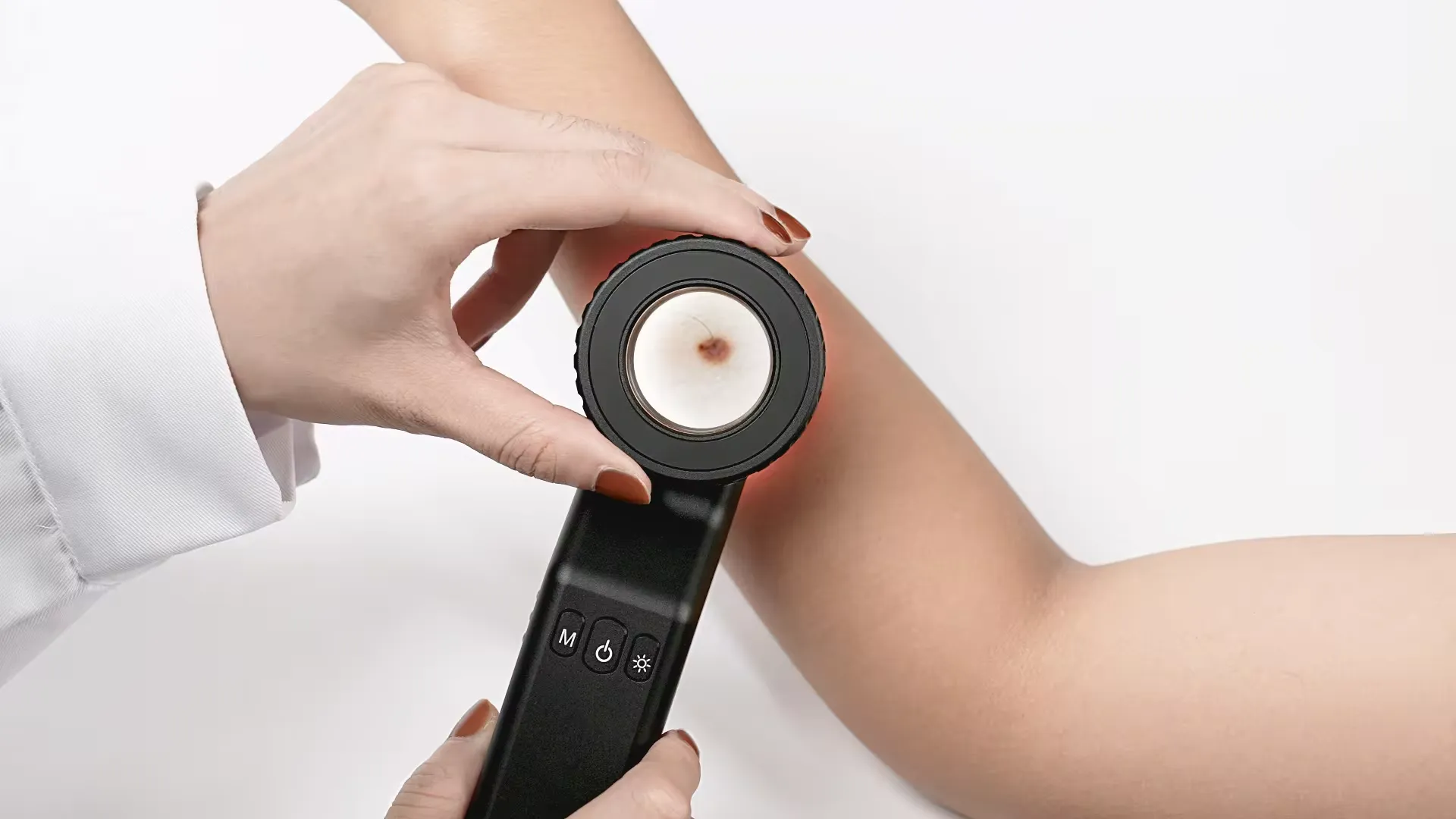
Professional Handyscope Suppliers China Manufacturers - IBOOLO

People May Ask

What Is The Standard Golf Performance Expected of A 75-Year-Old Male Golfer?

Golf Performance Standards Based on Age
Age Categories Median Golf Scores for Males
50s Range 96 to 101
60s Range 98 to 103
70s Range 100 to 105
80s Range 102 to 107
Additional Data Points•

Is It Possible for A Lazy Eye to Correct Itself Naturally?

No, amblyopia does not resolve spontaneously, and children are not able to naturally outgrow it. In the absence of treatment, amblyopia can potentially lead to long-term vision complications, including the potential loss of sight in the affected eye.

Is There Still A Chance to Correct My Strabismus?

Technically, there is no specific age restriction for amblyopia treatment. However, Dr. Borriello emphasizes that the optimal time for its management is during childhood.

Am I Considered to Have A Disability Solely Because I Wear Glasses?

Does the act of wearing glasses constitute a disability? The wearing of glasses, regardless of its corrective power, is not classified as a disability. Legally, visual impairment is evaluated based on the concept of "best corrected vision," which refers to an individual's optimal visual clarity achieved through the use of corrective lenses.

Methods for Determining Handicap?

Exploring the Mechanics of a Golf Handicap? Here's a Simplified Approach to Calculate Yours.
To determine your golf handicap, consider averaging the top eight scores from your last 20 rounds.
Employ this formula to assess your handicap for various courses: Course Handicap = (Handicap Index multiplied by (SR divided by 113)) plus (CR minus Par).
Published on

Should We Refer to Them As Individuals with Disabilities Or Those with Handicaps?

In essence, it is crucial to prioritize the individual before their disability. Those with disabilities are, ultimately, human beings at heart. Assigning a label that solely identifies a person with a specific condition can be deemed disrespectful and reductive of their humanity.

Is It Appropriate to Refer to Someone As Handicapped Or Disabled?

2. Preferred and Undesirable Vocabulary
Avoid Employ
Terms like (the) handicapped, (the) disabled The phrase 'disabled (people)'
Expressions such as afflicted by, suffers from, victim of Prefer 'has [specific condition or disability]'
Phrases like confined to a wheelchair, wheelchair-bound Instead, use 'wheelchair user'
Additional 10 examples•

Can We Consider Lazy Eye As A Form of Impairment?

Examples of visual impairments encompass, yet are not comprehensive to: total loss of sight, impaired vision, abnormalities in ocular movement or motility, difficulty in utilizing both eyes synergistically, a misalignment in the alignment of the eyes known as strabismus, a condition where one eye fails to develop properly known as amblyopia, issues with focusing such as accommodative disorders, as well as visual sensory...

What Is The Significance of The Term 'Handicap' When Referring to A Person?

The Britannica Dictionary's interpretation of HANDICAP is as follows: [quantifiable] 1. occasionally deemed insensitive: a bodily or cognitive state that can potentially constrain an individual's capabilities, encompassing both physical and mental impairments.

What Approach Is Most Effective in Defining A Handicap?

1. A hindrance that significantly complicates the attainment of goals and achievements. 2. Occasionally deemed offensive: a limitation in one's physical abilities.

Handyscope Products

The AmScope SE400-Z is a professional binocular stereo microscope with a boom arm mount, LED illumination, 10X and 20X magnification, 1X objective lens, and 110V–120V power supply.

TOMLOV Coin Microscope 1000, Model DM4

Amoper 7-inch 1600X Digital Microscope with Silicone Repair Pad, 28 Light Coin Microscope, 12-MP Sensor Soldering Electronic Repair Microscope with IPS Touch Control Panel, 32GB—

Handheld, compact, mini, recorder, camera with six LED lights, Bee-Bot Flexi-Scope Digital USB Kids Microscope 10X-200X

dimer-glass calcite

An essential tool for inspectors, surveyors, engineers, and architects is the AdirPro Pocket Stereoscope, a lightweight wearable microscope with two lenses that change in distance from 55 to 75 mm.

Eisco Labs Electroscope Demonstration

攰字听诊器 - 直接传输到蓝牙耳机 - 无系绳听诊自由 - 放大听诊器 - 电子听诊器

With eight adjustable LED lights, this 4.3-inch handheld USB microscope is perfect for adults PCB soldering and children using it outside. It has a 50X–1000X magnification coin microscope video camera.

DM9 7 LCD Digital Microscope 1200X, 1080P Coin Microscope Magnifier, 12MP Adult Ultra-Precise Focusing Soldering Microscope, PC View, 32GB

Top Reviews

Kirt Keltner

It is intended for the purpose for which I purchased it—close-up visual examination of minute flaws that are invisible to the unaided eye. In the deburr section, everyone is now requesting to utilize it, and I do allow them to do so if it improves their work.

Brandon Friedrich

About nine months ago, I made this transaction. After setting it up, I hadn't used it, and the Keypad was gone. After I wrote an email to their company's assistance, I received a brand-new, fully functional unit. To return it, all I need is the shipping label that they will provide. Excellent piece of gear. I solder solderless semiconductors onto PC boards with it. The company offers excellent customer support. Kevin Rea Lancaster, CA, United States

Pat C

For my nine-year-old granddaughter, I purchased this. It is quite simple to use. I adore it as much as she does. We are examining a grain of salt as well as insects, stones, and flowers. It's an excellent present. I heartily endorse it.

0

Have questions on gear or your order?

Our Gear Guides are here to help! Get personal advice from pro creatives

Name
Subject
Email address
How can we help?

Instant Answers

How Can Dermoscopy Images Be Captured?

Dermoscopy images can be captured and stored in different ways, such as: • Using a smartphone or tablet with dermoscopic adapter, which consisted in the package.• Using a digital camera

Dermoscopy images can be captured and stored in different ways, such as:

• Using a smartphone or tablet with dermoscopic adapter, which consisted in the package.
• Using a digital camera with dermoscopic adapter, there’s 49mm screw size camera adapter available to order now.

Compatible phone/tablet models:
All iPhone models, 95% Android phones, 90% tablet. For phone/tablet size in 5.25-14mm

Compatible camera models:
All camera with built 49mm filter screw, such as Canon EOS 70D, 80D, 90D; Canon EOS R7, R10, R50, R100; Canon M100, M200, M50, Mark II; Canon G7X Mark III, Sony ZV-1

How Can I Connect My Phone to My Dermatoscope?

There’s universal phone adapter for all our dermoscopes. Please check the installation procedure bellow or watch operation guide. Smartphone Connector (1) Place phone adapter screw in the center of smartphone’s

There’s universal phone adapter for all our dermoscopes. Please check the installation procedure bellow or watch operation guide.

Smartphone Connector

(1) Place phone adapter screw in the center of smartphone’s main camera.
(2) Screw magnet attachment on phone adapter.
(3) Put dermoscope’s back ring and magnet attachment together

Take The Best Images

You need to adjust the focus ring after the dermoscpe connected on smartphone to get the best images.

How Can I Clean My Dermoscopy after Usage?

Cleaning your dermoscopy after usage is important to prevent cross-contamination and infection. The cleaning method may vary depending on the type and model of your dermoscopy, so you should always

Cleaning your dermoscopy after usage is important to prevent cross-contamination and infection. The cleaning method may vary depending on the type and model of your dermoscopy, so you should always follow the manufacturer’s instructions. However, some general steps are:

• Turn off and disconnect your dermoscopy from any power source or device.

• Wipe off any visible dirt or debris from the dermoscopy with a soft cloth or tissue.

• Disinfect the dermoscopy with an alcohol-based wipe or spray, or a disinfectant solution recommended by the manufacturer. Make sure to cover all surfaces, especially the lens and contact plate.

• Let the dermoscopy air dry completely before storing it in a clean and dry place.

• Do not use abrasive or corrosive cleaners, solvents, or detergents that may damage the dermoscopy.

• Do not immerse the dermoscopy in water or any liquid, unless it is waterproof and designed for immersion.

You should clean your dermoscopy after each use, or at least once a day if you use it frequently. You should also check your dermoscopy regularly for any signs of damage or malfunction, and contact the manufacturer or service provider if needed.

Polarized VS Non-Polarized Dermoscopy

A dermoscopy is a device that allows the examination of skin lesions with magnificationand illumination. By revealing subsurface structures and patterns that are not visible tothe naked eye. It can

A dermoscopy is a device that allows the examination of skin lesions with magnificationand illumination. By revealing subsurface structures and patterns that are not visible tothe naked eye. It can improve the diagnose accuracy of skin lesions, such as melanoma,basal cell carcinoma, seborrheic keratosis, etc.

There are two main types of dermoscopy: Non polarized and polarized dermoscopy.We’ve fitted most of our dermoscopys with polarized and non-polarized light. They canbe used in multiple skin structures.

Non-polarized contact Mode

In non-polarized mode, the instrument can provide information about the superficialskin structures, such as milia-like cysts, comedo-like openings, and pigment in theepidemis.

The dermoscopy requires applying a liquid such as mineral oil or alcohol to the skin andplacing the lens in contact with the skin. This reduces surface reflection and enhancesthe view of subsurface structures.

Image with non-polarized light (DE-3100)

Polarized contact Mode

In polarized mode, the instrument allows for visualization for deeper skin structures,such as blood vessels, collagen, and pigment in the dermis.

The dermoscopy does not need to be in contact with the skin or use any liquid. Theirpolarized light can help to eliminate surface reflection and allow visualization ofvascular structures.

Image with polarized light (DE-3100)

Polarized non-contact Mode

The dermoscopy can also use polarized light to examine the skin without direct contact.

In polarized non-contact mode, the instrument allows for examination infected areasand lesions that are painful for the patient, or the difficult to contact pigmented lesions,such as nails and narrow areas.

The contact plate should be removed in this mode, and it does not require applying aliquid to the skin. As it doesn’t require pressure or fluid application on the skin, it canalso avoid cross-contamination and infection risk.

Image in polarized non-contact mode (DE-3100)

How Effectiveness Is Dermoscopy

Compared with visual inspection, the dermoscopy can be used to capture and store skin lesion photos, which play an important role in early skin cancer examination. The dermoscopy allows the

Compared with visual inspection, the dermoscopy can be used to capture and store skin lesion photos, which play an important role in early skin cancer examination.

The dermoscopy allows the examination of skin lesions with magnification and illumination. This can be greatly avoiding the factors that cause interference to visual detection. Such as lighting, skin color, hair and cosmetics.

Several studies have demonstrated that dermoscopy is useful in the identification of melanoma, when used by a trained professional.

It may improve the accuracy of clinical diagnosis by up to 35%
It may reduce the number of harmless lesions that are removed
In primary care, it may increase the referral of more worrisome lesions and reduce the referral of more trivial ones

A 2018 Cochrane meta-analysis published the accuracy of dermoscopy in the detection.

Table 1. Accuracy of dermoscopy in the detection of melanoma in adults
Detection Method Sensitivity, % Specificity, % Positive Likelihood Ratio NegativeLikelihood Ratio
Visual inspection alone (in person) 76 75 3.04 0.32
Dermoscopy with visual inspection (in person) 92 95 18 0.08
Image-based visual inspection alone (not in person) 47 42 0.81 1.3
Dermoscopy with image-based visual inspection (not in person) 81 82 4.5 0.23
ROC—receiver operating characteristic. *Estimated sensitivity calculated on the summary ROC curve at a fixed specificity of 80%.

As we can see, the dermoscope can improve the accuracy of diagnosis of skin lesions, especially melanoma.

Table 1. Accuracy of dermoscopy in the detection of melanoma in adults
Detection Method Sensitivity, % Specificity, % Positive Likelihood Ratio NegativeLikelihood Ratio
Visual inspection alone (in person) 79 77 3.4 0.27
Dermoscopy with visual inspection (in person) 93 99 93 0.07
Image-based visual inspection alone (not in person) 85 87 6.5 0.17
Dermoscopy with image-based visual inspection (not in person) 93 96 23 0.07
ROC—receiver operating characteristic. *Estimated sensitivity calculated on the summary ROC curve at a fixed specificity of 80%.

Characteristics of the dermatoscopic structure of the skin lesions include:

• Symmetry or asymmetry
• Homogeny/uniformity (sameness) or heterogeny (structural differences across the lesion)
• Distribution of pigment: brown lines, dots, clods and structureless areas
• Skin surface keratin: small white cysts, crypts, fissures
• Vascular morphology and pattern: regular or irregular
• Border of the lesion: fading, sharply cut off or radial streaks
• Presence of ulceration

There are specific dermoscopic patterns that aid in the diagnosis of the following pigmented skin lesions:

• Melanoma
• Moles (benign melanocytic naevus)
• Freckles (lentigos)
• Atypical naevi
• Blue naevi
• Seborrhoeic keratosis
• Pigmented basal cell carcinoma
• Haemangioma

No products in the cart.

No products in the cart.

Hot Search Terms